Plasmonic imaging and detection of single DNA molecules

a plasmonic imaging and single molecule technology, applied in the field of single molecule detection, can solve the problems of difficult to quantify the image intensity, study single molecules over a long time, and limited image contrast of optically transparent samples.

Active Publication Date: 2019-09-10
ARIZONA STATE UNIVERSITY
View PDF4 Cites 6 Cited by
  • Summary
  • Abstract
  • Description
  • Claims
  • Application Information

AI Technical Summary

Benefits of technology

[0005]In one embodiment, the present invention is a method of label-free imaging of single biomolecule, macromolecule, or other molecules with a differential surface plasmon resonance technique (DSPR). The plasmons may be created optically at the surface of a metal film, and the prorogating plasmonic waves are scattered by the sample molecules on the surface, creating a plasmonic image with contrast many orders of magnitude greater than the conventional bright field optical microscopy image. Noises and unwanted interference patterns from the optical system may be removed with the differential method, which further enhances the image contrast and make it possible to image and detect single molecules such as DNA or proteins.

Problems solved by technology

Contrast of imaging of optically transparent samples is limited when such imaging is carried out with traditional optical microscopy.
In comparison with imaging utilizing traditional optical microscopy, fluorescence imaging requires additional sample-preparation steps, which may distort the natural properties of the molecules of the object being imaged.
Fluorescence imaging is also subject to blinking and photobleaching, which shortcomings make it difficult to quantify the image intensity and study single molecules over a long time.
In addition, due to relatively weak fluorescence emission, the speed of the fluorescence imaging procedure is relatively slow, which is not suitable for imaging of fast biological processes.
The atomic force microscopy (AFM) is a powerful label-free technique for imaging single DNA molecules, but it is operationally slow (taking seconds to minutes to acquire a single image).
Additionally, the scanning of the AFM probe may perturb the DNA samples.
Despite of the advances, it remains a difficult challenge to image single molecules, such as DNA, proteins or any other biomolecules or macromolecules with surface plasmon resonance microscopy.
A primary reason is the background noise associated with the surface plasmon resonance microscope, including interference patterns arising from the coherence of light, dirt on and imperfection of the optical components, including objective and light sources, and non-uniform distribution of the light illumination.

Method used


Image

  • Plasmonic imaging and detection of single DNA molecules
  • Plasmonic imaging and detection of single DNA molecules
  • Plasmonic imaging and detection of single DNA molecules

Examples


Embodiment Construction

[0050]Materials used in experiments included λ-DNA (cIind 1 ts857 Sam 7) (48,502 bp), TE buffer (1×, pH 8.0), and Sma I digestion enzyme from Invitrogen (Carlsbad, Calif.); YOYO-1 dye (1 mM in DMSO) from Molecular Probes (Eugene, Oreg.). Thiol-PEG-Amine (HS-PEG-NH2, MW 1000) from Nanocs (Boston, Mass.), and 2-mercaptoethanol from Gibco (Grand Island, N.Y.).

[0051]Surface Modification.

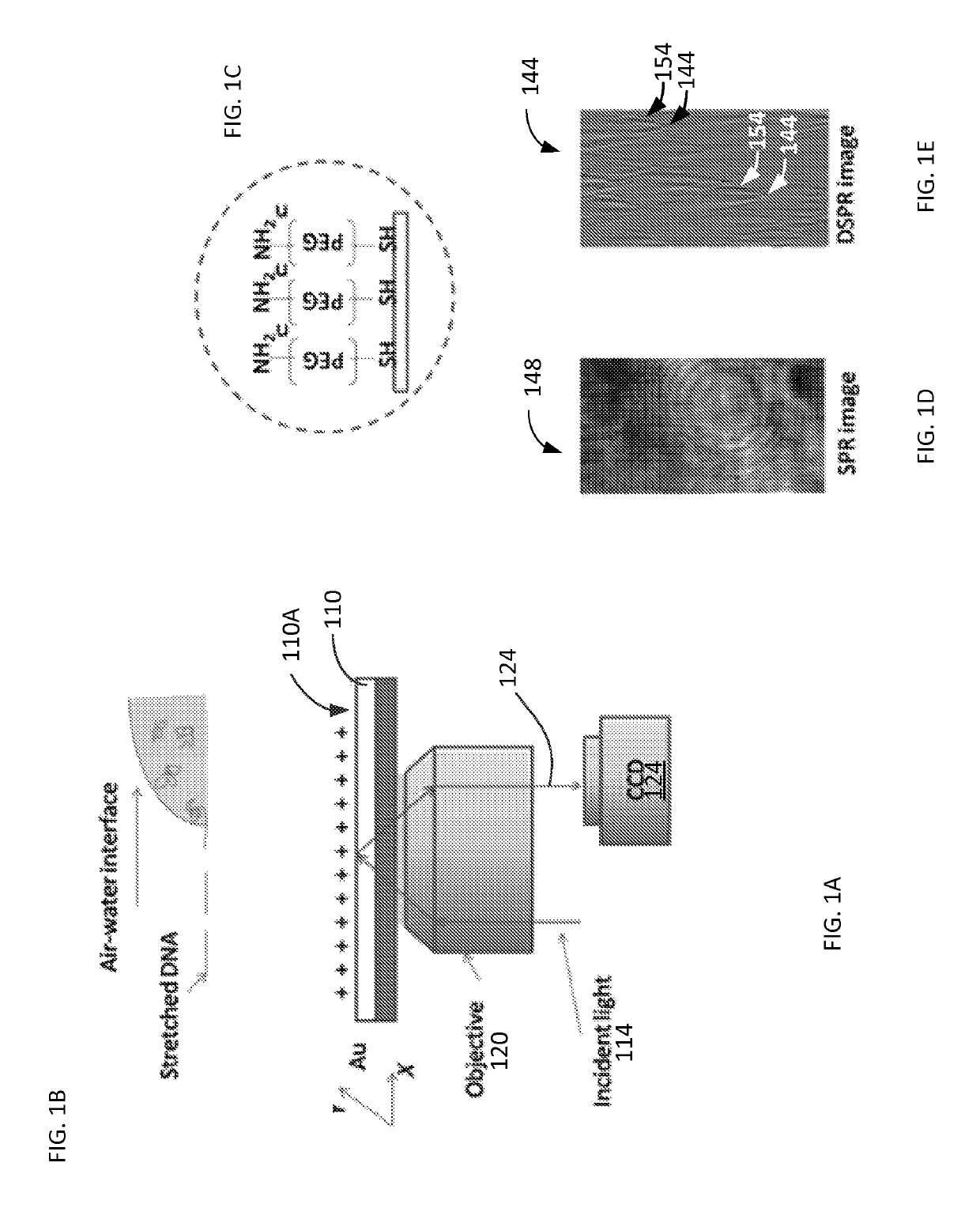
[0052]The SPR substrate 110 included BK7 glass cover slips (from VWR, www.vwr.com) coated with 2 nm thick layer of chromium and then with a 47 nm thick layer of gold. The gold surface 110A (of FIG. 1A) was covered with a SH-PEG-NH2 self-assembled monolayer. Each resulting substrate chip was rinsed with de-ionized water and ethanol, and then blown dry with nitrogen gas. The chip was then further cleaned with hydrogen flame and immediately submerged in 0.5 mM HS-PEG-NH2 water / ethanol (1:1) solution. After left in the solution for 24 h in the dark, the chip was taken out of the solution and rinsed with de-i...


PUM

PropertyMeasurementUnit
pHaaaaaaaaaa
Full-Widths Half-Maximumaaaaaaaaaa
thickaaaaaaaaaa

Abstract

Method and system to remove background noise with a differential approach in optical imaging is disclosed. The differential approach moves the sample position laterally over a small distance, and a differential image is generated from the images recorded before and after the lateral translation. This approach can significantly improve the image quality of objects, including single DNA molecules, for label-free optical imaging techniques, such as surface plasmon resonance imaging. Disclosed imaging technique provides high-resolution genome-wide restriction maps of single DNA molecules.

Description

CROSS-REFERENCE TO RELATED APPLICATIONS[0001]This patent application represents the national stage entry of PCT International Application No. PCT / US2015 / 010018 filed on Jan. 2, 2015 and claims priority from and benefit of the U.S. Provisional Patent Application No. 61 / 923,477 filed on Jan. 3, 2014 and titled “Plasmonic Imaging and Detection of Single DNA Molecules.” The disclosure of the above-identified patent document is incorporated herein by reference.BACKGROUND[0002]Optical microscopy has become an indispensable tool in research, clinical and industry labs. Contrast of imaging of optically transparent samples is limited when such imaging is carried out with traditional optical microscopy. To enhance imaging contrast, various strategies including fluorescence labeling have been developed. In comparison with imaging utilizing traditional optical microscopy, fluorescence imaging requires additional sample-preparation steps, which may distort the natural properties of the molecules...

Claims


Application Information

Patent Timeline
Patent Type & Authority Patents(United States)
IPC IPC(8): G01N21/552C12M1/34
CPCG01N21/553G01N21/552C12M41/46
Inventor TAO, NONGJIANYU, HUISHAN, XIAONANWANG, SHAOPENG
Owner ARIZONA STATE UNIVERSITY
Why Eureka
Try Eureka