System and method for rapid examination of vasculature and particulate flow using laser speckle contrast imaging

a contrast imaging and vasculature technology, applied in the field of vascular imaging technologies and methods, can solve the problems of increasing vision loss and progression toward blindness, difficult for surgeons to use preoperatively obtained images of the brain for anatomical landmarks, and no single method is considered adequate in all scenarios, so as to relieve carpel tunnel syndrome, improve the efficiency and effectiveness of cerebrovascular imaging, and reduce the risk of damage to the vascular anatomy.

Active Publication Date: 2021-06-01
VASOPTIC MEDICAL
View PDF5 Cites 2 Cited by
  • Summary
  • Abstract
  • Description
  • Claims
  • Application Information

AI Technical Summary

Benefits of technology

[0023]For high-resolution LSCI, speckle contrast is calculated using a temporal algorithm that requires the processing of a time-stack of several (typically N=80, but may be different) images. This reduces the temporal resolution of any functional information extracted from the data, and also the prolonged image acquisition time makes the system and method susceptible to motion artifact. Even if images were acquired at 150 frames per second (fps), generating an LSCI image from the 80 frames would require over 0.5 s. To avoid the slow output rate, speckle contrast can be calculated over a rolling time-stack of images—that is by including each new image frame to the processing image stack and removing the oldest image from the stack. This first in first out (FIFO) strategy coupled with fast implementation of speckle contrast calculations allows the output rate to be as high as the camera frame rate but the system would still suffer from latency (i.e., the output would trail the real-time event by a latency of at least the amount of time that is required to acquire the N=80 image frames). If the acquisition speed is 150 fps, the latency corresponding to the acquisition of 80 frames, would be 0.533 seconds.
[0035]In preparation for cerebrovascular surgery, the surgical team establishes a surgical plan, typically based on MRI and CT images of the patient's brain. The surgical plan involves a strategy for navigating to the specific surgical site in a way that minimizes risk of damage to the vascular anatomy. The surgical procedure often requires the use of an operating microscope to observe the surgical field and typically involves observation of vessel patency and vessel-specific physiological information in the surgical field. In an aneurysm surgery, for example, blood flow in perforator vessels associated with the aneurysm and its parent vessels are of great importance for clinical outcomes and, hence, an important monitoring target. Other types of neurosurgeries for which observation of vessel patency and vessel-specific anatomical and physiological information in the surgical field is important include vascular grafting surgeries in patients suffering from Moyamoya disease, arteriovenous malformation surgeries, brain tumor resection surgeries, awake craniotomies where functional mapping is desired, spinal cord surgeries, and surgeries performed to relieve carpel tunnel syndrome. The subject invention can be embodied to provide the surgeon with real-time or near-real-time assessment of vessel patency and vessel-specific anatomical and physiological information to improve the efficiency and effectiveness of cerebrovascular and other neurosurgical procedures.Plastic and Reconstructive Surgery:
[0039]Endoscopic surgeries typically involve manipulation or resection of vessels, tissue, and organs. As with other surgical procedures, the ability to identify the vessel anatomy and physiology in the surgical site is critical for navigation to prevent unintended injury or rupture of the vessels and to identify areas of disease. For example, mesenteric ischemia (where blood flow to the gastrointestinal system is decreased due to blood vessel blockage), intraoperative, real-time or near-real-time monitoring of blood flow in the intestinal tissue can facilitate identification of specific regions of decreased flow to improve, for example, the efficiency of plaque removal procedures or to minimize the portion of damaged tissue that must be removed. Likewise, the ability to evaluate the patency and viability of vessels and organs following a resection is critical to the patient's outcome. In some cases these procedures are performed manually, semi-automatically, or automatically using a computer-aided surgical system. The subject invention can be embodied to provide real-time or near-real-time visualization of anatomical and physiological information during an endoscopic surgical procedure through the integration of LSCI with endoscopic instruments, with or without computer-aided surgical equipment.
[0044]In various embodiments, the system and method are designed to compensate for motion artifact. Acquisition of multiple image frames makes LSCI susceptible to any motion artifacts. In such embodiments, the system is designed to reduce motion artifact by incorporating a stable surgical microscope or animal stand and rapid acquisition (less than 40 ms) of a small number of images (i.e., only 5 fast-acquired image frames in stLSCI) for every blood flow image generated. In some embodiments, the system comprises one or more motion compensation mechanisms (e.g., 3-axis accelerometer) to detect large or fast motion and a mechanism and method to tag any resulting speckle data as potentially inaccurate. In such embodiments, the system may comprise a mechanism to indicate to the user that the data is inaccurate, including through blanking of the display until the undesired motion ceases or the display of an appropriate message in the FOV. In some embodiments, the system employs a threshold for pixel intensity to eliminate noise from the displayed blood flow image.

Problems solved by technology

Vessel occlusion is typically followed by vessel proliferation (i.e., angiogenesis), which results in increased vision loss and progression toward blindness.
During brain surgery, when a craniotomy is performed, the brain often moves within the intracranial cavity due to the release of intracranial pressure, making it difficult for surgeons to use preoperatively obtained images of the brain for anatomical landmarks.
While non-LSCI methods of intraoperative real-time blood flow assessment are currently used, no single method is considered adequate in all scenarios.
The use of Indocyanine Green (ICG) Videoangiography has been assessed for this purpose but challenges still remain because of the potential for dye leakage.
However, angiography does not provide real-time assessment during the actual performance of surgery.
Furthermore, given the invasive nature of this technique, and despite advancements, the risk of complications is not eliminated.
However, real-time, quantitative imaging can add a tremendous benefit in assessing the adequacy of a bypass, revealing problems to the surgeon in real time to facilitate correction during surgery rather than postoperatively when either it is too late or the patient requires a reoperation.
These approaches have limitations of spatio-temporal resolution and availability of anatomical and physiological information on a real-time or near-real-time basis.

Method used


Image

  • System and method for rapid examination of vasculature and particulate flow using laser speckle contrast imaging
  • System and method for rapid examination of vasculature and particulate flow using laser speckle contrast imaging
  • System and method for rapid examination of vasculature and particulate flow using laser speckle contrast imaging

Examples


Embodiment Construction

[0057]The following detailed description of the present subject matter refers to the accompanying drawings that show, by way of illustration, specific aspects and embodiments in which the present subject matter may be practiced. These embodiments are described in sufficient detail to enable those skilled in the art to practice the present subject matter. The invention can assume various embodiments that are suitable to its specific applications.

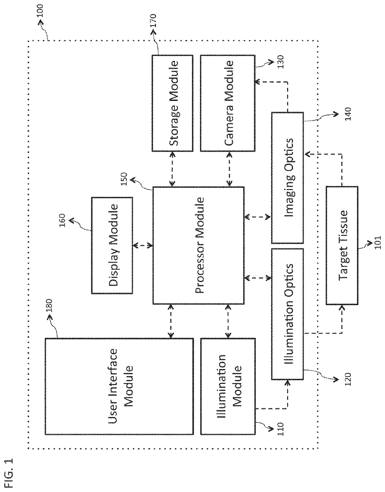
[0058]FIG. 1 is a block diagram illustrating an embodiment of a system 100 for rapid examination of particulate flow in a target tissue 101. In various embodiments, the target tissue 101 comprises any tissue, organ, or organ system of any human or animal biological system, including but not limited to the cornea, sclera, retina, epidermis, dermis, hypodermis, skeletal muscle, smooth muscle, cardiac muscle, brain tissue, the spinal cord, the stomach, large and small intestines, pancreas, liver, gallbladder, kidneys, endocrine tissue, and assoc...


PUM


Abstract

Examination of the structure and function of blood vessels is an important means of monitoring the health of a subject. Such examination can be important for disease diagnoses, monitoring specific physiologies over the short- or long-term, and scientific research. This disclosure describes technology and various embodiments of a system and method for imaging blood vessels and the intra-vessel blood flow, using at least laser speckle contrast imaging, with high speed so as to provide a rapid estimate of vessel-related or blood flow-related parameters.

Description

RELATED APPLICATIONS[0001]This application is a National Stage application under 35 U.S.C. 371 of PCT International Application No. PCT / US2016 / 055976, filed Oct. 7, 2016, which claims the benefit of U.S. Provisional Application No. 62 / 239,529, filed Oct. 9, 2015 the entire disclosure of which is hereby incorporated by reference herein.GOVERNMENT GRANT SUPPORT[0002]This invention was made with government support under grant 1R43EB019856-01A1 awarded by the National Institute of Biomedical Imaging and Bioengineering (of the National Institutes of Health). The government has certain rights in the invention.FIELD OF THE INVENTION[0003]The present invention relates generally to vascular imaging technologies and methods and, more particularly, to the rapid examination of vasculature and blood flow using laser speckle contrast imaging.BACKGROUND[0004]Blood vessels are the fundamental mechanism by which human and animal biological systems and related tissues, organs, and organ systems recei...

Claims


Application Information

Patent Timeline
Patent Type & Authority Patents(United States)
IPC IPC(8): A61B5/026G02B27/48A61B5/00A61B5/02A61B3/12A61B3/14A61B3/00
CPCA61B5/0261A61B3/1233A61B5/0066A61B5/02A61B5/721A61B5/7207A61B5/742G02B27/48A61B3/0075A61B3/0083A61B3/12A61B3/1208A61B3/14
Inventor REGE, ABHISHEKBROOKE, M. JASONMURARI, KARTIKEYALIU, YUSI
Owner VASOPTIC MEDICAL
Why Eureka
Try Eureka