
Devices, compositions and related methods for accelerating and enhancing bone repair

a technology of nanoparticles and devices, applied in the field of new therapeutic nanoparticles, can solve the problems of unrelenting effects, corrosive impact, significant morbidity, etc., and achieve the effects of preventing iron infiltration into the fracture site, enhancing and/or activating angiogenesis, and high degradability

Active Publication Date: 2021-06-29
UNIVERSITY OF KANSAS +1
View PDF5 Cites 0 Cited by
  • Summary
  • Abstract
  • Description
  • Claims
  • Application Information

AI Technical Summary

Benefits of technology

The present technology involves the development of a hyaluronic acid nanoparticle conjugated with an agent able to enhance and / or activate angiogenesis (e.g., DFO). The nanoparticle was shown to prevent iron infiltration into the fracture site and to have improved drug delivery and anti-inflammatory properties. The therapy can be used alone or in conjunction with internal fixation of bony fractures and is designed to maximize therapeutic effect.

Problems solved by technology

Although XRT has increased survival it also results in damage to adjacent normal tissues leading to significant morbidity.
The corrosive impact of these XRT-induced side effects can be unrelenting and their complex management is rarely remedial.
Severely problematic wound healing issues impact the reconstructive efforts to replace the bone and soft tissue removed by tumor extirpation and the options to treat XRT-induced pathologic fractures and osteoradionecrosis.
Attendant complications often lead to delays in initiation of therapy jeopardizing prognosis as well as quality of life.
XRT drastically impairs fracture healing, however, precluding the utilization of DO as a durable reconstructive method for HNC.

Method used


Image

  • Devices, compositions and related methods for accelerating and enhancing bone repair
  • Devices, compositions and related methods for accelerating and enhancing bone repair
  • Devices, compositions and related methods for accelerating and enhancing bone repair

Examples


example i

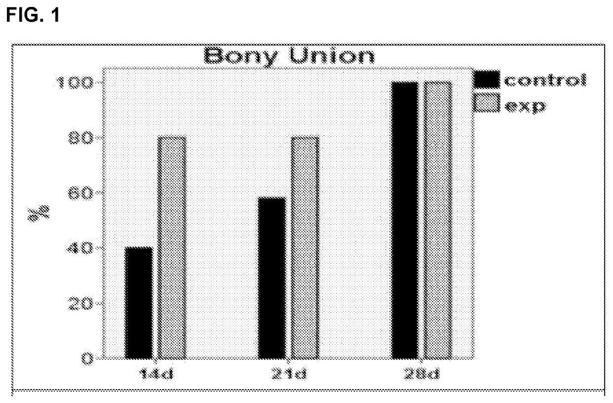
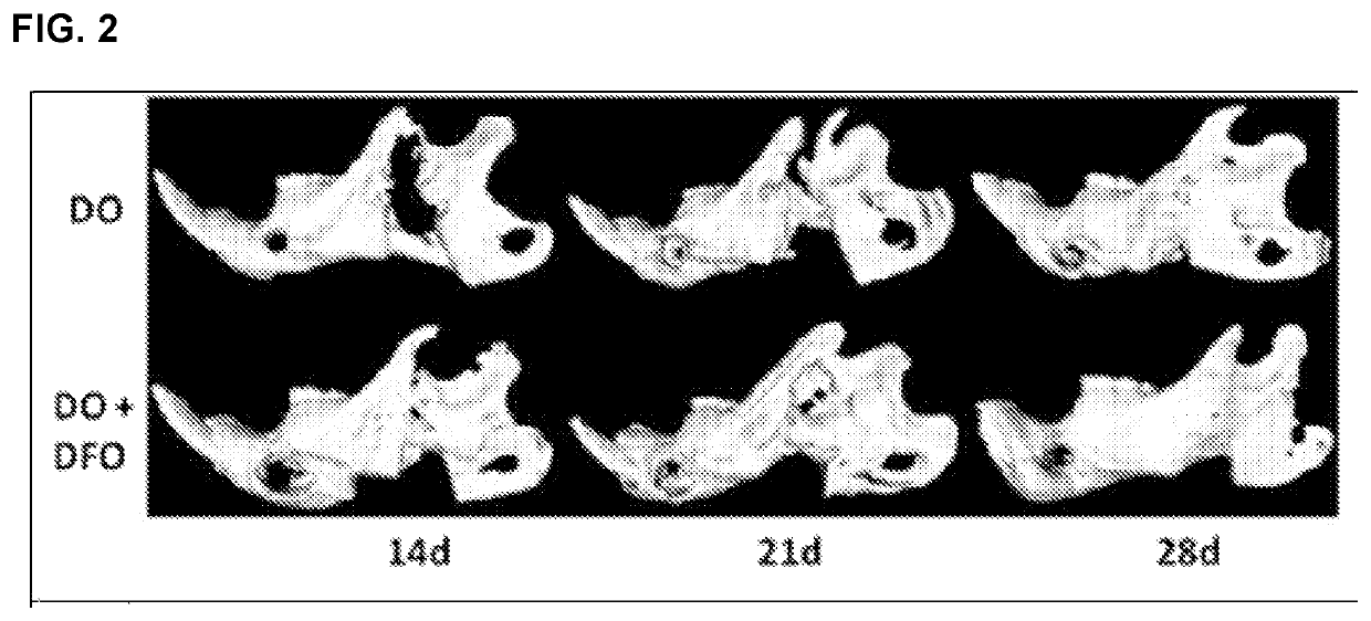
[0096]This example demonstrates that DFO accelerates bone regeneration in maxillofacial distraction osteogenesis.

[0097]The effectiveness of DFO in enhancing regenerate vascularity at a full consolidation period (28 days) in a murine mandibular DO model was established. To investigate whether this augmentation in vascularity would function to accelerate consolidation without compromising regenerate quality or strength, consolidation periods prior to μET imaging and biomechanical testing (BMT) were progressively shortened. Three time points (14 d, 21 d and 28 d) were selected and six groups of Sprague-Dawley rats (n=60) were equally divided into control (C) and experimental (E) groups for each time period. Each group underwent external fixator placement, mandibular osteotomy, and a 5.1 mm distraction. During distraction, the experimental groups were treated with DFO injections into the regenerate gap. After consolidation, mandibles were imaged and tension tested to failure. ANOVA was ...

example ii

[0098]This example demonstrates that DFO augments fracture healing in the irradiated mandible.

[0099]The effects of radiation on bone formation and healing are mediated through the mechanisms of vascular damage, direct cellular depletion and diminished function of osteocytes. Over time, this accumulated damage predisposes patients to the debilitating problem of late pathologic fractures and non-unions. Here, the use of DFO was employed to bolster the vascular response during bone healing in this setting. It was posited that the untoward effects of radiotherapy on vascular density and osteocyte count and ultimately the mineralization and biomechanical strength of our bone would be improved with the addition of DFO. 12 rats received fractionated radiotherapy to left hemi-mandibles. After recovery, fracture repair ensued with external fixator placement and mandibular osteotomy. DFO was injected into the callus site every other day from post-operative days 4-12. A 40-day healing period w...

example iii

[0108]This example demonstrates a real-time investigation of the angiogenic effect of deferoxamine on endothelial cells exposed to radiotherapy.

[0109]The effect of DFO on endothelial cells exposed to radiation in vitro was investigated. It was posited that radiation would significantly diminish the ability of endothelial cells to form tubules; and subsequently, that the addition of DFO would effect a restoration of tubule formation. Four groups of human umbilical vein endothelial (HUVEC) cells (control, radiated, radiated+low dose DFO, or radiated+high dose DFO) were incubated in Matrigel and video recorded in real-time over 12 hours. DFO groups received either 25 or 50 μM doses at the time of incubation. Tubule formation was photographed at 100× magnification every four hours. Tubule numbers between groups were compared using ANOVA with p≤0.05 considered statistically significant. A severe diminution in endothelial tubule formation after radiotherapy was observed. Specifically, tub...


PUM

PropertyMeasurementUnit
molecular weightaaaaaaaaaa

Abstract

The present invention relates to novel therapeutic nanoparticles. In particular, the present invention is directed to nanoparticles associated (e.g., complexed, conjugated, encapsulated, absorbed, adsorbed, admixed) with angiogenesis-activating-agents, methods of synthesizing the same, devices or compositions comprising such nanoparticles, as well as systems and methods utilizing the nanoparticles (e.g., in therapeutic settings for enhancing and / or activating angiogenesis at targeted tissue region).

Description

CROSS-REFERENCE TO RELATED APPLICATIONS[0001]This application is a Section 371 U.S. national stage entry of International Patent Application No. PCT / US2016 / 067320, International Filing Date Dec. 16, 2016 which claims priority to and the benefit of U.S. Provisional Application No. 62 / 268,926, filed Dec. 17, 2015, which are hereby incorporated by reference in their entireties.STATEMENT REGARDING FEDERALLY SPONSORED RESEARCH OR DEVELOPMENT[0002]This invention was made with government support under CA125187 and CA173292 awarded by the National Institutes of Health. The government has certain rights in the invention.FIELD OF THE INVENTION[0003]The present invention relates to novel therapeutic nanoparticles. In particular, the present invention is directed to nanoparticles associated (e.g., complexed, conjugated, encapsulated, absorbed, adsorbed, admixed) with angiogenesis-activating-agents, methods of synthesizing the same, devices or compositions comprising such nanoparticles, as well ...

Claims


Application Information

Patent Timeline
Patent Type & Authority Patents(United States)
IPC IPC(8): A61K47/69A61P19/00A61P19/08A61K9/51A61L27/50A61K47/61A61K31/16A61L27/20A61L27/54A61L27/58
CPCA61K47/6939A61K9/5169A61K31/16A61K47/61A61L27/20A61L27/50A61L27/54A61L27/58A61P19/00A61P19/08A61L2300/412A61L2300/624A61L2300/626A61L2400/12A61L2430/02
Inventor BUCHMAN, STEVEN R.COHEN, MARKDONNEYS, ALEXISNELSON, NOAHFORREST, LAIRDZHANG, TIYANG, QIUHONG
Owner UNIVERSITY OF KANSAS
Learn More