Enhancement of NMR and MRI in the presence of hyperpolarized noble gases

a noble gas and nmri technology, applied in the field of hyperpolarized noble gas enhancement in the presence of noble gas, can solve the problems of low sensitivity of mri and nmr spectroscopy for these molecules, persistent challenge in sensitivity of nmr, and inability to achieve the effect of reducing the growth of proton signals

Inactive Publication Date: 2003-01-23
RGT UNIV OF CALIFORNIA
View PDF2 Cites 17 Cited by
  • Summary
  • Abstract
  • Description
  • Claims
  • Application Information

AI Technical Summary

Benefits of technology

0041] FIG. 13. Schematic diagram of the pulse sequence used to obtain heteronuclear difference SPINOE spectra. The proton magnetization is saturated first by a series of .pi. / 2 pulses and a z-axis magnetic field gradient is applied in between the pulses to dephase the transverse components of the magnetization for optimal saturation. The .pi. pulses helps to reduce the growth of proton signal due to spin-lattice relaxations. A .pi. pulse is also applied to the .sup.129Xe resonance at the same time of the proton .pi. pulses so that the .sup.129Xe magnetization is inverted in synchronization with the proton magnetization. This synchronization ensures that the SPINOE signals will be accumulated during the entire mixing time. Both proton and xenon .pi. pulses are adiabatic pulses BIR4 of 1 ms in duration.

Problems solved by technology

Sensitivity poses a persistent challenge to the use of NMR, both in imaging and spectroscopy.
Thus, these agents are of little use in functional brain imaging.
Noble gases are of interest as tracers and probes for MRI and NMR spectroscopy (Middleton, H., et al., Magn. Res. Med. 33: 271 (1995)), however, the sensitivity of MRI and NMR spectroscopy for these molecules is relatively low.
A factor which contributes to the lack of sensitivity of these techniques for the noble gases is that the spin polarization, or net magnetic moment, of the noble gas sample is low.
Lower temperatures and higher fields, to the extent that these can be imposed, provide only limited benefit.
Although hyperpolarized noble gases have proven useful as probes in the study of the air spaces in lungs, the effectiveness or sensitivity of these methods is somewhat compromised for biological materials and organs, such as blood and the parts of the body that are accessible only through the blood.
During its residence time in the blood, the hyperpolarized gas is diluted considerably and the delay in transferring the gas from the lung space to the blood consumes much of the time (e.g., T.sub.1) required for the polarized gas to return to its non-hyperpolarized state.
Further complicating the situation, the penetration of the hyperpolarized gas into the interior of red blood cells dramatically reduces the T.sub.1 of the hyperpolarized gas and thus, sorely attenuates the temporal range over which the gas can serve as an effective probe.

Method used


Image

  • Enhancement of NMR and MRI in the presence of hyperpolarized noble gases
  • Enhancement of NMR and MRI in the presence of hyperpolarized noble gases
  • Enhancement of NMR and MRI in the presence of hyperpolarized noble gases

Examples


example 1

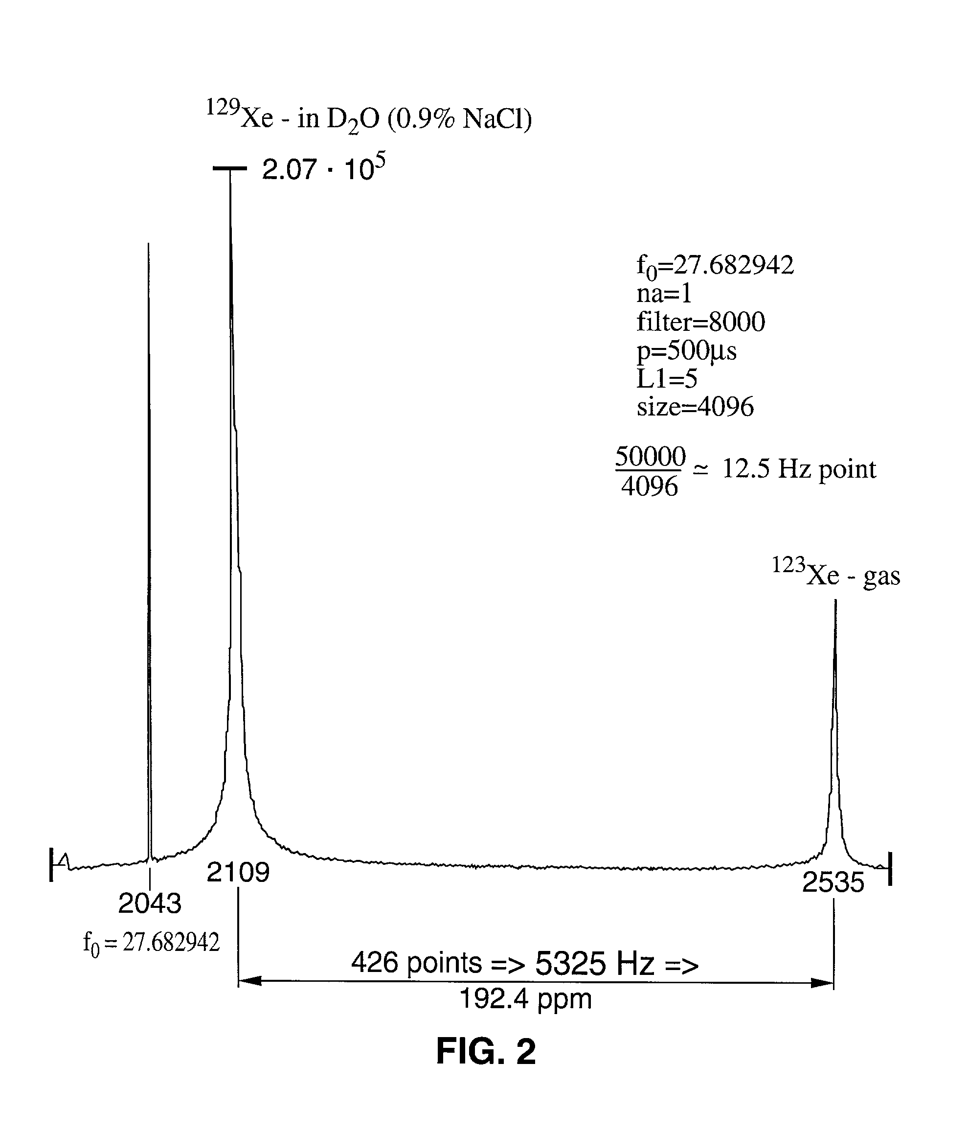
[0124] This example describes the .sup.129Xe NMR of a sample of hyperpolarized .sup.129Xe dissolved in aqueous saline. The T.sub.1 of xenon was measured in both H.sub.2O saline and D.sub.2O / saline.

[0125] In an NMR tube open to the atmosphere were combined saline and hyperpolarized .sup.129Xe. The saline used had a NaCl concentration of 0.9% by weight. .sup.129Xe was dissolved in saline as described in the materials and methods section above. Xenon has a low solubility in saline, with an Ostwald coefficient of only 0.0926 (the standard temperature and pressure volume of xenon dissolved in 1 liter of liquid at 1 atmosphere of gas pressure; 1 atm=101.3 kPa). In H.sub.2O / saline, the T.sub.1 of xenon is quite long (66 s at 9.4 T). The .sup.129Xe NMR spectrum of a solution of .sup.129Xe in D.sub.2O saline is displayed in FIG. 2. In saline made with D.sub.2O, the T.sub.1 of xenon is .apprxeq.1000 s. Thus, the shorter T.sub.1 of xenon in H.sub.2O saline is due to dipolar couplings between t...

example 2

[0127] This example demonstrates the use of xenon NMR to study the partition of xenon between the intracellular and extracellular compartments in a sample of human blood. The NMR of xenon in human blood was measured using both hyperpolarized and unpolarized xenon.

[0128] 2.1 Materials and Methods

[0129] A sample of human blood was prepared by allowing fresh blood from a volunteer to settle for a few hours and subsequently decanting off a portion of the plasma. The portion removed accounted for approximately 30% of the total volume of the blood sample. Following removal of a portion of the plasma, xenon saturated saline (1 mL) was injected into the red blood cell (RBC) sample (1 mL) and the .sup.129Xe NMR was measured. The NMR spectra were measured on a Bruker AM-400 spectrometer.

[0130] 2.2 Results

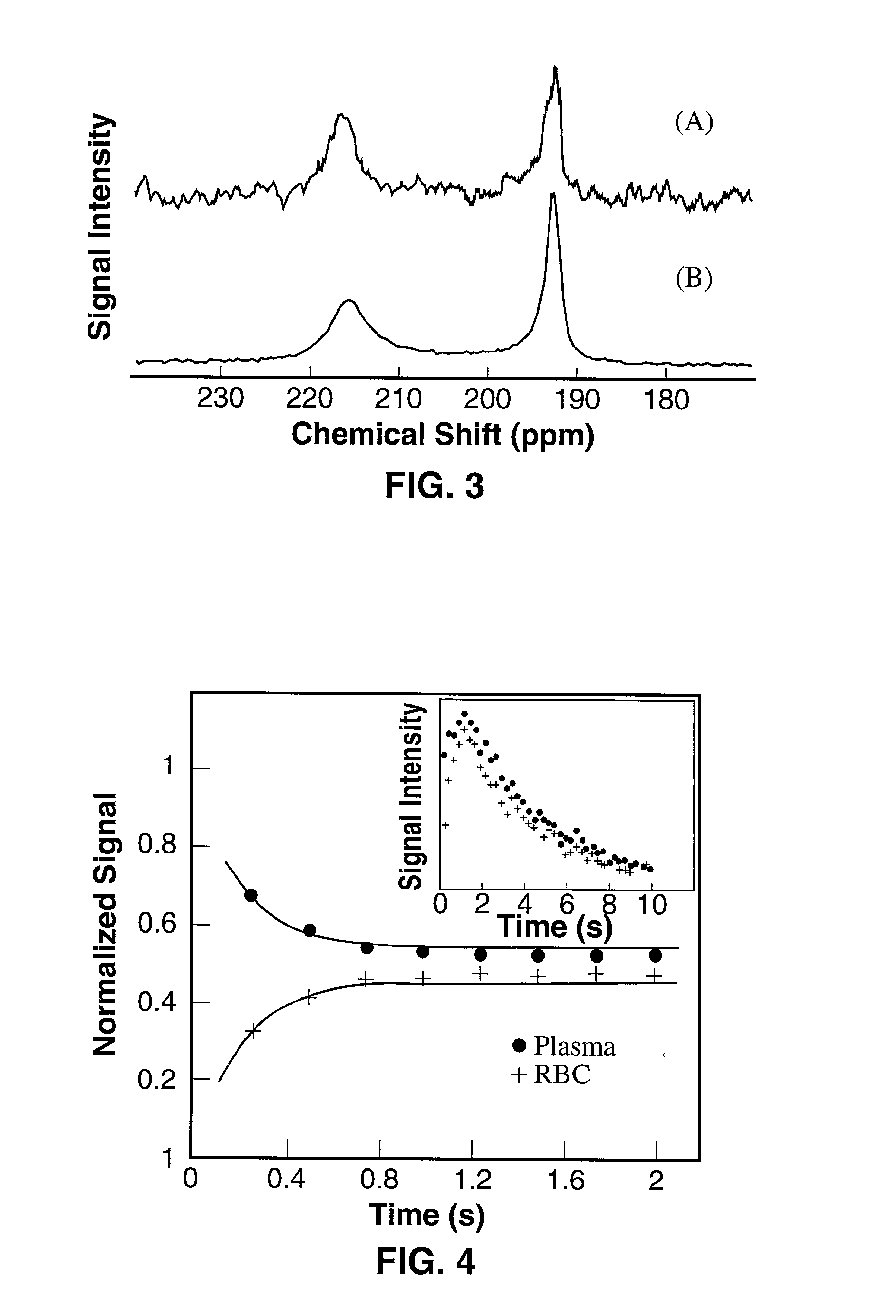
[0131] The NMR spectrum of non-polarized xenon was measured in an RBC sample (FIG. 3A). Considerable signal averaging was required in order to obtain a spectrum with an acceptable signal-to-n...

example 3

[0134] Example 3 illustrates the use of NMR spectroscopy to observe the dynamics of the mixing of laser polarized .sup.129Xe between the intracellular and extracellular compartments of a sample consisting of red blood cells and plasma.

[0135] 3.1 Materials and Methods

[0136] A sample of laser polarized xenon in saline and a RBC sample were prepared as described in Examples 1 and 2, respectively. By using short rf pulses of small tipping angle, .sup.129Xe NMR spectra were acquired as a function of time after injection of the xenon / saline mixture into the blood. NMR spectra were measured on a Bruker AM-400 spectrometer.

[0137] 3.2 Results

[0138] The results of this experiment are illustrated in FIG. 4. In FIG. 4, the main figure shows the time dependence of the xenon signal in the RBC and in the saline / plasma, normalized by the total signal. The initial rise of the RBC signal and decrease in the saline / plasma signal indicates the transfer of xenon from the saline / plasma water mixture to t...


PUM


Abstract

The present invention relates generally to nuclear magnetic resonance (NMR) techniques for both spectroscopy and imaging. More particularly, the present invention relates to methods in which hyperpolarized noble gases (e.g., Xe and He) are used to enhance and improve NMR and MRI. Additionally, the hyperpolarized gas solutions of the invention are useful both in vitro and in vivo to study the dynamics or structure of a system. When used with biological systems, either in vivo or in vitro, it is within the scope of the invention to target the hyperpolarized gas and deliver it to specific regions within the system.

Description

[0001] The present invention relates generally to nuclear magnetic resonance (NMR) techniques for both spectroscopy and imaging. More particularly, the present invention relates to the use of hyperpolarized noble gases (e.g., Xe and He) to enhance and improve NMR and MRI.[0002] Nuclear magnetic resonance (NMR) is an established technique for both spectroscopy and imaging. NMR spectroscopy is one of the most powerful methods available for determining primary structure, conformation and local dynamic properties of molecules in liquid, solid and even gas phases. As a whole-body imaging technique, Magnetic Resonance Imaging (MRI) affords images possessing such superb soft tissue resolution that MRI is the modality of choice in many clinical settings. MRI can produce images which allow the clinician to distinguish between a pathological condition and healthy tissue. For example, MR images clearly differentiate tumors from the surrounding tissue. Further, using MRI it is possible to image...

Claims


Application Information

Patent Timeline
Patent Type & Authority Applications(United States)
IPC IPC(8): A61B5/055G01R33/48A61K49/18G01R33/28G01R33/62
CPCA61K49/1815G01R33/282Y10T436/24G01R33/62G01R33/5601
Inventor PINES, ALEXANDERBUDINGER, THOMASNAVON, GILSONG, YI-QIAOAPPELT, STEPHANBIFONE, ANGELOTAYLOR, REBECCAGOODSON, BOYDSEYDOUX, ROBERTOROOM, TOOMASPIETRASS, TANJA
Owner RGT UNIV OF CALIFORNIA
Why Eureka
Try Eureka