Dual mode real-time screening and rapid full-area, selective-spectral, remote imaging and analysis device and process

a real-time screening and remote imaging technology, applied in the direction of spectroscopy, catheters, instruments, etc., can solve the problems of more difficult detection of flat polyps (sessile villous adenomas), increased risk of anesthesia, and increased scan time, so as to avoid the delay of analyzing areas and scan quickly

Inactive Publication Date: 2003-08-21
STI MEDICAL SYST
View PDF13 Cites 156 Cited by
  • Summary
  • Abstract
  • Description
  • Claims
  • Application Information

AI Technical Summary

Benefits of technology

0028] The process of this invention comprises providing an area that contains both normal and abnormal tissues with an exogenous fluorescent marker that selectively localizes in abnormal tissue and that fluoresces (preferably brightly) at a particular marker wavelength when illuminated by an excitation light. Imaging light (preferably white visible light) is then filtered to remove light of that marker wavelength to result in markerless imaging light. Of course, alternative means for creating markerless imaging light can also be used, such as using multiple broadband light sources that do not emit light at the marker wavelength, one emitting light at shorter wavelengths and the other emitting light at longer wavelengths. In a screening mode, the area is illuminated with both markerless imaging light and an excitation light, so that the exogenous fluorescent marker in the abnormal tissue is induced to fluoresce (preferably brightly enough that the fluorescent light can be detected even though the tissues are simultaneously illuminated with an imaging light) at the marker wavelength. Light at the marker wavelength is then separated to form a marker image, while the remaining light forms a background image, over which the marker image is preferably overlaid, displayed side by side, or displayed alternately. The user can thus rapidly scan for areas of potential interest (suspicious areas), avoiding the delay of analyzing areas that do not fluoresce at the marker wavelength.

Problems solved by technology

It is also more difficult to detect flat polyps (sessile villous adenomas) with such examinations.
In a colonoscopy, the entire length of the intestine can be screened, but the patient must be fully anesthetized, which introduces all the attendant risks of anesthetization.
UVB radiation is considered to be more dangerous than UVA and causes damage to genes, and UVC radiation is considered to be even more dangerous than UVB because of its shorter wavelength and higher energy.
Thus, the power and allowable time for exposing patients to light having wavelengths of less than 400 nm is restricted.
The intensity of autofluorescence is fairly weak, relative to induced fluorescence, and can be difficult to detect, requiring a longer time for exposure, much as a photograph in weak light requires a long time exposure.
It is not practical to use high intensity, long duration ultraviolet light to increase the intensity of autofluorescence because such high intensity, long duration ultraviolet light may damage a patient's tissues.
However a shorter wavelength light also may increase the risk of damage to a patient's tissues and may be subject to even more stringent regulatory requirements.
However, taking a tissue sample creates a risk of cramping, infection, excessive bleeding or perforation, and may be completely out of the question for certain people, such as hemophiliacs or others with impaired clotting ability or immune systems.
The differentiating portions of the autofluorescence spectra of normal and abnormal tissues may be faint, so a substantial amount of time is needed to acquire autofluorescence spectral data for analysis.
A disadvantage of spectroscopy of a single point is that areas must be spectrally analyzed point by point to delineate the spatial extent of any abnormal tissue.
The area of coverage for point spectroscopy therefore depends on the size of the fiber optic that abuts against the tissue, which may make determination of the extent (i.e., perimeter) of the abnormal tissue difficult.
However, Palcic indicates that one transformation which has been reported with tumor localizing drugs was found not to be useful for the imaging method.

Method used


Image

  • Dual mode real-time screening and rapid full-area, selective-spectral, remote imaging and analysis device and process
  • Dual mode real-time screening and rapid full-area, selective-spectral, remote imaging and analysis device and process
  • Dual mode real-time screening and rapid full-area, selective-spectral, remote imaging and analysis device and process

Examples


Embodiment Construction

[0063] While this invention is illustrated and described with respect to a presently preferred best mode with respect to screening and analysis of the intestine, devices according to this invention may be in many different configurations, forms, and materials, and applied to screening and analysis of other tissues, organs, materials and objects. The best mode disclosed herein is an exemplification of the principles of the invention and the associated functional specifications of the materials for its construction, and is not intended to limit the invention to the illustrated best mode. Those persons skilled in the art will envision many other possible variations within the scope of the present invention.

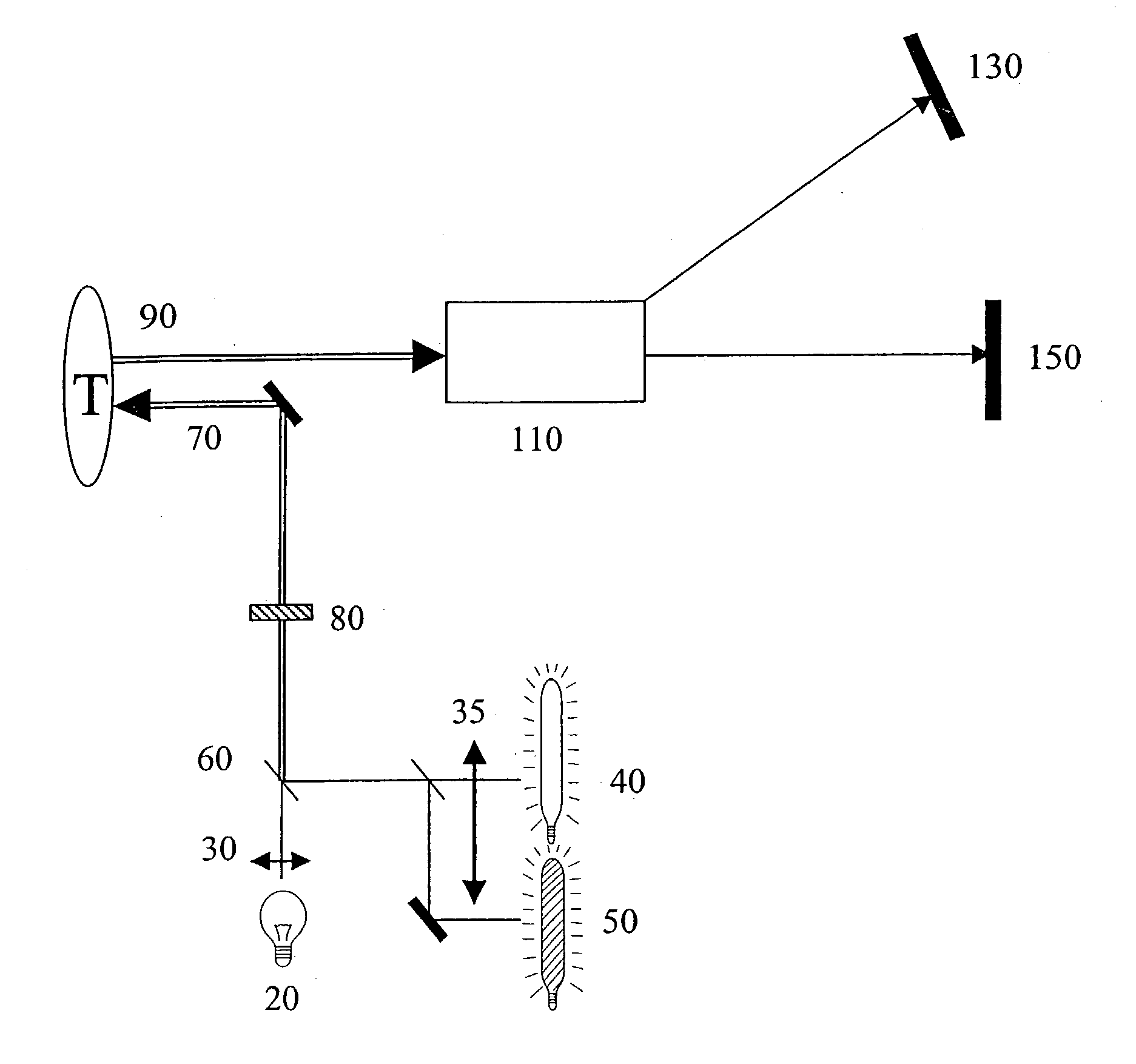
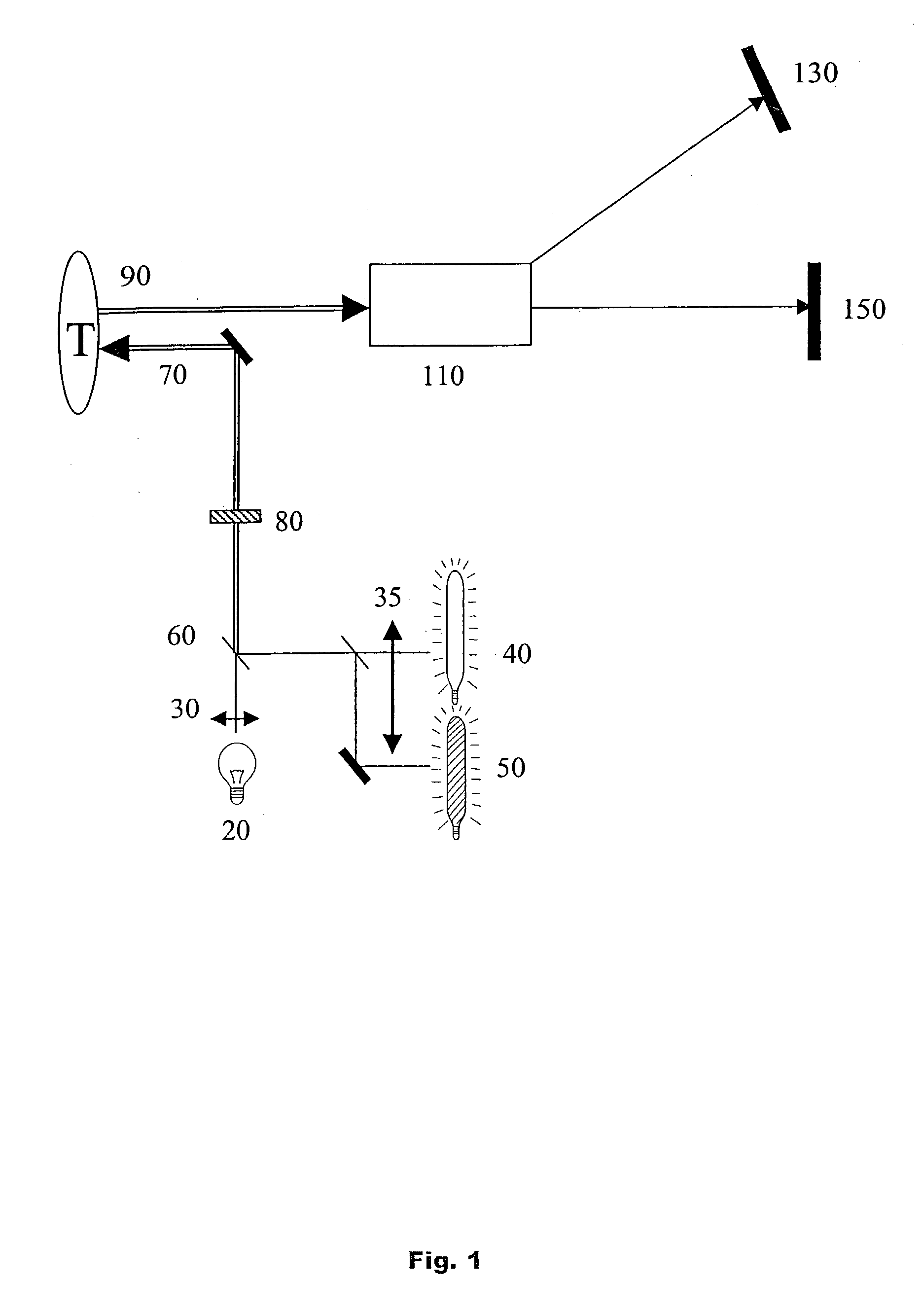
[0064] The presently preferred best mode for carrying out the present invention is illustrated by way of example in FIG. 1.

[0065] Referring to FIG. 1, the device includes an imaging light source 20 preferably emitting approximately spectrally flat broadband visible white light from U...


PUM


Abstract

The invention provides a device and process for real-time screening of areas that can be identified as suspicious either through image segmentation utilizing image processing techniques or through treatment with an exogenous fluorescent marker that selectively localizes in abnormal areas. If screening detects a suspicious area, then the invention allows acquiring of autofluorescence images at multiple selected narrow differentiating spectral bands so that a "virtual biopsy" can be obtained to differentiate abnormal areas from normal areas based on differentiating portions of autofluorescence spectra. Full spatial information is collected, but autofluorescence data is collected only at the selected narrow spectral bands, avoiding the collection of full spectral data, so that the speed of analysis is increased.

Description

[0001] This invention relates to a device and process that provides, in a screening mode, real-time screening and remote imaging, and in an analysis mode, rapid full-area, selective-spectral remote imaging and analysis. More specifically, this invention relates to a device for allowing real-time detection and rapid remote analysis of surfaces with differentiating spectral properties, such as potentially cancerous regions in the cervix, intestine, lungs or other organs.PRIOR ART[0002] Cancers, especially cancers in the intestine, cervix, lungs, and other hollow organs, need to be detected early for effective treatment. For example, intestinal cancers typically start with polyps, either protruding (pedunculated tubular adenomas) or flat (sessile villous adenomas). These polyps sometimes convert into cancer. Therefore, the detection or removal of polyps by colonoscopy significantly reduces the risk of getting colon cancer. Traditionally, polyps are detected using devices that allow a p...

Claims


Application Information

Patent Timeline
Patent Type & Authority Applications(United States)
IPC IPC(8): A61B5/00
CPCA61B1/043A61B1/0638A61B1/0653A61B1/00186A61B5/0075A61B5/0084A61B5/0071
Inventor WOLTERS, ROLF HOLGERSWEAT, JOSEPH ALLENDEWEERT, MICHAEL JAMESSEIPLE, ROBERT BENTON JR.WALTON, DAVID GEORGE SHIBASAKI
Owner STI MEDICAL SYST
Why Eureka
Try Eureka