Viral identification by generation and detection of protein signatures

a technology of protein signatures and viral identification, applied in the field of micrototal analysis systems, can solve the problems of non-specificity, laborious current methods, and difficulty in rapidly detecting and identifying viruses

Inactive Publication Date: 2005-01-20
SANDIA NAT LAB
View PDF47 Cites 89 Cited by
  • Summary
  • Abstract
  • Description
  • Claims
  • Application Information

AI Technical Summary

Benefits of technology

Certain aspects of the present invention provide for the collection and identification of macromolecules, such as biologically-derived macromolecules (e.g., proteins), by measuring the molecular weight distribution of macromolecular samples. The precision

Problems solved by technology

Research in this particular area is presently intense, reflecting some of the intrinsic difficulties of realizing miniaturized devices.
However, there still remains the difficult problem of rapidly detecting and identifying viruses; For example, current methods are laborious, time consuming and depend on specific reagents for detecting single viral isoforms.
Antibodies are difficult to produce in mass quantities and are non-specific, particularly for viral agents.
An outstanding problem is the detection of viruses using a micro analytical system that does not require

Method used


Image

  • Viral identification by generation and detection of protein signatures
  • Viral identification by generation and detection of protein signatures
  • Viral identification by generation and detection of protein signatures

Examples


examples

Preparing Microfluidic Chips

Microfluidic chips were generally fabricated from Corning 7980 fused silica wafers (100 mm diameter, 0.75 mm thickness using standard photolithography, wet etch, and bonding techniques. Fused Silica wafers were PECVD deposited with amorphous silicon (150 nm), which served as the hard mask. A 7.5-micron thick layer of positive photoresist was spin-coated and soft-baked (90° C., 5 minutes). The mask pattern was transferred to the photoresist by exposing it to UV light in a contact mask aligner. After exposure, the photoresist was developed and hard-baked (125° C., 30 minutes). Exposed silicon was etched in a plasma etch tool. Silicon etch process typically consisted of a 30 second oxygen ash @200W DC @25 mTorr, followed by 150 second SF6 @200W DC & 50 mTorr. The exposed glass was etched with a 49% HF solution. Via access holes were drilled in the cover plate (Corning 7980) with diamond-tipped drill bits. The etched wafers and drilled cover plates were cl...

example

Analysis of Eukaryotic Cells and Tissues

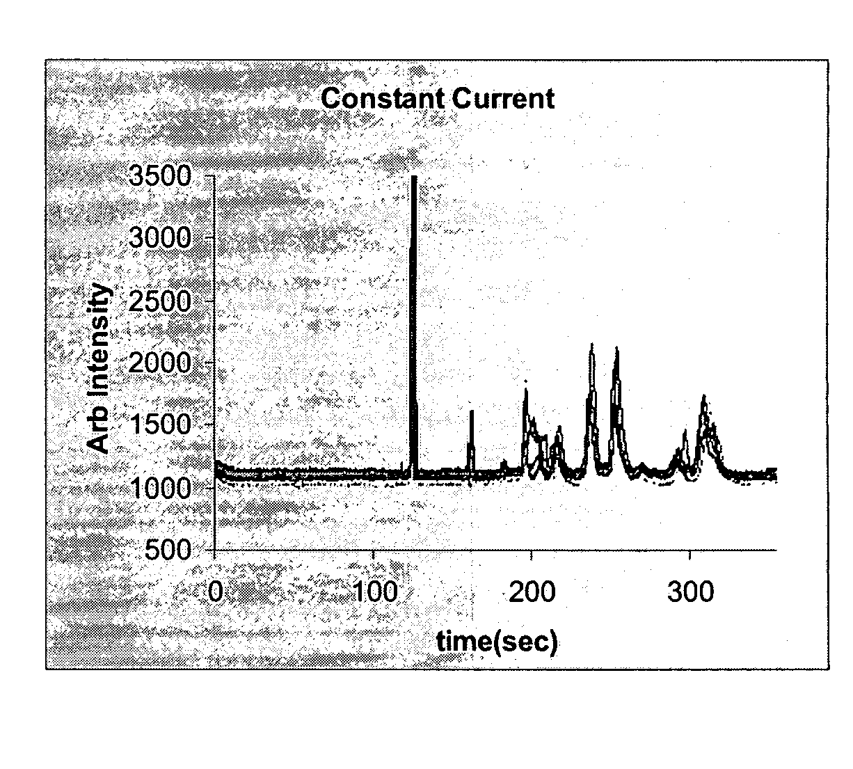
Samples of eukaryotic cells and tissues are first lysed and the proteins solubilized. Cell and tissue samples are first solubilized in a phosphate buffer, or an appropriate buffer containing detergent such as SDS, Triton-X or NP-40, at a neutral pH such as 7.4. Once these sample are dissolved in buffer, they are heated to approximately 100° C. for a period of five minutes or more. A fraction of this sample is then placed in the standard CGE separation buffer containing 5 mM boric acid, 5 mM sodium lauryl sulfate in water drop-wise adjusted to pH 8.5 with 1M NaOH for injection onto the microfluidic chip. The proteins are then labeled using an appropriate dye, such as fluorescamine. Labeled samples are then injected on to the microfluidic chip using a specially modified gas tight syringe as previously described. Subsequently an EK injection and separation are then performed as previously described in “Detection of Viral Signatures”.


PUM

PropertyMeasurementUnit
Timeaaaaaaaaaa
Timeaaaaaaaaaa
Timeaaaaaaaaaa

Abstract

The present invention provides systems and processes for the collection and identification of macromolecules, such as biologically-derived macromolecules (e.g., proteins and nucleic acids), by measuring and comparing the molecular weight signatures of macromolecular samples. Reproducible molecular weight signatures provides reliable sample identification. In the case of viruses, proteomic molecular weight signatures can be used for identifying viral agents.

Description

FIELD OF THE INVENTION The present invention is related to the field of micro-total analysis systems (μ-TAS). The present invention is also related to microanalytical instruments for analyzing and identifying biological substances and to methods of operating microanalytical instruments for analyzing and identifying biological substances. The present invention also relates to systems and methods for analyzing and identifying macromolecular substances and chemical compounds. BACKGROUND OF THE INVENTION The field of micro-Total Analysis Systems (μ-TAS) relates to miniaturized devices where all necessary parts and methods to perform a chemical analysis are integrated. More than simply shrinking down traditional bench top techniques, μ-TAS requires innovations in many fields, including chemistry, biology, electronics, optics, materials, fluid mechanics and microfabrication. μ-TAS devices arose as a consequence of a development when electronic circuits were miniaturized and integrated i...

Claims


Application Information

Patent Timeline
IPC IPC(8): C07K1/107G01N27/447G01N33/569G01N33/68
CPCG01N27/44791G01N33/56983G01N33/6803G01N27/44721C07K1/1077G01N27/44713G01N2550/00
Inventor WEST, JASON ANDREW APPLETONSTAMPS, JAMES FREDERICKSHOKAIR, ISAAC RAMZYRENZI, RONALD F.VANDERNOOT, VICTORIA A.WIEDENMAN, BOYD J.LANE, TODD WILLIAMFRUETEL, JULIA ANN
Owner SANDIA NAT LAB
Why Eureka
Try Eureka