
Method for culturing and expansion of mammalian undifferentiated epidermal kerainocytes exhibiting stem cell characteristics

a technology of stem cell characteristics and kerainocytes, which is applied in the field of culturing of mammalian e, can solve the problems of inability to achieve the maintenance of a substantial undifferentiated state with the intent of adding to a wound site, inability to obtain reliable supply, and inability to co-culturing the human dermal fibroblasts and human epidermal keratinocytes. to achieve the effect of increasing the quantity of hla

Inactive Publication Date: 2005-01-27
PHARMAGAP
View PDF0 Cites 5 Cited by
  • Summary
  • Abstract
  • Description
  • Claims
  • Application Information

AI Technical Summary

Benefits of technology

[0043] According to one embodiment of this aspect of the invention, we have found it advantageous to start the wound healing process as soon as possible with the addition of pre-screened allogenic human dermal fibroblasts and previously grown and stored stem cell-like epidermal keratinocytes exhibiting LDN as down regulated MHC molecules. Concurrently, the patients cells are harvested and expanded in the novel medium to ensure a homogeneous population of stem cell-like epidermal keratinocytes and dermal fibroblasts, to be subsequently applied to the wound site in order to promote optimal healing.
[0050] Another major advantage of the novel medium is that due to the absence of animal by-products, cross transmittance of disease is avoided.
[0052] The stem cell-like epidermal keratinocytes and dermal fibroblasts could also be added directly to the wound as a viable mixed culture of stem cell-like keratinocytes and dermal fibroblasts, the stem cell-like keratinocytes exhibiting increasing HLA quantities along with the fibroblasts ensures an accelerated and more complete epithelization of the wound site preventing a miss matched tolerance.

Problems solved by technology

Further, the co-culturing of the human dermal fibroblasts and human epidermal keratinocytes has proven very difficult up to now because of the different calcium requirements of the two phenotypes for survival.
Also, the in vitro growth of massive numbers of stem like keratinocytes and their maintenance in a substantially undifferentiated state with the intent to add to a wound site has not been accomplished.
However, this requires a reliable source of supply, and intense screening for human infections.
Further, if cells from the patient were to be used there would be a time delay in expanding those cells for transplantation.

Method used


Image

  • Method for culturing and expansion of mammalian undifferentiated epidermal kerainocytes exhibiting stem cell characteristics
  • Method for culturing and expansion of mammalian undifferentiated epidermal kerainocytes exhibiting stem cell characteristics
  • Method for culturing and expansion of mammalian undifferentiated epidermal kerainocytes exhibiting stem cell characteristics

Examples


example 1

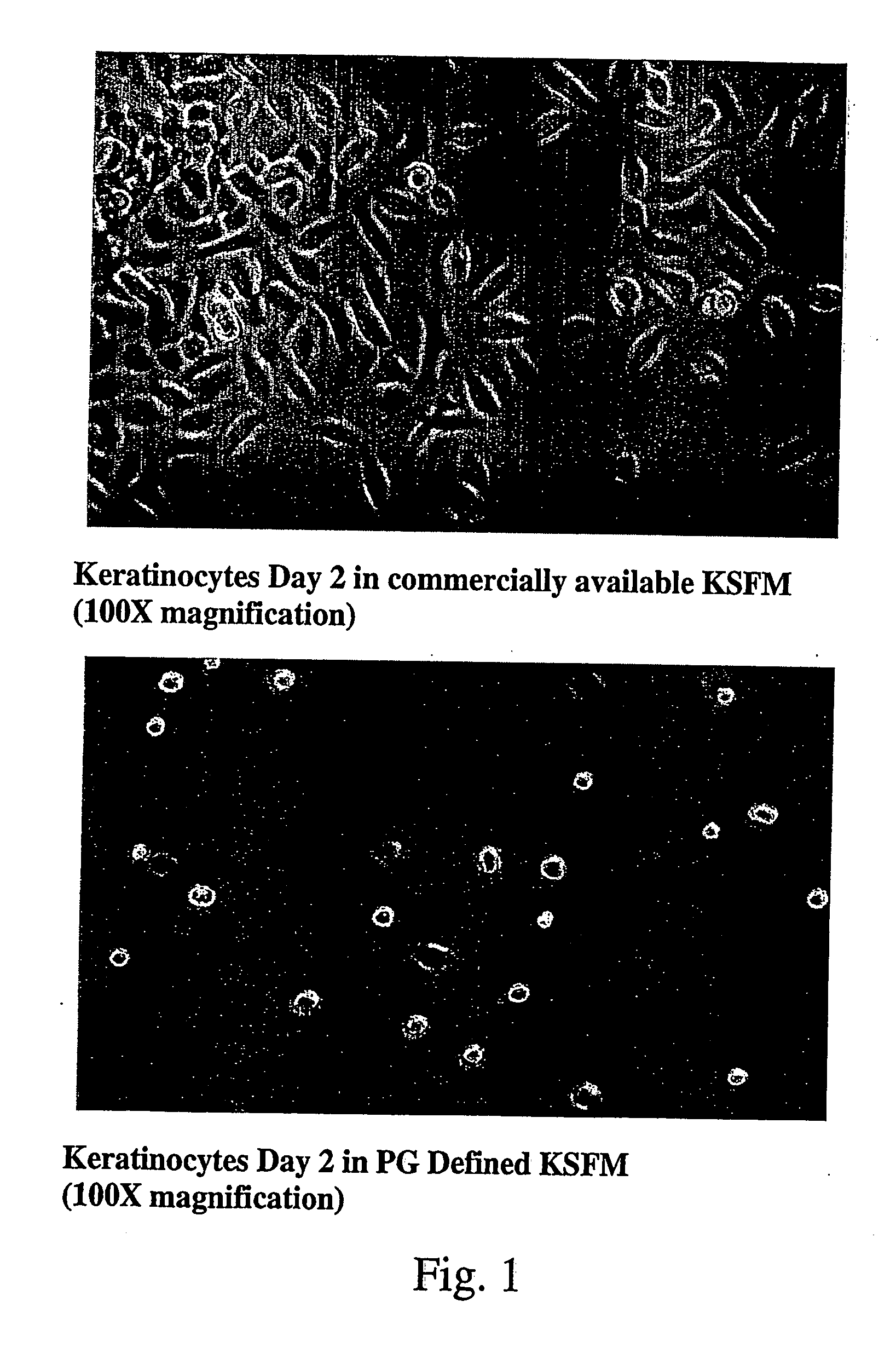
[0064] Experiments adding various amounts of FGF7 / KGF to the basal GIBCO® KSFM catalogue no. 17005-042, were completed to indicate the optimal concentration of KGF addition to the basal medium. The amounts added were 10, 5, 2.5 and 1 ng / ml of medium. After an 8 day period, the 10 ng / ml addition provided superior growth of the stem cell-like keratinocytes. The 5 ng / ml version showed good cell viability, but slower stem cell-like keratinocyte growth. The lower concentrations showed inadequate stem cell-like keratinocyte growth. We did not go above 10 ng / ml, because of the high cost of the compound. However, at this point, the graph is levelling off in any case.

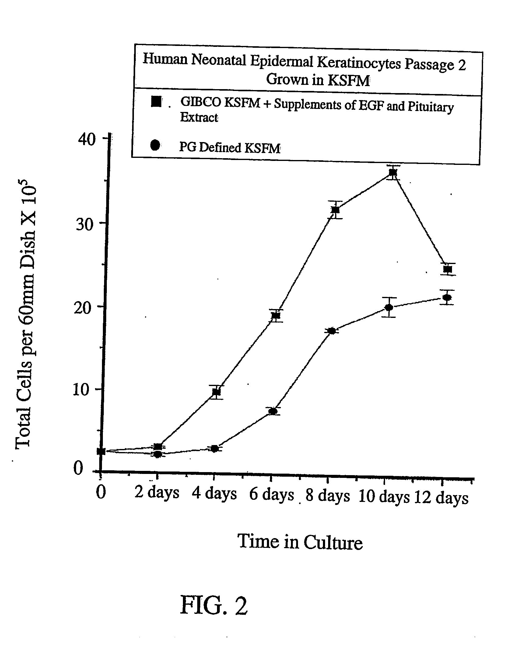
[0065]FIG. 2 is a graph illustrating the comparison of cultures grown in commercially available KSFM ie GIBCO® KSFM, supplemented with growth promoting additives including EGF and BPE, and cultures grown in the novel PG Defined KSFM according to the invention, including 10 ng / ml of medium of FGF7 / KGF. The growth curve shows tha...

example 2

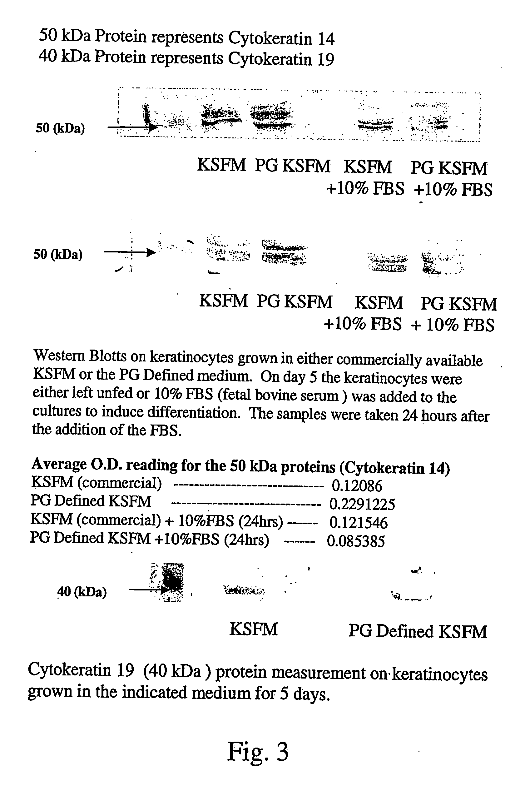
[0068] Allogenic stem keratinocytes have been isolated utilizing a small number of cells, using methods described by Jones et al. 19952, the disclosure of which is incorporated herein by reference. After isolation of a small number of the stem keratinocytes, the cells are grown in the PG Defined KSFM including 10 ng / ml of medium of FGF7 / KGF, into a homogeneous culture of keratinocytes with stem cell-like characteristics, therefore reducing the risk of immunogenicity. 90+% of the viable cells were found to exhibit keratin 5-14 expression pattern of keratinocytes progenitors.

[0069] Autologous keratinocytes were expanded in the novel PG Defined KSFM on either a commercially constructed imatrix or in a culture, giving a population of keratinocytes with stem cell like characteristics and then transferred to the wound site using a variety of methods of application.

[0070] This method, using the novel medium (PG Defined KSFM) amplifies small numbers of either allogenic or autologous kerat...

example 3

[0072] Experimental results obtained by (Jones and Watts4), the disclosure of which is incorporated herein by reference, indicate that the total number of stem keratinocytes in a primary epidermal cell population is approximately 20%. Their method for the isolation of stem keratinocytes is a timed adherence of the cells to a 10% collagen coating using 5 minutes as a maximum adherence time. When adhesion tests were performed on keratinocytes grown in the GIBCO KSFM+EGF+Pituitary Extract, only 2-5% of the keratinocytes complied whereas 60-65% of the keratinocytes appeared on the same collagen base after 5 minutes when grown in PG Defined KSFM, including 10 ng / ml of FGF7 / KGF.

[0073] When keratinocytes were grown in the commercially available GIBCO® KSFM+supplements (Epidermal Growth Factor and Pituitary Extract) and then transferred after sub-culturing to either their original growth medium GIBCO KSFM+supplements or PG Defined KSFM including 10 ng / ml of FGF7 / KGF, the results were as fo...


PUM


Abstract

The invention disclosed relates to the culturing of mammalian e.g. human cells, and in particular to the culturing and expansion of substantially undifferentiated human epidermal keratinocytes exhibiting stem cell-like characteristics, and in co-culture with human dermal fibroblasts utilizing a low calcium serum free and animal by-product free medium derived from a commercially available medium. The medium consists of a commercially available basal medium, and a single fibroblast growth factor (FGF) or a mimic thereof e.g. FGF7 / KGF. A method is also disclosed for treating mammalian e.g. human skin wound injuries, by applying to the wound an effective amount of substantially pure mammalian e.g. human epidermal stem cell-like keratinocytes in a substantially undifferentiated state, and optionally additionally, dermal fibroblasts e.g. human dermal fibroblasts. Both cell types can be grown separately or in a co-culture for application purposes.

Description

BACKGROUND OF THE INVENTION [0001] This invention relates to the culturing of mammalian e.g. human cells, and in particular to the culturing and expansion of substantially undifferentiated human epidermal keratinocytes exhibiting stem cell characteristics, and in co-culture with human dermal fibroblasts utilizing a serum free and animal by-product free medium derived from a commercially available medium. [0002] Up to this point in wound healing and other areas that utilize the growth of keratinocytes and fibroblasts or the co-culturing of both, no one has initiated the growth of human keratinocytes that exhibit stem cell like characteristics, for use in wound healing and / or for studies using a true skin model consisting of a complete dermis and epidermis. [0003] Moreover, Keratinocyte Serum Free Medium used by others has contained animal by products such as dialyzed serum (bovine usually), or additives such as pituitary extract (bovine) or other animal derived compounds. [0004] Two ...

Claims


Application Information

Patent Timeline
IPC IPC(8): A61K35/12A61K35/36A61P17/00C12N5/071
CPCA61K35/12A61K35/36C12N5/0629C12N2502/1323C12N2501/117C12N2502/094C12N5/0698A61P17/00
Inventor ISAACS, RICHARD JOHN
Owner PHARMAGAP
Learn More