Prenatal screening

a technology for prenatal screening and tissue, applied in the field of prenatal screening, can solve problems such as unworkable approaches to da

Inactive Publication Date: 2005-03-10
BRANDEIS UNIV
View PDF8 Cites 0 Cited by
  • Summary
  • Abstract
  • Description
  • Claims
  • Application Information

AI Technical Summary

Benefits of technology

[0066] The advantages of the present invention include, but are not limited to, facilitating prenatal screening by optimizing conditions for nuclear activation, which causes the nucleus of a fetal cell to either swell, replicate nucleic acid, and / or form metaphase chromosomes. Important information regarding nucleic acid sequences or chromosome morphology can be readily obtained from these various stages of activation, for example by using DNA probes or visualizing the produced metaphase chromosomes. Because some fetal cells, such as trophoblasts, erythrocytes, and leukocytes can be obtained from a maternal source, an advantage of the invention is a non-invasive procedure to detect the presence of genetic defects in such cells.

Problems solved by technology

After sporadic reports of success, recent articles appear to indicate that these markers are insufficiently specific and actually are attached to maternal cells frequently enough to make this approach unworkable to date.”

Method used


Image

  • Prenatal screening
  • Prenatal screening
  • Prenatal screening

Examples


example 1

Further Induction Optimization

[0239] This example describes additional experiments carried out to further determine the optimal induction time for an activating egg extract. Protein synthesis during the early part of the first cell cycle in activated eggs or egg extracts is required for preparation of an activating extract capable of efficient and complete genome replication. The required proteins can either be synthesized in intact eggs before preparation of extracts or in extracts including frozen / thawed extracts. As noted above, activating egg extract should be prepared from extracts induced for more than 10 minutes to enhance DNA synthesis activation activity.

[0240] It was found that proteins synthesized during the first 28-30 minutes in intact eggs (incubated at 20° C.) or during the first 60-80 minutes in a freshly prepared and activated extract (incubated at 25° C.), promote subsequent DNA replication. In contrast, proteins synthesized later in the first cell cycle, i.e., a...

example 2

cAMP Supplemented Activating Egg Extract

[0245] The affect of cAMP on DNA replication in activated Xenopus red blood cells was determined. Xenopus nuclei were isolated and pretreated by a method based on Coppock et al., Developmental Biology 131: 102 (1989), as follows: Xenopus blood was obtained from females by cardiac puncture and collected using a syringe half-filled with Barth's solution (88 mM NaCl, 2.3 mM KCl, 0.82 mM MgCl2 and 10 mM Hepes, pH 7.4) containing heparin (10 mg / ml); the blood was immediately diluted into 10 ml of ice-cold 0.6×SSC (1×SSC is 0.15 M NaCl, 0.015 M Sodium citrate, pH 7.0) containing 0.1 mg / ml heparin, 0.1 mM TPCK (N-tosyl-L-phenylalanine chloromethyl ketone), 0.1 mM TLCK (Na-p-Tosyl-L-lysine chloromethyl ketone), 0.05 mM PMSF (phenylmethylsulfonyl fluoride), 5 μg / ml leupeptin, and 31.25 mM Na2S2O5; bleeds containing clots, even small ones, were rejected; diluted blood was underlaid with 0.5 volumes of ice cold Metrizamide• (refractive index of 1.3660 i...

example 3

Caffeine Supplemented Activating Egg Extract

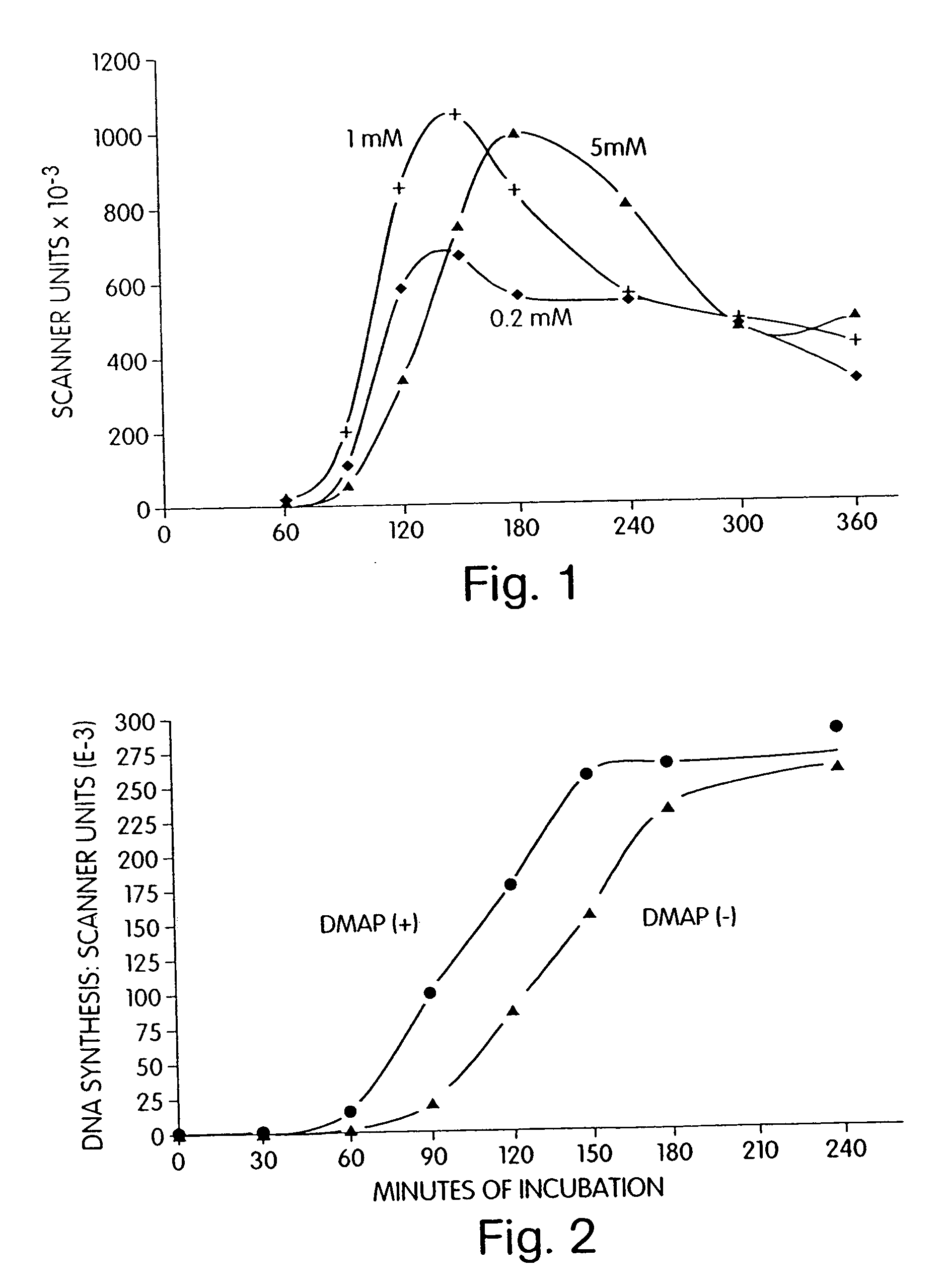
[0249] The effect of caffeine on DNA replication in activated Xenopus red blood cells was determined. The experimental conditions used were as described in Example 1 with the following changes: the concentration of cAMP was set at 0.3 μM and caffeine was added to the activating egg extract to a concentration of either 0.2 mM, 1.0 mM, or 5.0 mM.

[0250] As illustrated by FIG. 1, caffeine at 1.0 mM in the presence of 0.3 μM cAMP gave the highest initial rate and extent of DNA replication in activated nuclei. Thus, caffeine can increase the activation activity of an activating egg extract.


PUM

PropertyMeasurementUnit
volumeaaaaaaaaaa
volumeaaaaaaaaaa
volumeaaaaaaaaaa

Abstract

The present invention concerns products and methods Particularly useful for activating and analyzing non-dividing cell nuclei. The featured products include activating egg extracts, cytostatic factor (CSF) extracts, kits containing these extracts, and a microchamber microscope slide useful in analyzing nucleus activation.

Description

RELATED APPLICATIONS [0001] This application is a continuation of U.S. Ser. No. 10 / 798,061, filed Mar. 10, 2004, which is a continuation of U.S. Ser. No. 09 / 226,766, filed Jan. 6, 1999, now U.S. Pat. No. 6,753,457, which is a continuation of U.S. Ser. No. 09 / 050,380, filed Mar. 30, 1998, which is a continuation of U.S. Ser. No. 08 / 455,981, filed May 31, 1995, now U.S. Pat. No. 5,773,217, which is a division of U.S. Ser. No. 08 / 190,771, filed Feb. 1, 1994, now U.S. Pat. No. 5,651,992, which is a continuation-in-part of U.S. Ser. No. 08 / 013,039, filed Feb. 3, 1993, now U.S. Pat. No. 5,480,772, each of which is incorporated herein by reference.FIELD OF THE INVENTION [0002] This invention concerns products, methods, and apparatus for analysis of non-dividing mammalian cell nuclei, such as human fetal cell nuclei and mammalian sperm cell nuclei. BACKGROUND OF THE INVENTION [0003] Jackson, Seminars in Perinatology 15: 49 (1991, describes various procedures to diagnose diseases. These proc...

Claims


Application Information

Patent Timeline
Patent Type & Authority Applications(United States)
IPC IPC(8): C12M3/04C12N5/02C12N5/076C12N15/873C12Q1/68C12Q1/6841C12Q1/70
CPCA01K2217/05C12N5/061C12N15/873C12N2500/80C12Q1/70C12N2501/70C12N2517/10C12Q1/6841C12N2501/01
Inventor WANGH, LAWRENCE J.
Owner BRANDEIS UNIV
Why Eureka
Try Eureka