
Using liquid crystals to detect affinity microcontact printed biomolecules

a technology of affinity microcontact and liquid crystals, applied in the field of analytical chemistry and biochemistry, can solve the problems of radioactive labels that require special handling, expensive labeling, and radioactive materials that are also hazardous

Active Publication Date: 2005-04-14
WISCONSIN ALUMNI RES FOUND
View PDF41 Cites 81 Cited by
  • Summary
  • Abstract
  • Description
  • Claims
  • Application Information

AI Technical Summary

Benefits of technology

In the method, the receptor is bound to the affinity substrate via one or more linking moieties. Further, the method also provides for quantification of amount of ligand present in the sample.
In another embodiment, the method provides the affinity substrate comprising an array of receptors located in distinct locations. Gener

Problems solved by technology

Labeled antibodies can be expensive, especially for assays requiring radioactive labels.
Additionally, radioactive labels require special handling as radioactive materials are also hazardous.
The labeling of a compound, which is the main drawback of these methods, may alter the binding affinity of antibody to analyte.
This can lead to an increase in background noise, loss of sensitivity, and potentially a false positive test result.
Additionally, the immobilization of proteins on the solid support can affect the conformation of the binding sites, leading to a decrease in sensitivity, and possible increase in non-specific binding.
This can lead to possible inactivation of the binding sites.
Introduction of an antibody specific to the adsorbed protein can link the beads, leading to agglutination.
Visual agglutination assays, however, are not sensitive and measurement by quasi-elastic light scattering requires complex apparatus and is not suitable for use in locations remote from central labs.
Furthermore, it is not possible to perform highly multiplexed agglutination assays using microarrays because of the bulk solution methodology of this type of assay.
This temperature stability requirement makes SPR unsuitable for most field applications.
In addition, non-specific adsorption of molecules on to or near the sensor surface can lead to false changes in signal, requiring a surface which minimizes non-specific interactions.
Therefore, surface plasmon reflectivity is more complex than ELISA, requires laboratory based equipment, and the preparation of a well defined surface.
The binding of a bivalent molecule to both the ion channel and membrane spanning lipid restricts the mobility of the outer ion channel, leading to a measurable decrease in conductivity.
However, if a large amount of protein adsorbs to the outer layer, the ion channel mobility presumably would be restricted and a false decrease in conductance could be observed due to non-specific interactions.
Additionally, this method requires sensitive devices for detecting the change in conductance.
The procedure for fabricating the membranes requires several hours and the membrane stability is limited (must be immersed in solution).
In addition, a change in the effective optical thickness of the film was reportedly observed upon introduction of streptavidin, however they could not differentiate between specific interactions and non-specific adsorption.
This method does not require labeled molecules, however, the porous silicon surface is susceptible to oxidation and non-specific adsorption.
A significant disadvantage of this method, however, is that it requires multi-valent analyte.
Furthermore, Langmuir-Blodgett deposition is a process which is difficult to translate from laboratory to commercial scale.
However, research based on vesicles, reveals the usefulness of the system to be limited because it is insensitive to the analyte at concentrations below 0.1 mg / ml.
Although the disclosed liquid crystal-based assay devices which use anisotropic gold films are suitable for use in determining whether a target protein is present in a sample, the preparation of the anisotropic gold film by oblique deposition is difficult.
For example, the preparation of the obliquely deposited gold films requires complicated cleaning steps and high vacuum deposition.
Because a single surface is used for both capture and detection, the surface cannot be optimized to perform both functions.
Although many of the conventional assay methods described above work very well to detect the presence of target species, many conventional assay methods are expensive and often require instrumentation and highly trained individuals, which makes them difficult to use routinely in the field.

Method used


Image

  • Using liquid crystals to detect affinity microcontact printed biomolecules
  • Using liquid crystals to detect affinity microcontact printed biomolecules
  • Using liquid crystals to detect affinity microcontact printed biomolecules

Examples


example 1

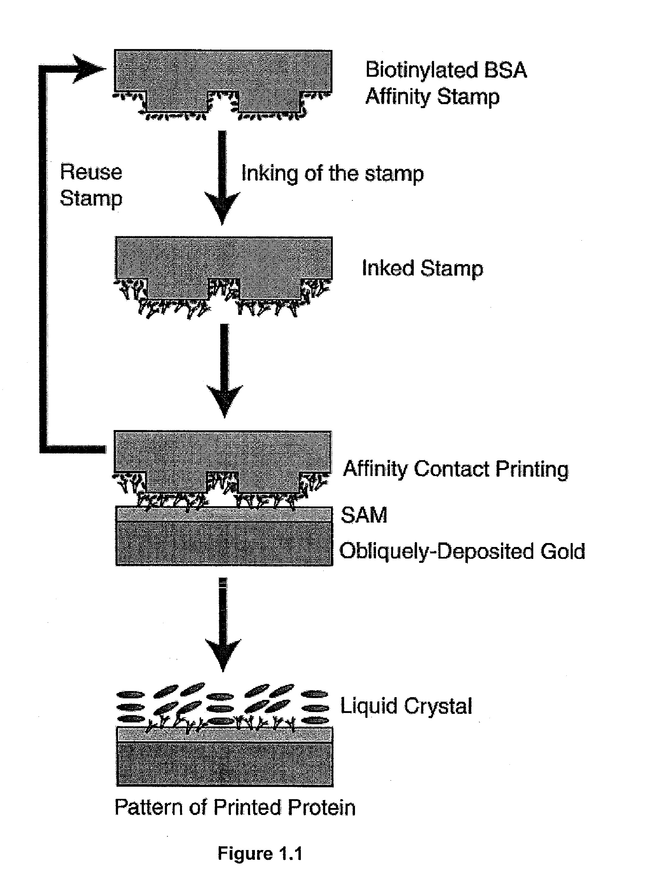
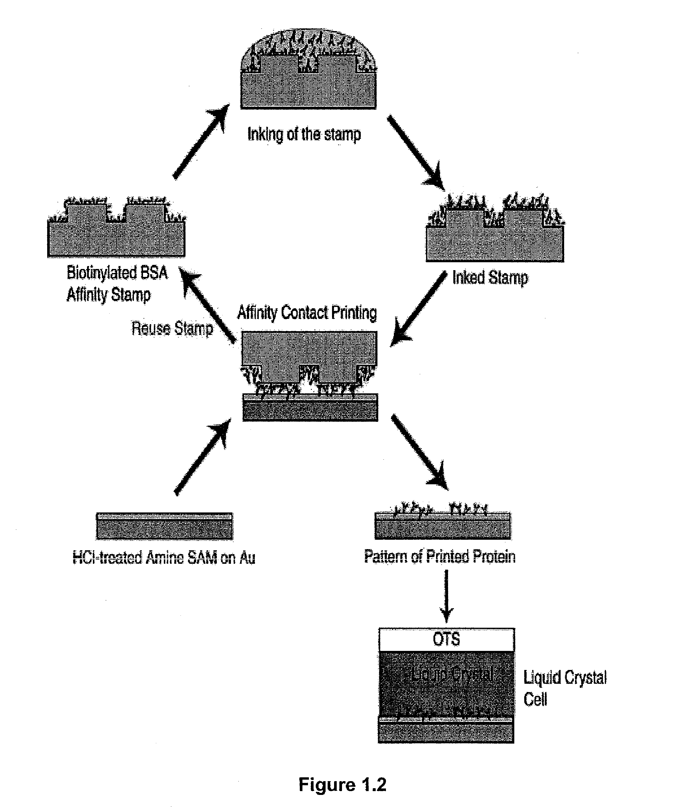
Design of Surfaces for Affinity Microcontact Printing. The present example used a detection surface having a SAM that possessed the following characteristics: (I) hydrophilic, (II) uniformly aligns liquid crystals, (III) protein will not desorb from the SAM when contacted with liquid crystal or an aqueous buffer. Amine-terminated SAMs are hydrophilic. Harnett, et al. Appl Phys Lett 2000, 76, 2466-2468, Dulcey, et al. Science 1991, 252, 551-554. Prior work has also shown that surfaces coated with primary amines non-specifically adsorb biological materials from solution. Accordingly, amine terminated SAMs were used in the present example. The final characteristic sought for the detection surface was uniform alignment of liquid crystal. To determine if the amine-terminated SAM would uniformly align the liquid crystal 5CB, a sandwich cell with the liquid crystal 5CB sandwiched between two substrates (liquid crystal thickness: ˜12 μm) was created. The bottom substrate was an amine-termi...

example 2

Using Ellipsometry to Confirm Affinity Microcontact Printing of Proteins. The transfer of protein from the affinity stamp to the amine-terminated SAM pretreated with 0.1 N HCl was confirmed by ellipsometry. For ellipsometry, flat PDMS were used stamps. The stamp was first oxidized with an O2 plasma and then functionalized with a primary amine using silane chemistry (aminopropyltriethoxysilane). Biotinylated BSA was then covalently attached to the stamp through Bis[sulfosuccinimidyl] suberate (BS3). The stamp was inked by placing a drop of anti-biotin IgG (1 mg / ml in PBS) on the stamp surface for 5 hours then rinsed with water for 15 seconds. The stamp was then contacted with the amine-terminated SAM for 30 seconds. A change in ellipsometric thickness of 10.8±0.4 nm was measured for stamping the inked anti-biotin IgG from the affinity stamp. Prior work has reported a similar change in ellipsometric thinkness (10 nm) for the binding of anti-biotin IgG to immobilized biotinylated BSA....

example 3

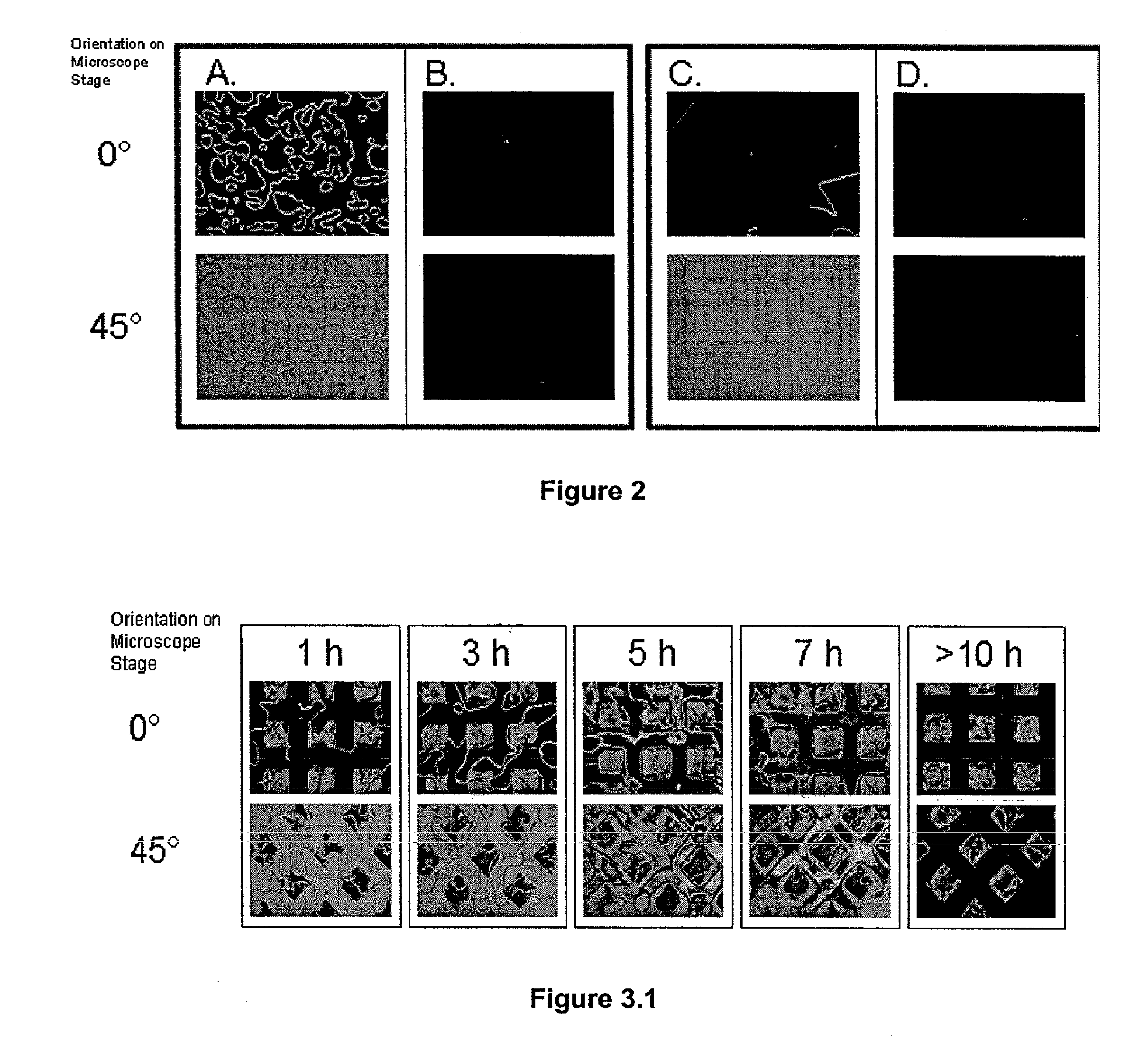
Orientations of Liquid Crystals on Affinity Microcontact Printed Proteins. The orientations of liquid crystals were next investigated on proteins that were deposited onto amine-terminated SAMs by affinity microcontact printing. The PDMS stamp used for the liquid crystal experiments possessed an array of 300×300 μm square pegs. Using the same procedure described above, the stamp was oxidized and then functionalized with a primary amine. Biotinylated BSA was covalently attached to the stamp. The stamp was then inked by placing a drop of anti-biotin IgG (1 mg / ml in PBS) on the stamp surface for 5 hours then rinsed with water for 15 seconds. The stamp was then contacted with the amine-terminated SAM for 30 seconds. The liquid crystal 5CB was then sandwiched between two surfaces for detection of the protein. The bottom surface of the liquid crystal cell was the amine-terminated SAM pretreated with 0.1N HCl that had been affinity microcontact printed with antibody. The top surface was OT...


PUM

PropertyMeasurementUnit
Affinityaaaaaaaaaa

Abstract

The present invention provides methods, devices and kits for detecting a ligand. The methods involve capturing a ligand from a sample with an affinity substrate that includes a receptor for a ligand, transferring captured ligand to a detection surface and detecting the ligand on the detection surface with a liquid crystal. Accordingly, the capture step is decoupled from the detection step.

Description

BACKGROUND Methods for detecting the presence of biological substances and chemical compounds in samples has been an area of continuous development in the field of analytical chemistry and biochemistry. Various methods have been developed that allow for the detection of various target species in samples taken from sources such as the environment or a living organism. Detection of a target species is often necessary in clinical situations before an illness may be diagnosed and a prescribed method of treatment may be undertaken. Several types of assay currently exist for detecting the presence of target species in samples. One conventional type of assay is the radioimmunoassay (RIA). RIA is a highly sensitive technique that can detect very low concentrations of antigen or antibody in a sample. RIA involves the competitive binding of radiolabeled antigen and unlabeled antigen to a high-affinity antibody. Typically, the labeled antigen is mixed with the antibody at a concentration tha...

Claims


Application Information

Patent Timeline
IPC IPC(8): B01J19/00C12Q1/68C12Q1/70C40B40/06C40B40/10C40B40/18C40B60/14G01N33/53G01N33/542G01N33/543
CPCB01J19/0046G01N33/54373B01J2219/00596B01J2219/00659B01J2219/0072B01J2219/00722B01J2219/00725B01J2219/0074B01J2219/00743B01J2219/00745B01J2219/0075B82Y15/00B82Y30/00C40B40/06C40B40/10C40B40/18C40B60/14B01J2219/00382
Inventor ABBOTT, NICHOLAS L.TINGEY, MATTHEW L.CLARE, BRIAN H.JANG, CHANG-HYUN
Owner WISCONSIN ALUMNI RES FOUND
Learn More