
Methods for accelerating bone, cartilage, and connective tissue growth

a technology of connective tissue and bone, which is applied in the direction of skeletal/connective tissue cells, parathyroid hormones, osteogenic factors, etc., can solve the problems of individual at risk of skeletal failure, slow process, and requiring years to reunite, and achieve the effect of enhancing bone and cartilage repair

Inactive Publication Date: 2005-08-25
UNIV OF SOUTHERN CALIFORNIA
View PDF30 Cites 4 Cited by
  • Summary
  • Abstract
  • Description
  • Claims
  • Application Information

AI Technical Summary

Benefits of technology

The present invention provides methods, kits, and compositions for improving bone and cartilage repair, as well as methods for attaching and fixing cartilage to bone or other tissues. These methods involve the use of angiotensinogen, angiotensin I, angiotensin II, and their analogues thereof. Additionally, the invention also includes methods for promoting the growth of chondrocytes, which are important for bone and cartilage health. These methods may involve the use of angiotensin II AT2 type 2 receptor agonists. Overall, the invention provides new ways to enhance bone and cartilage regeneration, which could be useful in treating bone and cartilage injuries or degenerative diseases.

Problems solved by technology

This process is very slow however, often requiring years to reunite a large segmental defect.
Inadequate organic matrix mass places an individual at risk of skeletal failure such that bone fractures can result from the minimal trauma of everyday life.
Such fractures cause significant illness, or morbidity, inasmuch as there is insufficient repair or healing of the fractures.
Such accelerated osteoclastic activity leads to excessive release of calcium from the inorganic mineral in bone, with a concomitant net loss of skeletal mass, often with an attendant disturbance in calcium homeostasis in the form of elevated blood levels of calcium.
As such, they are not as effective in patients with established osteoporosis of either type (decreased bone density with fractures of the vertebrae and / or hip), or in patients with Type II osteoporosis.
In addition, the most accepted preventive agent for osteoporosis currently in use is estrogen therapy, which is not an acceptable therapeutic agent for women with a history of breast cancer or endometrial cancer or for men with osteoporosis.
An estimated ten percent of bone and joint prosthetic devices that are placed in people fail to function due to non-bonding of the bone to an implant.
The resulting disability often requires reoperation and reimplantation of the device.
Furthermore, five to ten percent of all bone fractures are never repaired.
Although many methods have been proposed to cure these non-healing bone fractures, none has yet proven to be satisfactory.

Method used


Image

  • Methods for accelerating bone, cartilage, and connective tissue growth
  • Methods for accelerating bone, cartilage, and connective tissue growth
  • Methods for accelerating bone, cartilage, and connective tissue growth

Examples


example 1

Method for Culture of Rabbit Chondrocytes

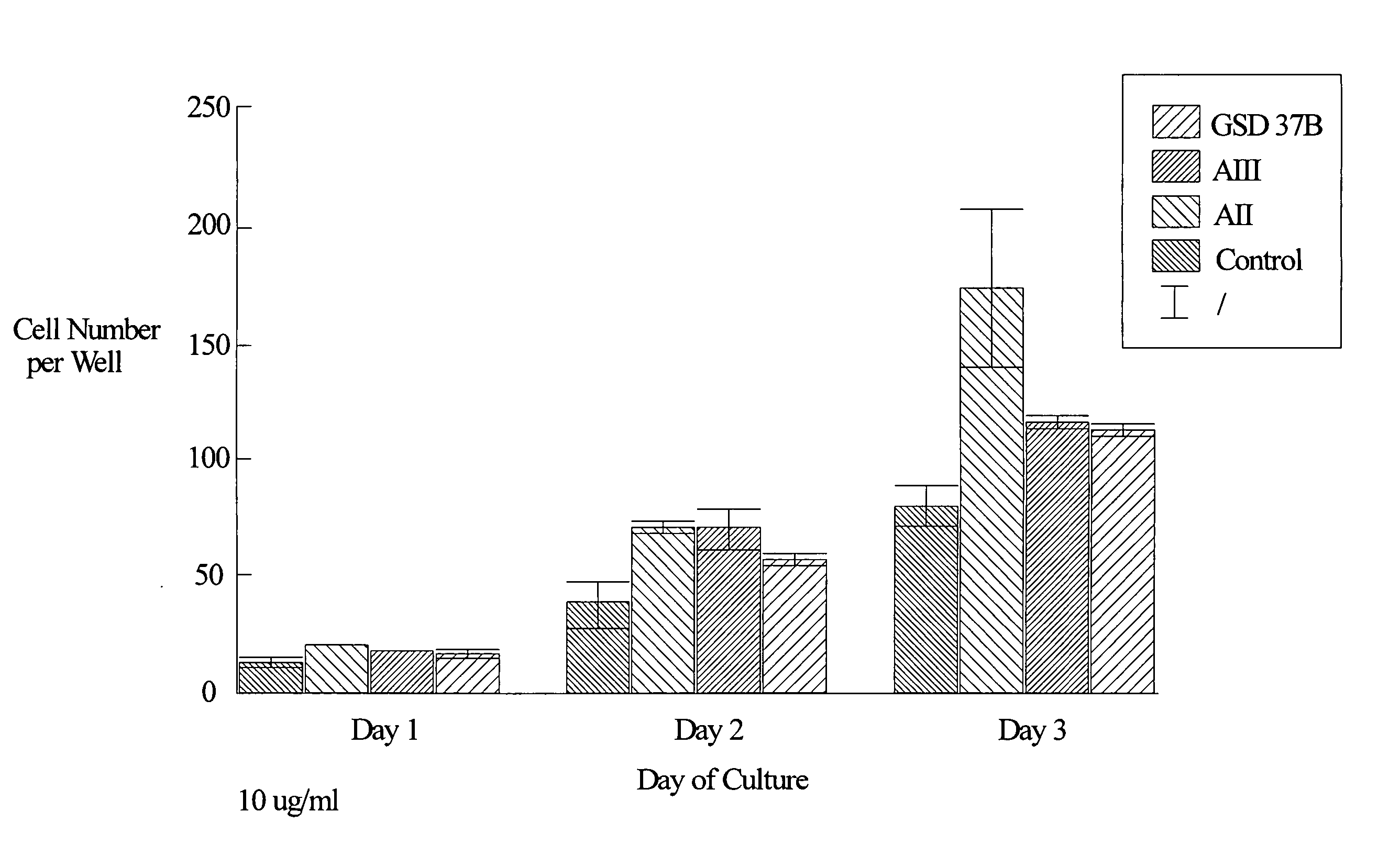
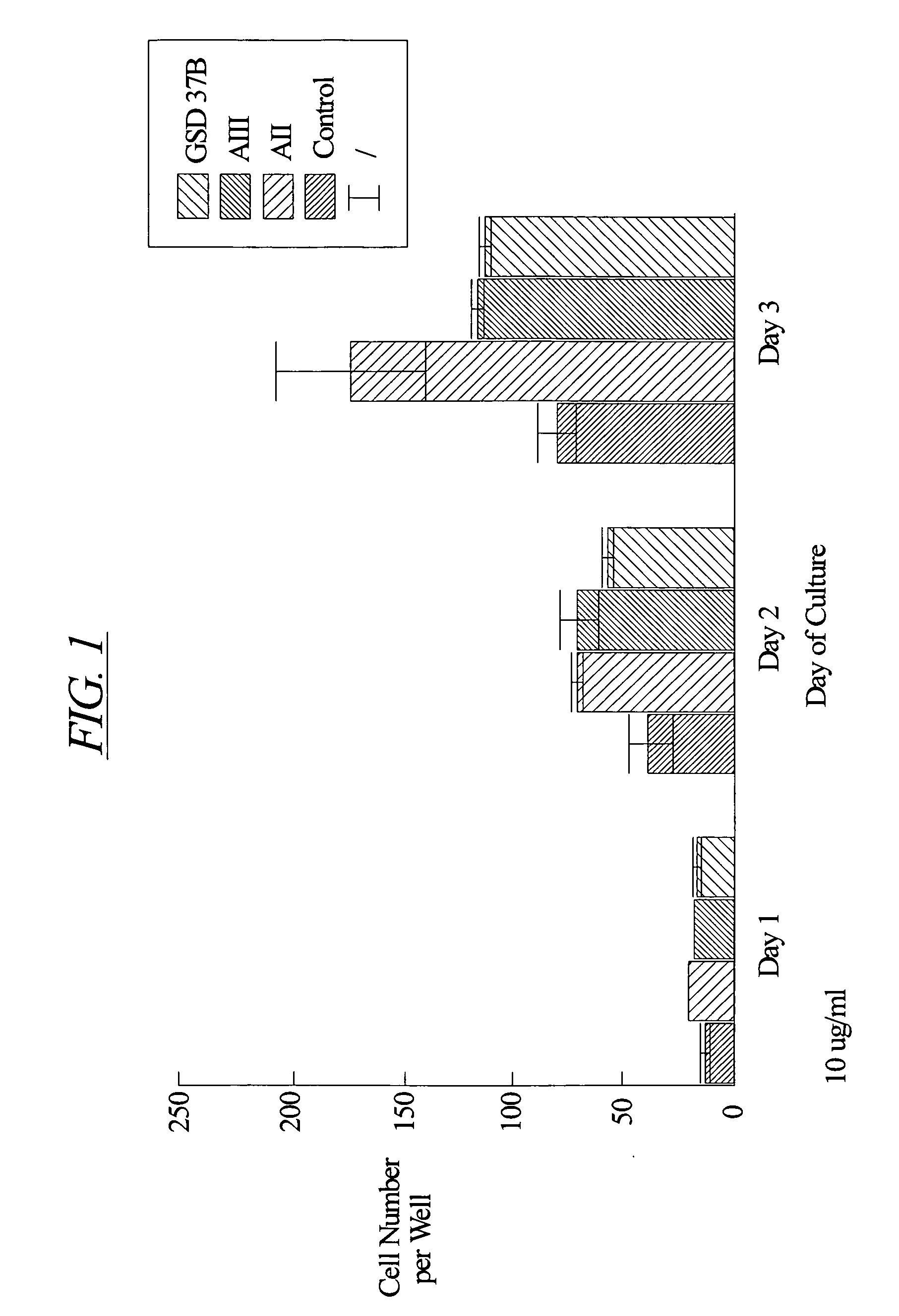
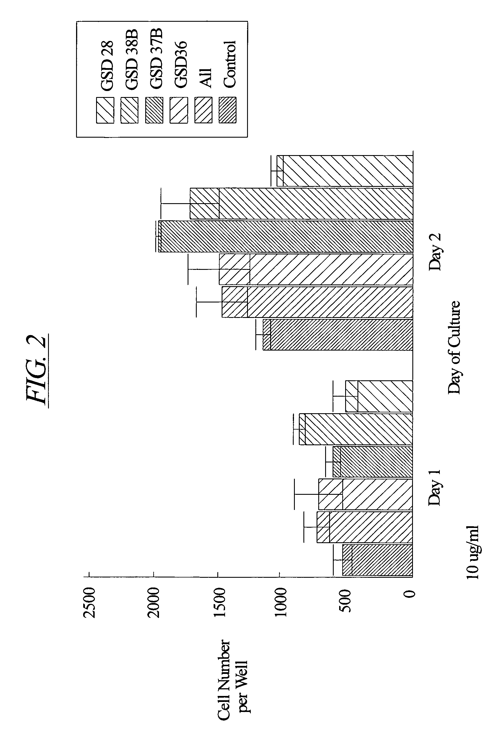
[0126] Chondrocytes were isolated from the cartilage of the knee joints of adult rabbits and cultured according to the method of Okazaki et al. (Ann. Rheum. Dis. 55:181-186, 1996). Briefly, cartilage explants were minced into small pieces (approximately 1.5 mm by 1.5 mm). The tissue pieces were digested at 37° C. sequentially in 0.05% hyaluronidase (415 U / mg protein) for 10 minutes, 0.2% Type III trypsin (10,000 U / mg protein, Sigma) and 0.53 mM EDTA for 15 minutes, and 0.2% Type I collagenase (136 U / mg protein, Sigma) for 30 minutes. The samples were then washed and incubated overnight at 37° C. in 5% CO2 in air in Dulbecco's modified Eagle's medium enriched with 10% fetal calf serum, 3.5 mg / ml glucose, 0.2% Type I collagenase and antibiotics (100 U / ml penicillin and 100 μg / ml streptomycin).

[0127] After this incubation, the cells were harvested and washed once with phosphate buffered saline (pH 7.2). The cells were then counted with a hemac...

example 2

Bone Healing

[0128] Female, Sprague Dawley rats underwent intramuscular anesthesia with ketamine / rompum and were prepared for sterile surgery by shaving the surgical site and scrubbing with Betadine scrub followed by 70% ethanol. The rat was then placed on a sterile field in a lateral decubitis position facing the surgeon. The shaved legs were then covered with Betadine solution and draped aseptically. A skin incision was performed parallel to the long axis of the right medial diaphysis. The muscle was separated along fascial planes to expose the tibia. A defect of 1.4 mm in diameter was then drilled from the lateral side of the midshaft cortex so that the defect extended from one cortical side to the other, through the bone marrow. Sterile saline (0.9% NaCl) for injection was then used to clean the surgical area of tissue debris and bone fragments. Either hydron polymer solution (vehicle: 10% Hydron, 60% ethanol, 1% polyethylene glycol polymer) or peptide (AII, AII(1-7), or 9GD at ...


PUM

PropertyMeasurementUnit
densityaaaaaaaaaa
concentrationaaaaaaaaaa

Abstract

The present invention provides improved methods, kits, and compositions for enhancing bone, cartilage and cartilage repair, bone and prosthesis implantation, and attachment and fixation of cartilage and cartilage to bone or other tissues, and chondrocyte proliferation comprising the administration of an effective amount of angiotensinogen, angiotensin I (AI), AI analogues, AI fragments and analogues thereof, angiotensin II (AII), AII analogues, AII fragments or analogues thereof or AII AT2 type 2 receptor agonists.

Description

CROSS REFERENCE [0001] This application is a continuation-in-part of U.S. Provisional Application Ser. No. 60 / 092,653 filed Jul. 13, 1998 and a continuation in part of U.S. Provisional Application Ser. No. 60 / 130,855 filed Mar. 8, 1999.FIELD OF THE INVENTION [0002] The present invention relates to methods, compositions, and kits for the repair, regeneration, and implantation of bone and cartilage. BACKGROUND OF THE INVENTION [0003] Natural mechanisms of repair, healing and augmentation are similar for bone and cartilage. (U.S. Pat. No. 5,686,116) Although repair, healing and augmentation require a complex series of events that are not well defined, it is known that specific, naturally occurring factors are required to achieve these objectives. Such factors are released or migrate into the injured area, and stimulate osteoblasts and chondrocytes and odontoblasts in bone and cartilage to stimulate matrix formation and remodeling of the wounded area. (ten Dijke et al., Bio / Technology, ...

Claims


Application Information

Patent Timeline
Patent Type & Authority Applications(United States)
IPC IPC(8): A61K35/12A61K38/00A61K38/18A61K38/29A61K38/30C07K7/14C12N5/077
CPCA61K35/12A61K38/1841C12N2501/32C12N2500/34C12N5/0655C07K7/14A61K38/30A61K38/29A61K38/1875A61K2300/00
Inventor RODGERS, KATHLEENDIZEREGA, GERE
Owner UNIV OF SOUTHERN CALIFORNIA
Learn More