

Method and apparatus for measuring changes in cell volume
a technology of cell volume and measurement method, applied in biochemistry apparatus and processes, instruments, material analysis, etc., can solve the problems of limited sensitivity of o'connor devices, difficult detection of small changes in resistance, and considerable time for measuring, if detected at all., to achieve small intracellular volume changes
- Summary
- Abstract
- Description
- Claims
- Application Information
AI Technical Summary
Benefits of technology
Problems solved by technology
Method used
Image

Examples
experiment # 1
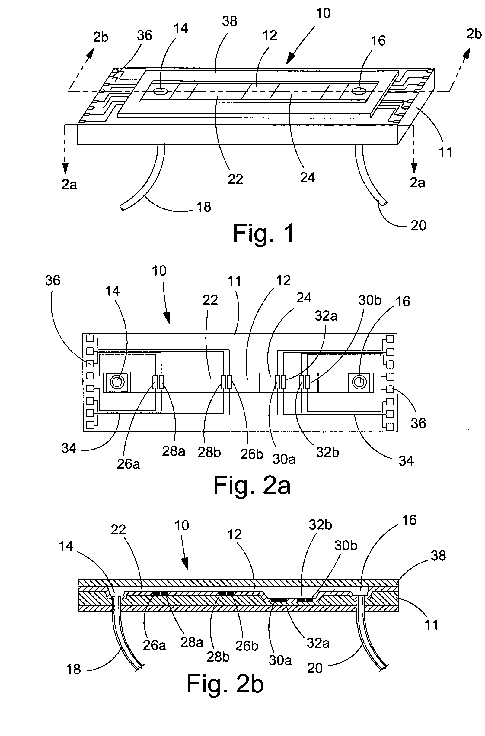
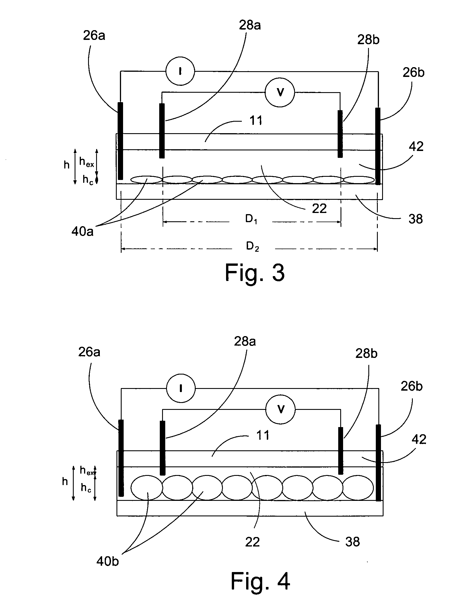
[0073] In a first experiment, a 1.5 mm wide, 25 μm deep fluidic channel 12 connecting inlet 14 and outlet 16 reservoirs carried test solutions and reagents. Two measuring chambers 22, 24 with different depths were located along the length of the fluidic channel. First chamber 22 was designed as the cell testing chamber and had a depth of 25 μm. Second chamber 24 was designed as the control / calibration chamber for extracellular fluid resistivity and had a depth of 55 μm. Four platinum electrodes, each 50 μm wide, were located within each the first and second chambers to form a four-point probes for electrical impedance measurements. A device similar to that of FIGS. 1-3 was secured glued to an acrylic platform (not shown). The acrylic platform contained a three way fluid input connection, which aligned with inlet 14 for changing testing solutions, and fluid outlet tubing 20 connected with the outlet 16. For testing, astrocytes were cultured on normal glass cover slips and placed on t...
experiment # 2
Experiment #2
[0077] A microfluidic chip comprising a channel 15 μm deep and 1.5 mm wide was fabricated an connected to a fluid inlet and a fluid outlet. Along the length of the channel, there were two chambers (labeled 22 and 24 in FIGS. 1-3). Chamber 22 was configured for measuring cell volume and comprised a depth of 15 μm. Chamber 24 was deeper (55 μm) and served as a calibrating chamber for monitoring solution resistivity. Thin film platinum electrodes disposed in each chamber formed a four-point probe for measuring the chamber impedance. The chip was mounted on an acrylic platform to mate with external fluid connections. For testing adherent cells, the cells were cultured on glass coverslips and inverted on top of the chip so that the cells faced chamber 22. The coverslip was pressed against the chip with a clamp that applied a uniform force of approximately 50N. For the electrical measurements, an active current source provided 1 μA of a 50 Hz sinusoid was applied to the two o...
experiment # 3
Experiment #3
[0082] A microfluidic chip was tested by screening peptides isolated from the tarantula Gammostola spatulata. The peptides were added to a hypotonic perfusate and their effects on astrocyte RVD were examined. The solid black curve of FIG. 13 shows a control RVD with a 188 mOsm stimulus. RVD was blocked by a small inhibitory cysteine knot (ICK) peptide called GsMT×1. This peptide was previously known to block swelling-induced Ca2+ uptake in GH3 cells. In this experiment, GsMT×1 completely blocked RVD at 1 μM, 10 nM, and 1 nM, as shown in FIG. 13. At 100 pM it reduced RVD by about 50%. This high affinity suggests that GsMT×1 is an antagonist to a key component of RVD, perhaps the volume sensing ability of the cell itself. GsMT×1 inhibition was striking in that it seemed to affect the set point of regulation rather than the rate of regulation.
PUM
 Abstract
Description
Claims
Application Information