

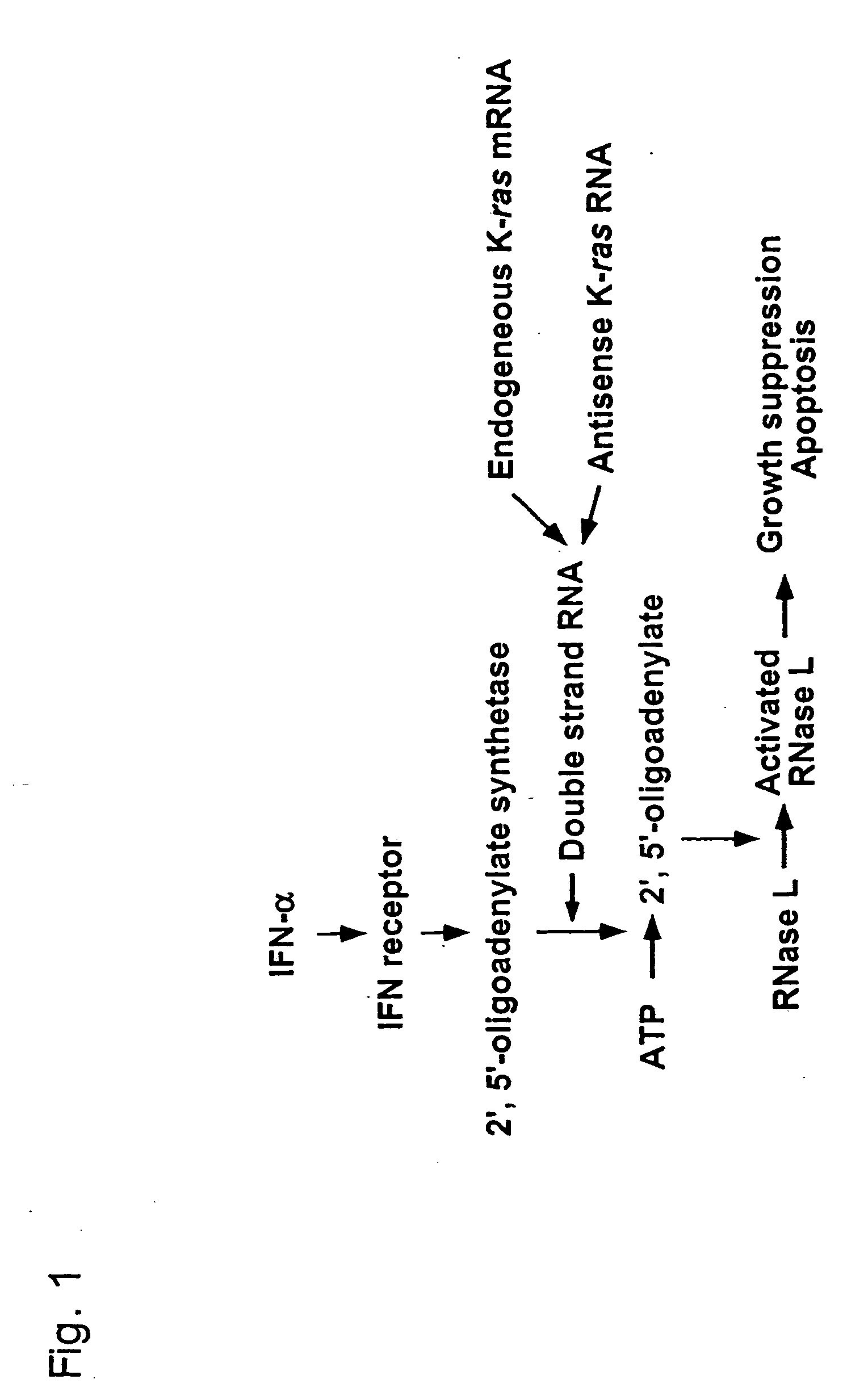
Interferon alpha and antisense K-ras RNA combination gene therapy
a technology of interferon and kras, which is applied in the direction of biocide, drug composition, genetic material ingredients, etc., can solve the problems of inability to document sufficient evidence for enlistment, and the study of ifn- gene transfer against pancreatic cancer has not been reported, so as to enhance ifn--induced cell death, enhance ifn--induced rnase l activation, and enhance ifn--induced
- Summary
- Abstract
- Description
- Claims
- Application Information
AI Technical Summary
Benefits of technology
Problems solved by technology
Method used
Image

Examples
example 1
Methods
[0055] A. Cell Lines and Culture Conditions
[0056] Five human pancreatic cancer cell lines (AsPC-1, MIAPaCa-2, Panc-1, PSN-1, 766T), 4 human non-pancreatic cancer cell lines (hepatocellular carcinoma cell line: HepG2, non-small cell lung cancer cell line: A549, prostate cancer cell line: PC-3, bladder cancer cell line: T24) and 3 primary cultures of human normal cell lines (hepatocytes, human umbilical vascular endothelial cells: HUVEC, smooth muscle cells: SMC) were used in this study. All cancer cell lines except for PSN-1 were obtained from American Tissue Culture Collection, and PSN-1 was established in our laboratory (36). All cancer cell lines except for 766T and HepG2 cells were maintained in an RPMI-1640 medium with 10% fetal bovine serum (FBS), and the 766T and HepG2 cells were cultured in Dulbecco's modified eagle's medium with 10% FBS. Primary cultures of normal human cells were purchased from Dainippon Pharmaceutical Co. (Osaka, Japan) and were cultured according...
example 2
Gene Transduction into Subcutaneous Tumor: I
[0070] AsPC-1 cells were used in an in vivo gene transfer model in Five week-old male Balb / c nude mice obtained from Charles River Japan (Kanagawa, Japan), and kept in a specific-pathogen-free environment. AsPC-1 cell suspensions (5×10⁶ cells per 50 μl) were injected subcutaneously into the left flank, and a total of 5×10⁸ or 1×10⁸ pfu of viral solution was injected once into the tumor with a 27-gauge hypodermic needle, when the tumor mass was established (˜0.6 cm in diameter). Control tumor-bearing mice were injected with 5×10⁸ or 1×10⁸ pfu of ADV-AP alone. Three groups of mice were injected with AxCA-AS and ADV-AP, AxCA-IFN and ADV-AP, or AxCA-IFN and AxCA-AS (2.5×10⁸ or 0.5×10⁸ pfu each), respectively. One mouse from ADV-AP control group and AxCA-IFN / ADV-AP (5×10⁸ pfu) injected group was sacrificed for a histological examination of the subcutaneous tumor by hematoxylin-eosin staining and TUNEL assay, another 3 mice from each group were...
example 3
Cytotoxic Effect of Interferon-α Gene Transduction into Pancreatic Cancer Cells
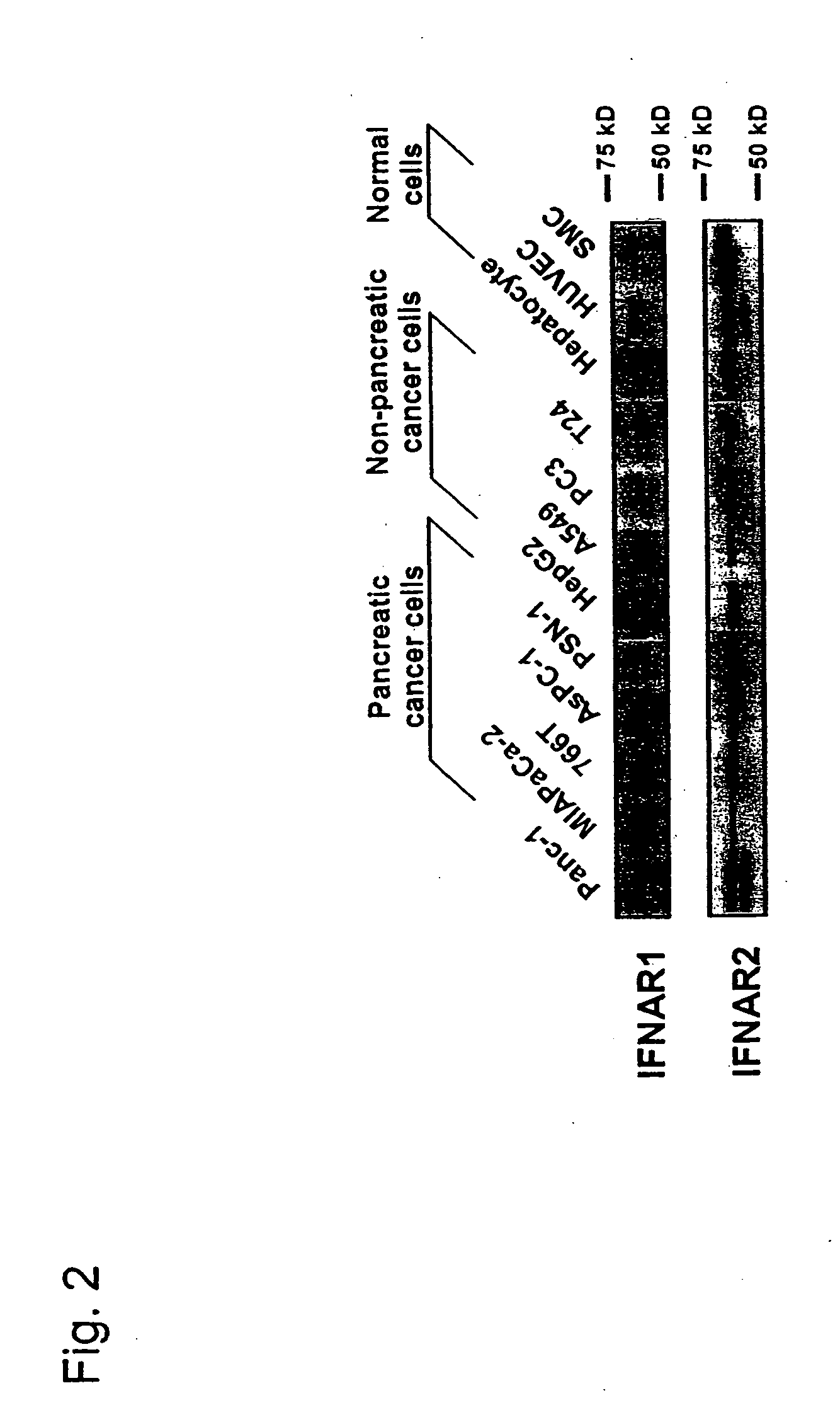
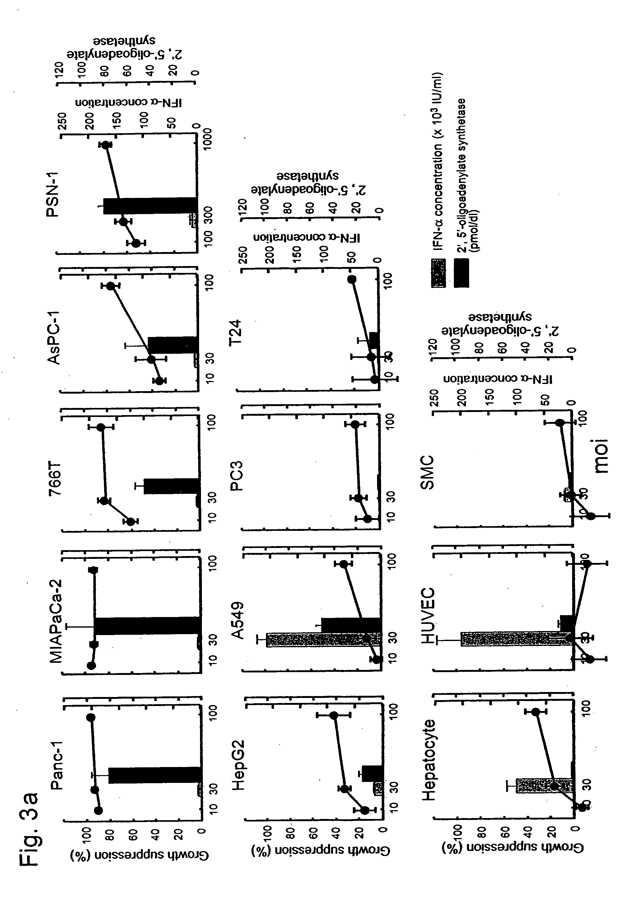
[0071] To study whether the expression of the human IFN-α gene effectively inhibits cell growth, 5 pancreatic cancer cell lines, 4 non-pancreatic cancer cell lines and 3 primary cultures of human normal cells were infected with the AxCA-IFN adenoviral vector. Western blot analysis showed that all cell lines express two subunits of interferon α / β receptor (IFNAR1 and IFNAR2) in varying degrees (FIG. 2). The amounts of IFN-α secreted from AxCA-IFN-infected cells were dependent on the adenoviral dose but varied substantially among the cell lines (Table 1). The concentrations in the culture medium of infected pancreatic cancer cells were rather lower than those of HepG2, A549, hepatocytes and HUVEC. The infection with AxCA-IFN showed some growth suppression of non-pancreatic cancer cells and hepatocytes, whereas the vector inhibited cell growth and induced cell death in pancreatic cancer cells much more effe...
PUM
| Property | Measurement | Unit |
|---|---|---|
| wavelength | aaaaa | aaaaa |
| wavelength | aaaaa | aaaaa |
| diameter | aaaaa | aaaaa |
Abstract
Description
Claims
Application Information