

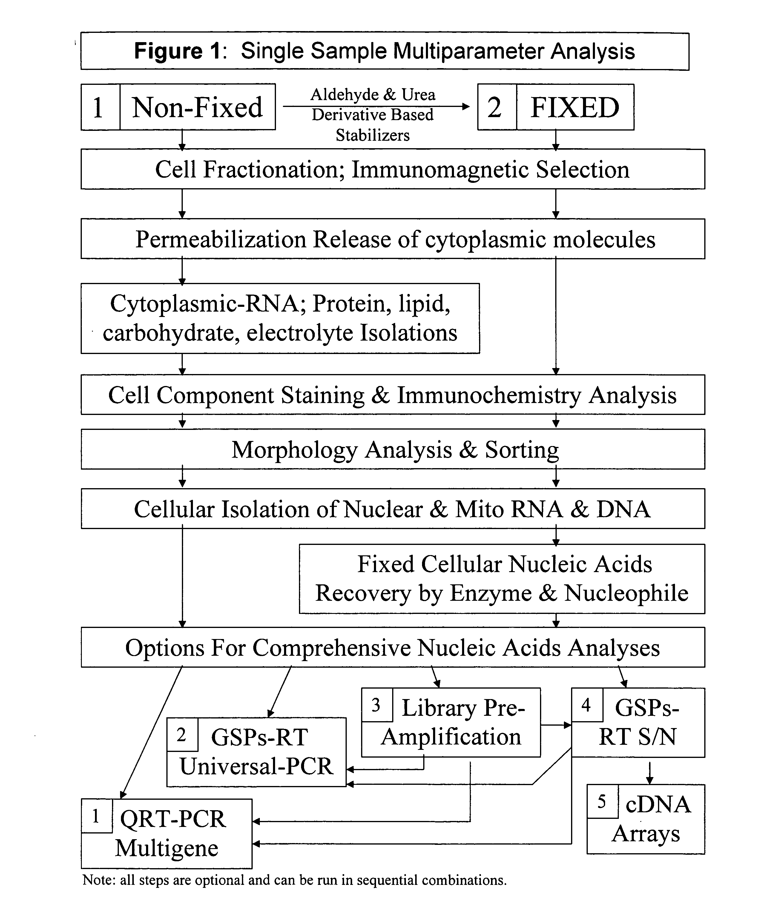
Multiparameter analysis of comprehensive nucleic acids and morphological features on the same sample
a morphological feature and multi-parameter analysis technology, applied in the field of gene specific amplification, analysis and profiling of cytosolic biomolecules, can solve the problems of slow and time-consuming analysis of several hundred thousand gene-specific probes, decreased signal-to-noise, and more difficult treatment of activated b cells
- Summary
- Abstract
- Description
- Claims
- Application Information
AI Technical Summary
Benefits of technology
Problems solved by technology
Method used
Image

Examples
example 1
Isolation of Cytoplasmic RNA
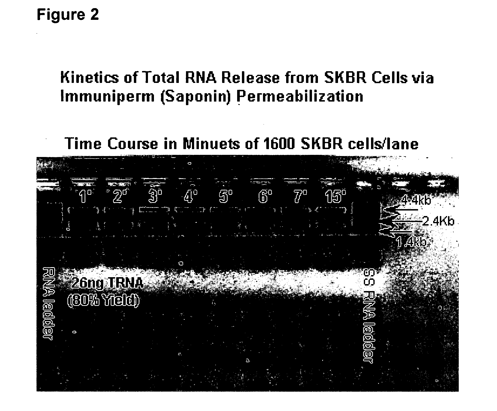
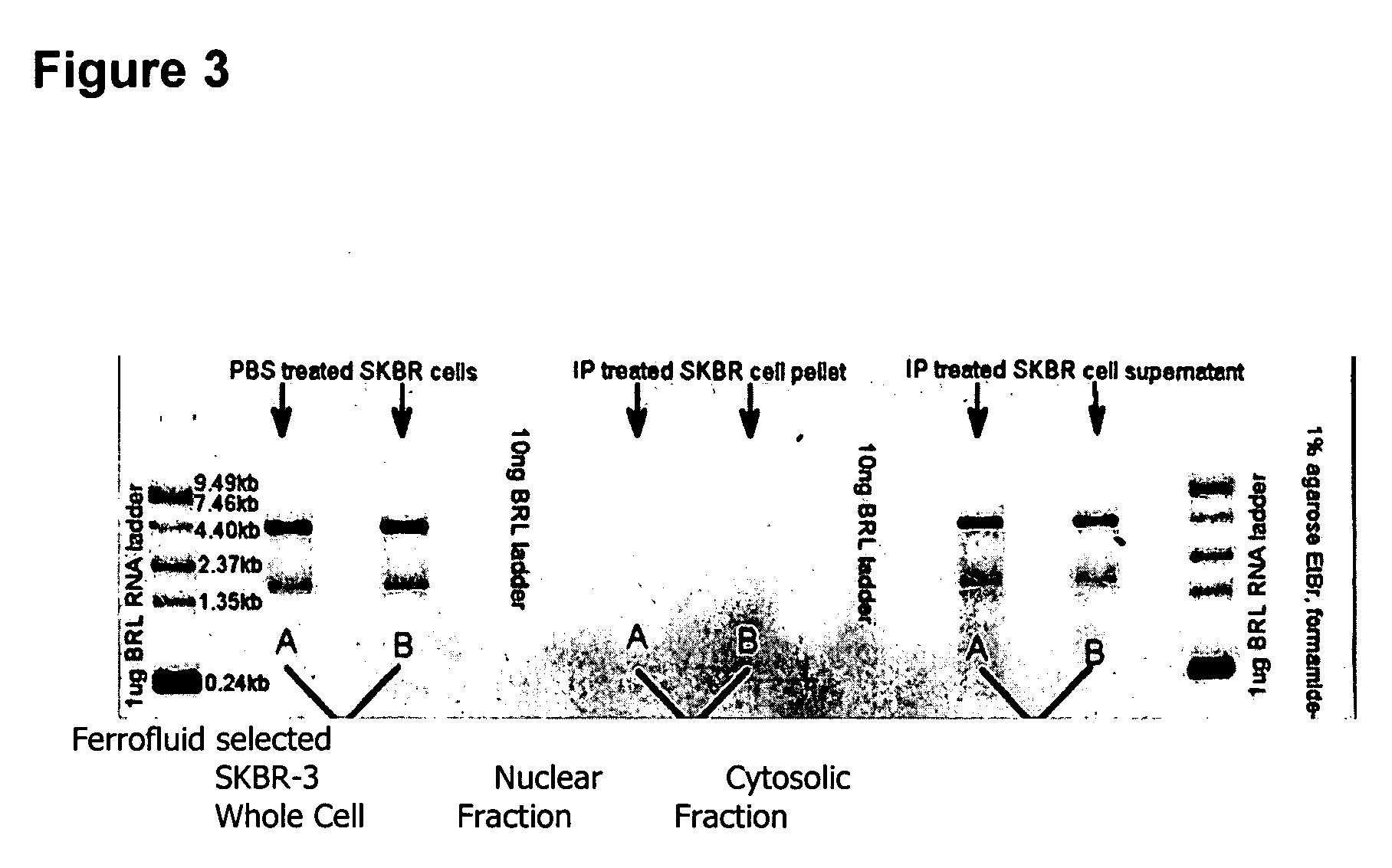
[0106] The supernatant obtained from ferrofluid selected unfixed cells that are permeabilized with Immuniperm, a phosphate buffered solution containing 0.05% saponin and 0.1% sodium azide was found to contain greater than 80% of the cellular total RNA residing in the cytoplasm of the cells. The RNA isolated from this supernatant showed no evidence of degradation as judged by native and denaturing agarose gel electrophoresis and ethidium bromide staining. This supernatant solution, which is normally discarded after intracellular staining of the ferrofluid selected cells, was unexpectedly found to contain the RNA in an intact or undegraded full-length form thus providing an mRNA profile of the same cells that were also used for morphologic analysis. FIG. 2 illustrates these findings showing that total RNA release occurs in less than one minute and that about 95% of the cytoplasmic total RNA can be readily and reproducibly isolated.
[0107] In dramatic cont...
example 2
Isolation of Circulating Tumor Cells from Peripheral Blood
[0117] Isolation of circulating tumor cells from peripheral blood followed by cell analysis by flowcytometry and gene expression analysis by RT-PCR can be performed as follows: EDTA-anticoagulated blood (7.5 ml) is transferred into a 15 ml conical tube and 6.5 ml of System Buffer (PBS also containing 0.05% sodium azide, Cat #7001, Immunicon Corp., Huntingdon Valley, Pa.) is added. The tube is securely capped and mixed by inverting several times. The blood-buffer mixture is centrifuged at 800×g for 10 minutes at room temperature. The supernatant is carefully removed by aspiration taking care not to disturb the buffy coat layer. Some supernatant can be left in the tube. The aspirated supernatant can be discarded. AB Buffer (System Buffer containing streptavidin as a reversible aggregation reagent, Immunicon Corp., Huntingdon Valley, Pa.) is added to the tube to a final volume of 10 ml. The tube is capped and mixed by invertin...
example 3
Cell Analysis of Circulating Tumor Cells from Peripheral Blood
[0118] The cells from Example 2 are resuspended in 200 ul of CellFix (PBS based buffer containing biotin as a de-aggregation reagent and cell preservative components, Immunicon Corp., Huntingdon Valley, Pa.) and incubated for 5 minutes. The sample is transferred to a 12×75 mm flow tube and 300 ul of PBS are combined, followed by the addition of the nucleic acid dye thioflavin T (Sigma # T3516, 10 ul) and about 10 ul of fluorescent beads (10,000 beads; Flow-Set Fluorospheres, Cat. # 6607007 Coulter, Miami, Fla.). The sample is mixed by vortexing. Preferably, the fluorescent beads tube is mixed by vortexing before pipetting the beads. The sample is then analyzed on a flowcytometer.
PUM
| Property | Measurement | Unit |
|---|---|---|
| temperature | aaaaa | aaaaa |
| time | aaaaa | aaaaa |
| time | aaaaa | aaaaa |
Abstract
Description
Claims
Application Information