Method of examing staphylococcus aureus

a staphylococcus and staphylococcus technology, applied in the field of staphylococcus aureus testing, can solve the problems of not being able to detect bacterium in a minute amount in blood, patients are often too serious to utilize the test, and the identification of bacterium is needed a long time, so as to achieve convenient and rapid testing, high sensitivity

Inactive Publication Date: 2006-03-09
SEKISUI MEDICAL CO LTD
View PDF1 Cites 14 Cited by
  • Summary
  • Abstract
  • Description
  • Claims
  • Application Information

AI Technical Summary

Benefits of technology

[0012] The present invention has enabled to test protein A at a high sensitivity without being affected by the IgG and the like in the specimen, and to conveniently and quickly test whether or not the patient is infected by S. aureus. The present invention can also determine the infection even if the S. aureus were MRSA.

Problems solved by technology

The conventional test methods, however, all required cultivation of the bacteria, and a substantial time was needed before the identification of the bacterium.
When the results of the identification were obtained, patients were often too serious to utilize the test results.
In addition, there has been a fair possibility that the bacterium present in a minute amount in blood is left undetected in patients receiving antibacterials.
However, protein A is known to have a site which binds with the Fc domain of immunoglobulin (IgG) from mammals, and accordingly, the immunoassays as described above had the problem of interaction of the IgG used in both or either one of the immobilized antibody and the labeled antibody with the site of the protein A that binds to the Fc domain.
Since S. aureus is an indigenous bacterium, its detection is associated with the risk shared by indigenous bacteria that those not infected are also detected as positive by the minute contamination of the bacterial gene.
This mecA gene, however, is sometimes found in Staphylococcus spp. such as CNS other than S. aureus, and detection of mecA gene has been associated with the difficulty of clearly differentiating between MRSA and CNS.

Method used


Image

  • Method of examing staphylococcus aureus
  • Method of examing staphylococcus aureus

Examples


example 1

(1) Preparation of a Plate Having Primary Antibody Immobilized Thereto

[0044] Mouse IgG 1 anti-protein A monoclonal antibody diluted with 25 mM phosphate buffer solution (PBS) containing 150 mM NaCl to 10 μg / mL was dispensed in the wells of a plate at 50 μL / well, and the plate was allowed to stand overnight at 4° C. for immobilization of the antibody. The content was removed from the plate the next day, and after adding 280 μL / well of BlockAce (manufactured by Dainippon Pharmaceutical) diluted to 4 folds with purified water, the plate was allowed to stand overnight or longer at 4° C. In use, the plate washed three times with PBS containing 0.05% Tween20 (PBS-T) was used for the reaction.

(2) Preparation of Biotinylated Secondary Antibody

[0045] 500 μL (1 mg / mL) of goat anti-protein A antibody F(ab!)2 obtained by digesting goat anti-protein A polyclonal antibody with pepsin was dialyzed against 0.1M sodium hydrogen carbonate. A solution of 0.05 mg of Sulfo-NHS-LC-Biotin (manufactur...

example 2

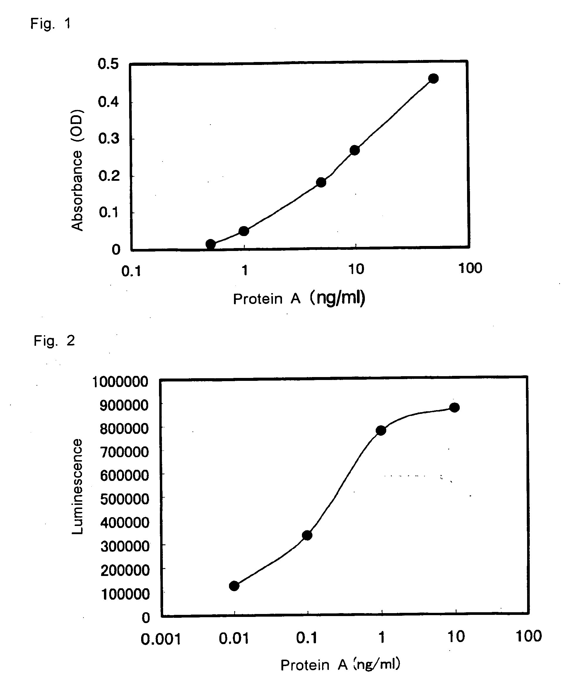
(1) Measurement of Protein A by Sandwich Enzyme Immunoassay (1)

[0046] 50 μL / well of purified protein A diluted with diluted BlockAce (BlockAce diluted to 10 folds with purified water) to 0.5, 1, 5, 10, and 50 ng / mL was added to the wells of the microtiter plate having the mouse IgG 1 anti-protein A monoclonal antibody immobilized thereto produced in Example 1(1). After allowing the reaction to take place overnight at 4° C., the wells were washed with PBS-T, and 50 μL / well of biotinylated secondary antibody of Example 1 diluted with diluted BlockAce to 2.9 μg / mL was added. The reaction was then allowed to proceed at room temperature for 1 hour, and the wells were washed with PBS-T. In the meanwhile, avidinated HRP (manufactured by Zymed) was diluted with 0.2% BSA-PBS to 4000 folds, and 50 μL / well of this dilution was added to the well, and the reaction was allowed to proceed at room temperature for 1 hour. After washing, 50 μL / well of 0.1M citrate buffer solution, containing 2 mg / m...

example 3

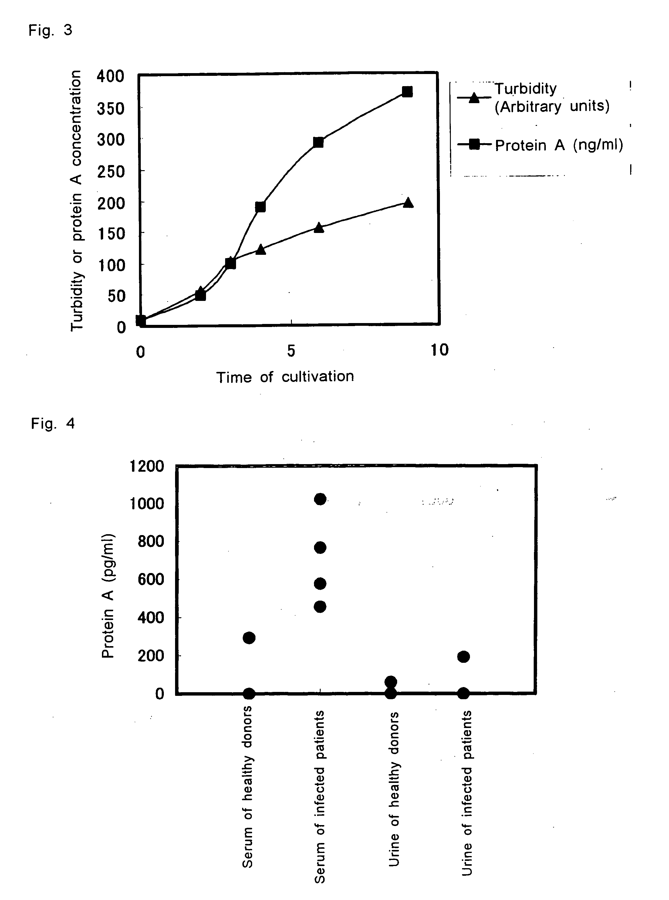
Measurement of the Amount of Protein A in the Culture Supernatant Over Time

[0050] To 10 mL of heart-infusion broth was added 80 μL of overnight culture of the clinically isolated strain of S. aureus (MRSA1932), and the cultivation was continued. 1 mL portion of the culture was collected over time, and the collected culture was centrifuged for 2 minutes. The supernatant was filtered with 0.2 μm membrane, and the filtrate was stored at −30° C. until measurement. In order to measure the amount of the protein A secreted in accordance with the growth of the bacterium, the sample was diluted 25 to 200 folds with diluted BlockAce for measurement by ELISA. The results are shown in FIG. 3.

[0051] As demonstrated by the results, production of protein A increased corresponding to with the growth phase of the bacterium, namely, at a faster rate in the logarithmic phase compared to the stationary phase.


PUM

PropertyMeasurementUnit
temperatureaaaaaaaaaa
wavelengthaaaaaaaaaa
concentrationaaaaaaaaaa

Abstract

This invention relates to a method for testing S. aureus in a specimen by an immunoassay using an antibody against protein A wherein at least one mouse IgG1 monoclonal antibody is used in the immunoassay, and a test kit for testing S. aureus in a specimen wherein the kit at least comprises a mouse IgG1 anti-protein A monoclonal antibody and a reagent for detecting the labeled protein A.
Use the present invention enables detection of S. aureus in the sample in a short time and at a high sensitivity.

Description

TECHNICAL FIELD [0001] This invention relates to a method for testing Staphylococcus aureus in a specimen at a high sensitivity without cultivating the Staphylococcus aureus. BACKGROUND ART [0002]Staphylococcus aureus (S. aureus) is a bacterium of Staphylococcus spp. which is gram-positive and which usually appears under the microscope as irregular grape-like clusters, and it is also a major pathogenic bacterium which produces various toxins and enzymes that induce purulent diseases in human and mammals. S. aureus is also a typical bacterial strain which causes food poisoning inhuman. MRSA (methicillin-resistant S. aureus) is also a species of S. aureus, and it has attracted special attention since it is a causal bacterium of nosocomial infection. [0003] A popular procedure that has often been used in detecting S. aureus is a procedure wherein pure cultivation is conducted in a selective isolation medium, and suspicious colonies are chosen for further identification tests of the bac...

Claims


Application Information

Patent Timeline
Patent Type & Authority Applications(United States)
IPC IPC(8): G01N33/554G01N33/569
CPCG01N33/56938
Inventor HORII, TOSHINOBUKONDO, AKIRAKANNO, TAKASHIMAEKAWA, MASATO
Owner SEKISUI MEDICAL CO LTD
Why Eureka
Try Eureka