Nano-sized optical fluorescence labels and uses thereof

a fluorescence label and nano-sized technology, applied in the field of fluorescence/luminescent probes, can solve the problems of photobleaching (loss of signal), single-molecule fluorescence microscopy studies present an extreme limit in which weak signals must be observed on essentially zero background, and the effect of reducing the number of experiments

Inactive Publication Date: 2006-03-09
GEORGIA TECH RES CORP
View PDF11 Cites 27 Cited by
  • Summary
  • Abstract
  • Description
  • Claims
  • Application Information

AI Technical Summary

Problems solved by technology

Single molecule fluorescence microscopy studies present an extreme limit in which weak signals must be observed on essentially zero background.
Relying on the introduction of artificial labels to identify the particular protein or structure of interest, fluorescence based methods suffer from two additional problems—photobleaching (loss of signal due to probe destruction) and autofluorescence (naturally occurring background fluorescence from native species within biological media).
Not only can protein function be altered both by the size and point of attachment of the fluorescent label, but also often because of the coupling chemistry used, the site of the fluorescent labeling is not accurately known.
Additionally, in biological systems, the autofluorescent background from flavins, porphyrins, and all other weakly fluorescent naturally occurring species can produce a large background that interferes with laser-induced fluorescence signals.
Unfortunately, single molecule sensitivities are as of yet difficult to attain in such high background in vivo studies and are often difficult to observe even in lower background in vitro studies.
Because of signal to noise constraints, single molecule studies are limited to fluorescence-based assays with all its associated difficulties.
While single molecule methods have been effective in “peeling back” the ensemble average to examine environmental and mechanistic heterogeneity, current techniques require expensive laser-based equipment, specialized synthetic methods, and are still fundamentally limited by the poor optical properties or bioincompatibility of available fluorescent labels.
Unfortunately in all single molecule studies, researchers are relegated to artificial fluorescent labeling of proteins of interest and limited to in vitro observation.
Re-introduction of labeled proteins into the cell has yet to produce viable in vivo single molecule signals due to the large autofluorescent background and poor photostability of organic fluorophores.
Single molecules also have many inherently undesirable properties that limit the timescales of experiments.
While this can be a very large amount of data on very biologically relevant timescales, many of the excitation cycles end up being consumed by finding the molecules of interest before collecting data.
Clearly, while reduction of oxygen can often increase the time before photobleaching, the photostability and overall brightness of the organic dyes limit all biological single molecule experiments.
While quite promising due to their bright and very narrow size-dependent emission, multiple problems with using CdSe as biological labels still exist.
Their synthesis requires high temperature methods using highly toxic precursors, they are comparable to the size of proteins that they may label (2-6 nm in diameter), and they suffer from the same need to externally label proteins of interest and possibly re-introduce the labeled proteins into cells.
Thus, while the strong oscillator strengths enable quantum dots to be easily observed with weak mercury lamp excitation, thereby avoiding much of the more weakly absorbing autofluorescent background, they are still not an ideal solution to in vivo or in vitro single molecule studies.
Unfortunately, while GFP can be specifically attached to the N or C terminus of any protein and expressed in vivo as a highly fluorescent label, problems, especially on the single molecule level, still remain.
In addition, emission only occurs once GFP has folded into its final conformation, a process that can take up to ˜1 hour and, while examples of GFP labeling have been reported within all regions of different cells, sometimes GFP does not properly fold under a given set of conditions (Heim, Proc. Nat. Acad. Sci, USA 1994, 91:12501-04; Cubitt et al., Trends in Biochem. Sci. 1995, 20:448-55).
Additionally, when considering single molecule studies, its emission significantly overlaps with the autofluorescent background, but its emission intensity is only comparable to standard exogenous organic dyes, thereby making in vivo single molecule studies very challenging.
In summary, while current labeling methods and materials have enabled myriad bulk studies and many in vitro single molecule experiments, single molecule experiments remain limited by the disadvantages of even the best fluorescent probes.

Method used


Image

  • Nano-sized optical fluorescence labels and uses thereof
  • Nano-sized optical fluorescence labels and uses thereof
  • Nano-sized optical fluorescence labels and uses thereof

Examples


example 1

Generation of Dendrite-Encapsulated Silver Nanoclusters

Methods

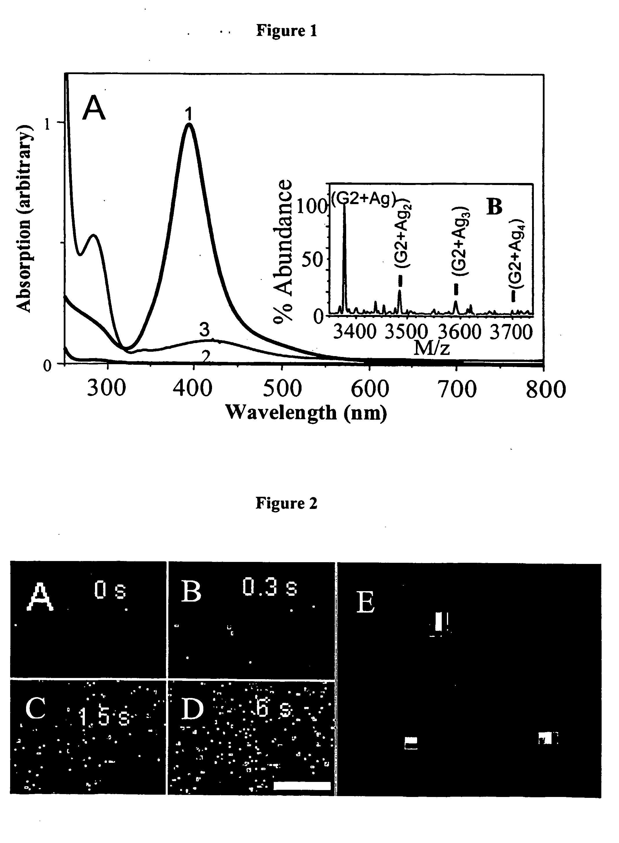
[0087] PAMAM is known to sequester metal ions from solution (Crooks et al., Accounts Chem. Res. 2001, 34:181; Ottaviani et al., Macromolecules 2002, 35:5105; Zheng et al., J. Phys. Chem. B 2002, 106:1252; Varnavski et al., J. Chem. Phys. 2001, 114:1962). PAMAM G4-OH and G2-OH dendrimers (4th and 2nd-generation OH-terminated poly(amidoamine), respectively, Aldrich) were therefore utilized to concentrate, stabilize, and solubilize Ag nanoclusters in both aerated and deaerated aqueous solutions. By dissolving 0.5 μmol G4-OH and 1.5 μmol AgNO3 into 1 ml distilled water (18 MΩ) and adjusting the solution to neutrality with 160 μmol acetic acid, silver ions readily interact with the dendrimer. Usually used to create small nanoparticles (>3 nm diameter), literature preparations generally add small amounts of reducing agents such as NaBH4 (Crooks et al., Accounts Chem. Res. 2001, 34:181; Ottaviani et al., Macromolecules 2002,...

example 2

Characterization of Individual Ag Nanodots

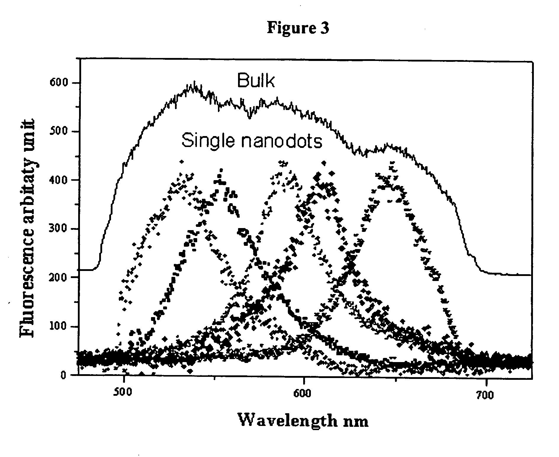
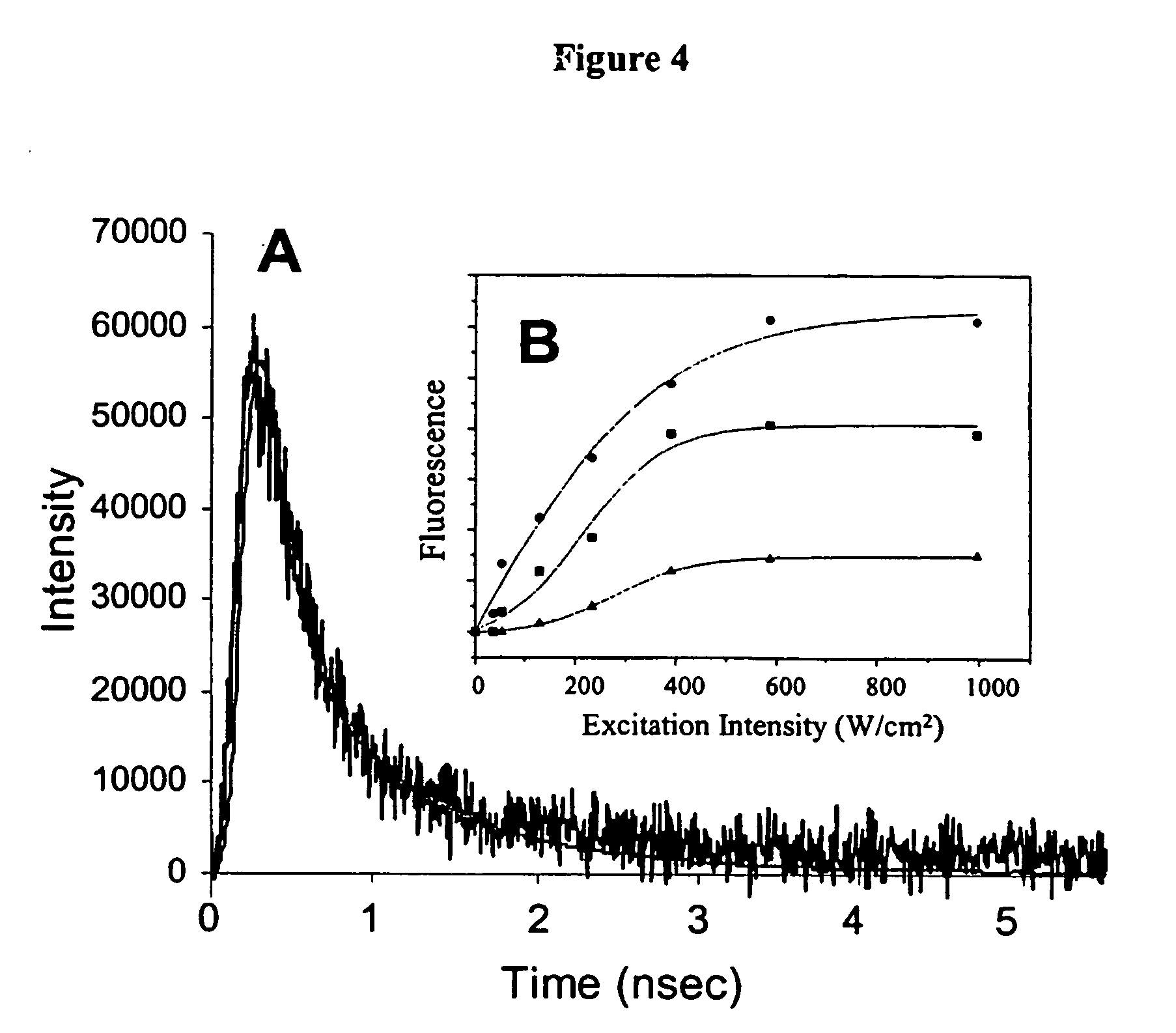
[0093] A range of PAMAM dendrimer generations (G0-OH through G4-OH with diameters (MW) ranging from 1.5 nm (517 g / mol) to 4.5 nm (14,215 g / mol) were used to yield highly fluorescent Agn nanodots. Very bright fluorescence was observed over a pH range of 8.0 to 3.0. These different generations of PAMAM enabled a measure of control over nanocluster distributions: nanodots created with smaller dendrimer generations exhibited different emission spectra than nanodots created with higher dendrimer generations. Not only was nanodot emission extremely stable in spectrum and intensity, but they also exhibited highly polarized emission with very clear and stable dipole emission patterns (FIG. 2E). The observation of emission patterns enabled to employment of the three-dimensional orientational methods developed to follow orientational dynamics either in solution or of immobilized features, as described in Bartko, & Dickson, J. Phys. Chem. B 1999, 103...

example 3

Generation of Dendrite-Encapsulated Gold Nanoclusters

[0097] Previous studies have yielded fluorescent, surface passivated gold nanoclusters ranging in size from 28 atoms to smaller particles (F, relative to that of bulk gold films (Mooradian, A. Phys. Rev. Lett. 1969, 22:185-187), have been created, the 10−3 to 10−4 quantum yields and polydisperse nanoparticle size distributions have precluded them from being good fluorophores (Link et al., J. Phys. Chem. B 2002, 106:3410-3415; Huang & Murray, J. Phys. Chem. B 2001, 105:12498-12502). The present invention discloses water-soluble, monodisperse, blue-emitting Au8 nanodots that when encapsulated in and stabilized by biocompatible PAMAM dendrimers (Tomalia, Sci. Am. 1995, 272:62-66), exhibited a fluorescence quantum yield of 41±5%, a more than 100-fold improvement over other reported gold nanoclusters (Link et al., J. Phys. Chem. B 2002, 106:3410-3415; Huang & Murray, J. Phys. Chem. B 2001, 105:12498-12502). Larger Aun nanodots have a...


PUM

PropertyMeasurementUnit
fluorescence quantum yieldaaaaaaaaaa
diameteraaaaaaaaaa
temperatureaaaaaaaaaa

Abstract

A composition is disclosed which is capable of being used for detection, comprising an encapsulated noble metal nanocluster. Methods for preparing the encapsulated noble metal nanoclusters, and methods of using the encapsulated noble metal nanoclusters are also disclosed. The noble metal nanoclusters are preferably encapsulated by a dendrimer or a peptide. The encapsulated noble metal nanoclusters have a characteristic spectral emission, wherein said spectral emission is varied by controlling the nature of the encapsulating material, such as by controlling the size of the nanocluster and / or the generation of the dendrimer, and wherein said emission is used to provide information about a biological state.

Description

BACKGROUND OF THE INVENTION [0001] 1. Field of the Invention [0002] The present invention relates to the creation of new classes of fluorescent / luminescent probes based on metal cluster fluorescence, methods of preparing such probes, and methods of use thereof. [0003] 2. Background Art [0004] Single molecule fluorescence microscopy studies present an extreme limit in which weak signals must be observed on essentially zero background. Such optical methods relying on high-intensity laser excitation of highly emissive and robust fluorophores require extremely efficient background rejection. Relying on the introduction of artificial labels to identify the particular protein or structure of interest, fluorescence based methods suffer from two additional problems—photobleaching (loss of signal due to probe destruction) and autofluorescence (naturally occurring background fluorescence from native species within biological media). Even with these problems, fluorescence microscopy remains th...

Claims


Application Information

Patent Timeline
Patent Type & Authority Applications(United States)
IPC IPC(8): G01N33/543G01N21/78C07K7/06C12N15/09C12Q1/04G01N33/58
CPCB82Y15/00G01N33/582G01N33/588Y10S436/805
Inventor DICKSON, ROBERTZHENG, JIE
Owner GEORGIA TECH RES CORP
Why Eureka
Try Eureka