
Medicine for detecting lipid components in vivo and vascular endoscope

Inactive Publication Date: 2006-03-23
UCHIDA YASUMI
View PDF5 Cites 2 Cited by
  • Summary
  • Abstract
  • Description
  • Claims
  • Application Information

AI Technical Summary

Benefits of technology

[0006] An object of the present invention is to provide a medicine and an apparatus which can discriminate lipids in vivo and detect the kind and spreading of the lipids not by a wavelength but by a two-dimensional color image, thus enabling diagnosis of diseases caused by accumulation of the lipids.

Problems solved by technology

These processes induce atherosclerotic plaques vulneable and resultant plaque disruption occurs.
All these methods, however, are applicable only in vitro and there are no available tools for measurement and accordingly discrimination of lipid components in the vascular wall or in circulating blood in vivo.
However, it is not known whether blue dyes, yellow dyes such as Methanyl yellow and Lissamin fast yellow, and Homidium evoke fluorescences characteristic of lipid components including oxLDL and lyso PC.

Method used


Image

  • Medicine for detecting lipid components in vivo and vascular endoscope
  • Medicine for detecting lipid components in vivo and vascular endoscope
  • Medicine for detecting lipid components in vivo and vascular endoscope

Examples


example 1

[0048] Microscopic Examination of Fluorescences Characteristic of Major Substances Composing Atherosclerotic Plaques

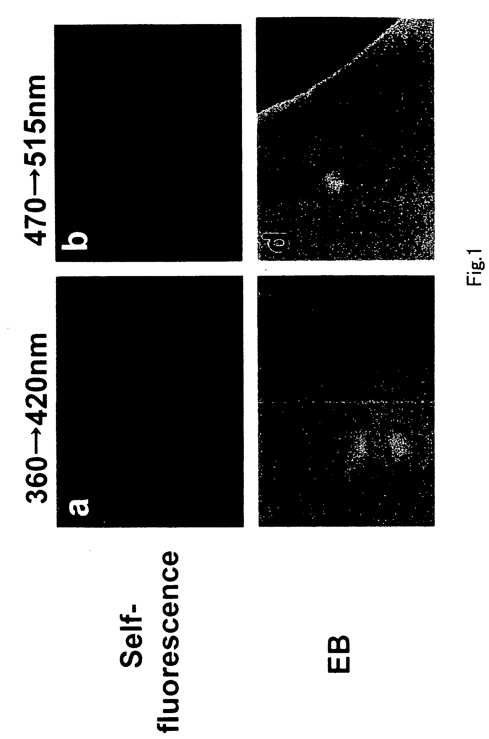
[0049] Self-fluorescences and dye-evoked fluorescences of the major substances composing the atherosclerotic plaque were examined by fluorescent microscopy.

1) Materials and Methods

[0050] a) Commercially obtainable substances composing atherosclerotic plaques were added to one of the dyes and the evoked fluorescences were examined by fluorescent microscopy (Olympus). The substances used are as follows: oxidized low density lipoprotein (oxLDL; Biogenesis LTd, Kingston), low density lipoprotein (LDL; Carbochem Co, LaJolla), very low densitiy lipoprotein (VLDL; Athens Res & Tech, Athens), intermediate density lipoprotein (IDL; personally isolated), high density lipoprotein (HDL; Chemicon Int.), phosphatidylcholine (PC; Sigma Co, St. Louis), L(a)-lyso phosphatidylcholine (lyso PC; Sigma Co, St. Louis), apolipoprotein B100 (apoB 100; ICN Biomedicals, Ohio), Triglyceride ...

example 2

[0062] Scanning Microscopic Observation of Dye-evoked Fluorescences Characteristic of Lipid Components in the Atherosclerotic Plaques of Human Coronary Artery

1) Materials and Methods

[0063] Atherosclerotic plaques of coronary artery removed from human cadavers by permission of family was cut into 2×2 mm2 segments, dipped into 10−5 M dye solution for 5 min, and they were washed with saline to mounted on deck glass for fluorescent microscopic observation.

2) Results

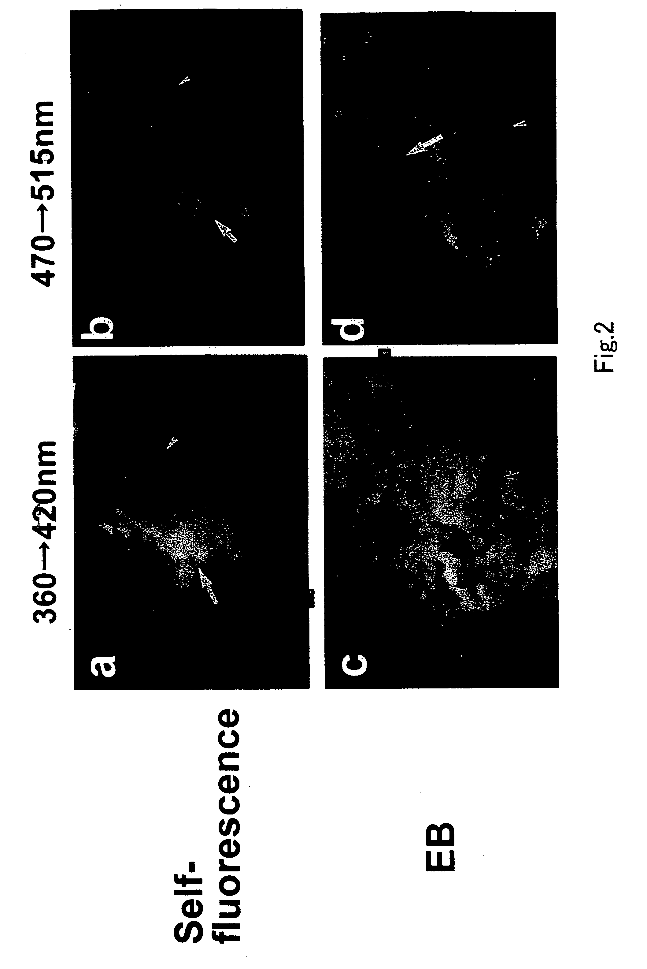
[0064]FIG. 2 shows Evans blue-evoked color fluorescence of human coronary plaque. Before dye administration, the plaque exhibited white to yellow self-fluorescence (arrow in a) by exciting at 360 nm and emitting at 420 nm and yellow to orange fluorescence by exciting at 470 nm and emitting at 515 nm (arrow in b), indicating existence of calcium phosphate, free cholesterol and beta-Carotene. After Evans blue administration, violet fluorescence was evoked in the plaque (arrow in c) by exciting at 360 nm and emitting at 42...

example 3

[0066] Observation of Dye-evoked Fluorescence Characteristic of Lipid Components in Atherosclerotic Plaques of Human Coronary Artery by Color Fluorescent Image Angioscopy System

1) Materials and Methods

[0067] a)Color fluorescent image angioscopy system

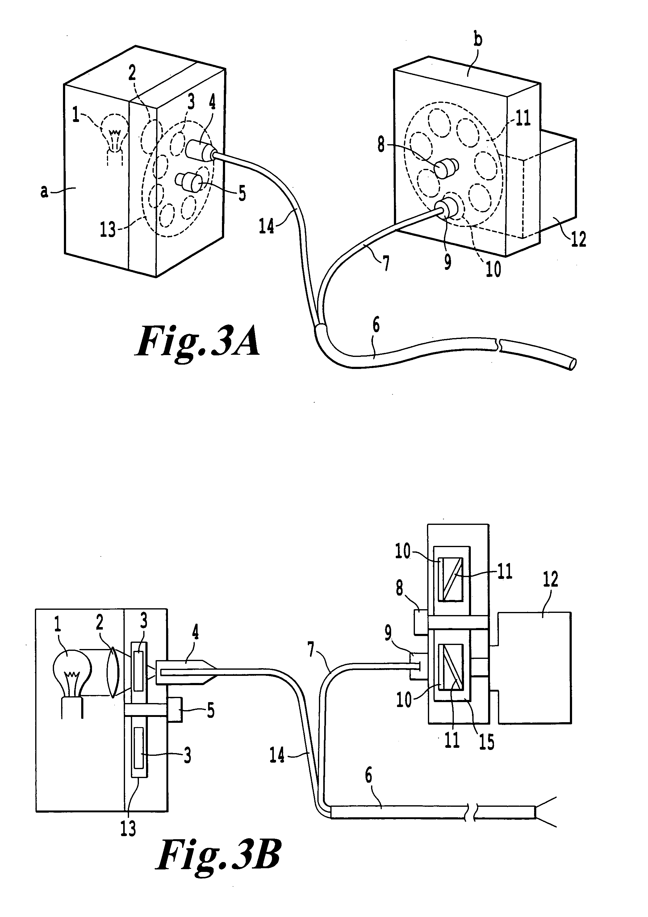
[0068] The color fluorescent image angioscopy system is composed of a fiberscope with 200 quartz fibers for light guide and 7000 quartz fibers for image guide incorporated in a 5 F balloon guiding catheter, fluorescence exciting part, fluorescence emitting part, 3CCD color digital camera (C37780, Hamamatsu Photo, Co, Hamamatsu), DVD and monitor. The exciting part is composed of Xe—Hg lamp, lens, and 7 exciting filters imbedded in a rotating disk. The emitting part is composed of 7 emitting filters imbedded in a rotating disk (FIG. 3). The wavelength of the exciting and emitting filters is shown in Table IV. By changing filters by rotation of the disks, a wide range of fluorescent wavelength can be examined.

[0069] b) Perfusion circu...


PUM


Abstract

The present invention provides a medicine and an apparatus which can detect the sites and spreading of in vivo lipids by a two-dimensional image, thereby allowing diagnosis of diseases due to accumulation of lipids. According to the present invention, provided are a medicine for detecting in vivo lipids, comprising one or more components such as oxLDL and lyso PC, as an effective component, selected from a sulfonic acid blue dye having a radical —SO3−, a homidium, Metanyl yellow and Lissamin fast yellow, and an angioscopic system for obtaining a fluorescent image, which comprises a mercury-xenon lamp light source, a quartz fiberscope wherein the light guide part has 100 to 200 fibers and the image guide part has 7000 to 9000 fibers, a rotational exciting filter, an emitting filter, and a high-sensitive digital camera for picking up the fluorescence as a two-dimensional color image.

Description

FIELD OF THE INVENTION [0001] The present invention relates to a medicine and an apparatus for detecting lipids existing in a living body (in vivo lipids). BACKGROUND OF THE INVENTION [0002] Serious obstructive arterial diseases such as acute coronary syndromes (acute myocardial infarction, unstable angina and sudden cardiac death), cerebral infarction and peripheral arterial obstruction are caused by disruption of atherosclerotic plaques and consequent thrombosis. Lysophosphatidylcholine (lyso PC) is produced by oxidization of phosphatidylcholine (PC), is a chemoattractant and accelerates migration of monocytes and macrophages into the vascular wall (McMurray HF, et al: J Clin Invest 92: 1004-1008, 1993; Yang LV, et al: Blood 105: 1127-1134, 2004; Sonoki K, et al: Metabolism 52: 308-3014, 2003). Oxidized low density lipoprotein (oxLDL) is produced by oxidation of low density lipoprotein (LDL) and accelerates proliferation of macrophages (Sakai M, et al: Atherosclerosis 133: 51-59, ...

Claims


Application Information

Patent Timeline
IPC IPC(8): A61K49/00A61B10/00
CPCA61B1/043A61B1/3137A61B5/0071A61K49/006A61B5/02007A61K49/0021A61K49/0028A61B5/0084
Inventor UCHIDA, YASUMIUCHIDA, HARUKO
Owner UCHIDA YASUMI
Learn More