
Methods and compositions for analyzing nucleic acids

a nucleic acid and composition technology, applied in the field of molecular biology, can solve the problems of labor-intensive and time-consuming northern blot and related methods, inconvenient detection methods, and insufficient or inconvenient detection of very short rnas, and achieve the effect of reducing the problem of bias

Inactive Publication Date: 2006-04-13
ASURAGEN
View PDF21 Cites 124 Cited by
  • Summary
  • Abstract
  • Description
  • Claims
  • Application Information

AI Technical Summary

Benefits of technology

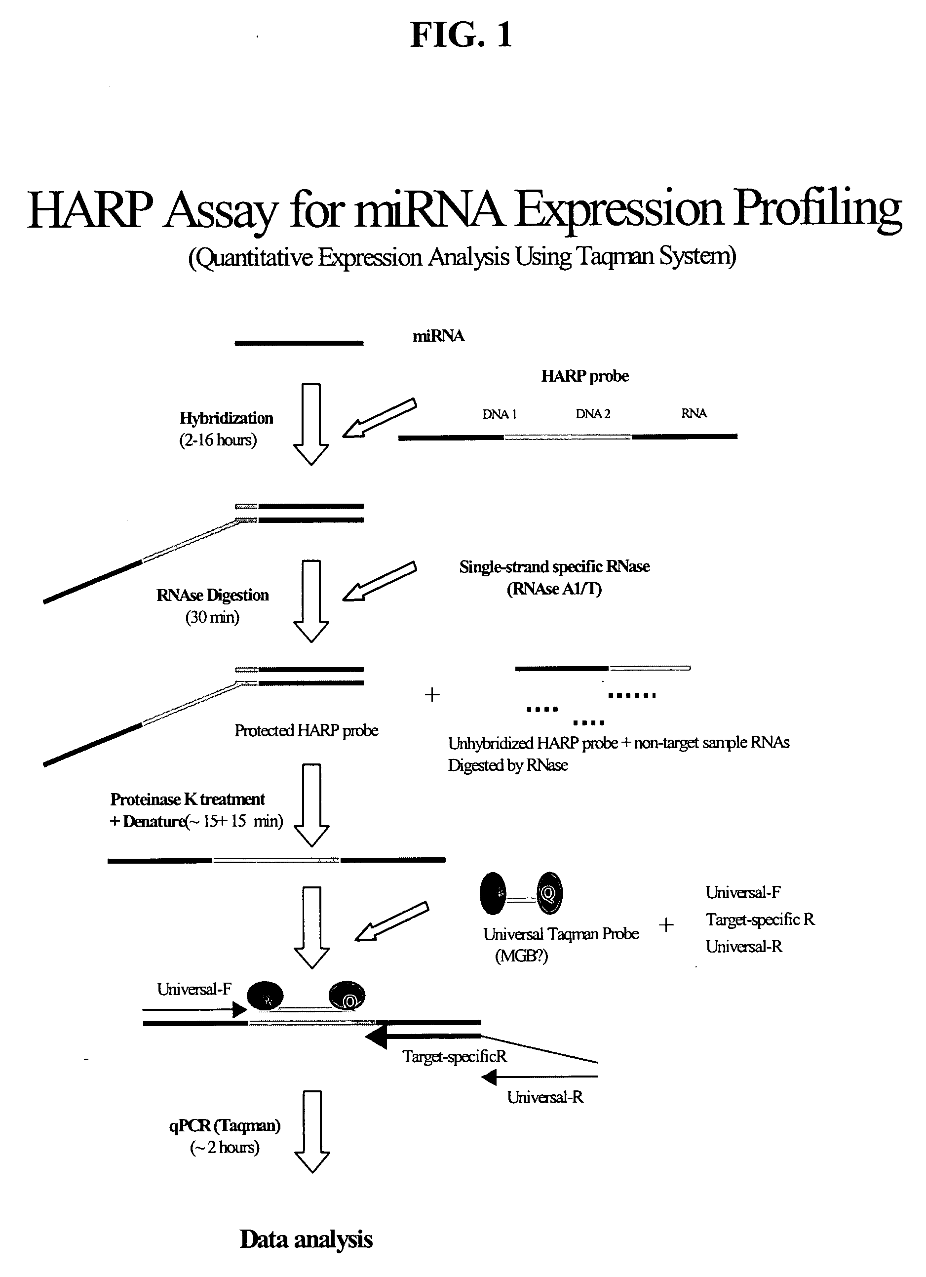
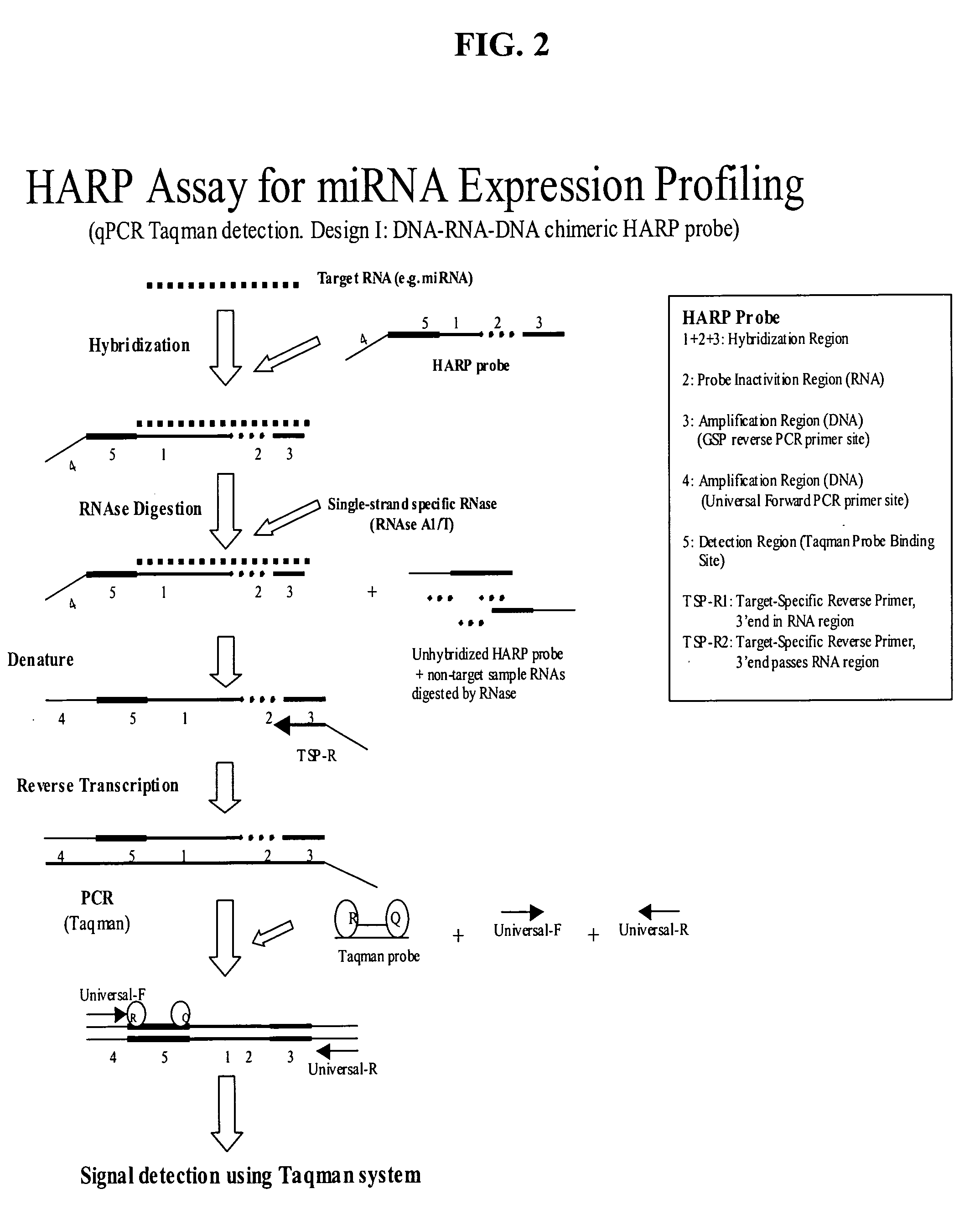
[0018] In some embodiments, methods for amplifying a target nucleic acid sequence involve, but are not limited to: a) contacting, under hybridization conditions, a nucleic acid comprising the target nucleic acid sequence with a HARP probe comprising i) a Target Hybridization Region that contains an Probe Inactivation Region and ii) at least one Amplification Region; b) exposing the HARP probe to one or more inactivating agents that modifies one or more residues in the Probe Inactivation Region of the probe if not hybridized to the target nucleic acid sequence; c) then amplifying the HARP probe that remains intact after treatment with the inactivating agent. In terms of the invention, the inactivating agent acts to “inactivate” unhybridized probe by eliminating or significantly reducing the ability of the unhybridized HARP probe to be amplified. In many embodiments, this inactivation will involve cleavage of the unhybridized probe.
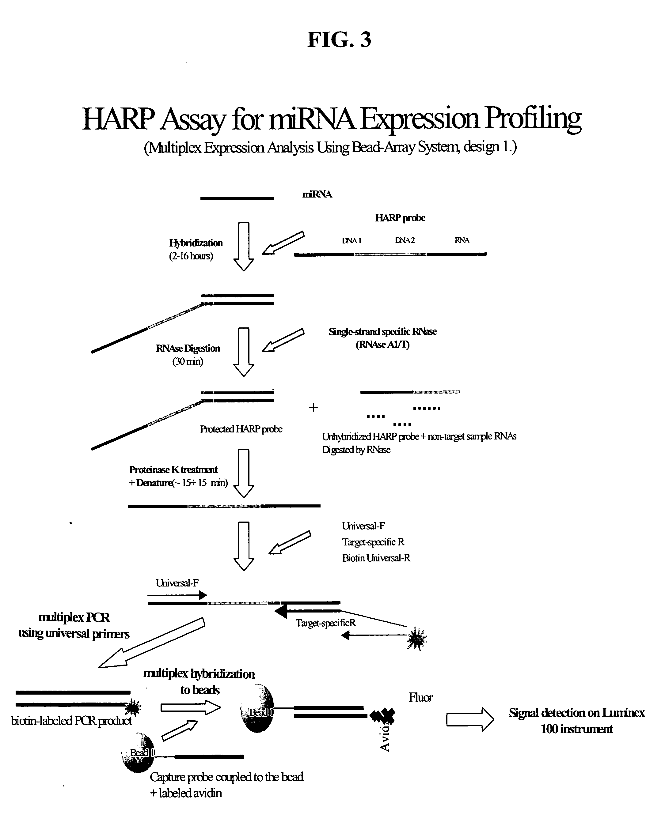
[0032] In some embodiments, the invention relates to multiplex HARP assays. The invention allows for a significant improvement over prior multiplex methods for the amplification of specific targets, which typically use a different primer pair for each target sequence to be amplified. The present invention permits multiplex amplification of many distinct target nucleic acids using a common pair of PCR™ primers, which avoids the problems cited above. This is made possible since the binding sites for the PCR™ primers are located in the external regions of the HARP probe, rather than within the target-complementary region itself. In prior multiplex assays, which employ different primer pairs for each target sequences, differences in annealing efficiency of different primer pairs result in a strong bias in the amplification of the different amplicons thereby strongly reducing the fidelity of a quantitative multiplex assay. Furthermore the presence of a large number of different primers results in a strongly increased risk of primer dimer formation diminishing the possibility of reproducible amplifying small amounts of target nucleic acids.
[0034] Some embodiments of the HARP assay relate to the specific design of the First-strand primer, which is the oligonucleotide used to prime synthesis of the complementary strand of the HARP probe; said synthesis comprises the initial step of the amplification of the HARP probe. One advantageous design for the First-strand primer is that in which the 3′ end of said primer traverses the RNA segment comprising the Probe Inactivation Region of the HARP probe. Such design avoids the need to use a reverse transcriptase enzyme or DNA polymerase enzyme with reverse transcriptase activity, for synthesis of the HARP-complementary strand, since the template for the initial nucleotide and all subsequent nucleotides incorporated into the complementary strand are DNA bases instead of RNA bases. In many embodiments of the HARP assay, the First-strand primer has a sequence which is different from that of the Reverse primer in the PCR™ amplification step. The First-strand primer and the Reverse primer are however related in that the product generated by the Forward PCR™ primer and the First-strand primer are able to hybridize to the Reverse primer, in order to permit amplification by subsequent cycles of PCR™. A consequence of this is that the sequence comprising the 5′ side of the First-strand primer is completely or substantially identical to the sequence which occurs at least at the 3′ side of the Reverse primer used for PCR™; that is, the 5′ end of the First-strand primer overlaps the 3′ end of the Reverse PCR™ primer. Particularly advantageous is a design in which the 5′ side of the First-strand primer is entirely identical to the Reverse primer. The sequence of the Reverse primer in such cases is a subset of the sequence of the First-strand primer. The advantage of using a Reverse primer which is different from the First-strand primer is that a common Reverse primer (also known as a Universal Reverse Primer) may then be used for amplification of multiple HARP probes designed to detect distinct targets; this will minimize the problems of bias in relative levels of amplified products introduced during amplification, and of unwanted primer interactions.

Problems solved by technology

These methods are however inadequate or inconvenient for detection of very short RNAs, including miRNA and siRNA targets, because such RNA species, being only ˜21-23 bases in length, are too short to be specifically primed (as is required for RT-PCR-based detection) and are poorly resolved on the gel matrices typically used for Northern blot and RPA detection.
Northern Blot and related methods are labor-intensive and time-consuming, as each step (gel electrophoresis, blotting, prehybridization, hybridization, and detection) typically requires one to several hours or longer.
This requirement precludes the use of samples that may be compromised due to lag time in harvesting the RNA or due to prior treatments such as aldehyde fixation, which are known to damage RNA.
Another major drawback to Northern blotting and other methods that do not include a target amplification step is that they are relatively insensitive.
Northern blotting may lack the sensitivity required for detection of low-abundance targets and for detecting targets in very small mass amounts of sample RNA such as those obtained from microdissected tissue.
The lack of a post-treatment amplification step results in lower sensitivity for detection of the target nucleic acid.
Some other variations of nuclease protection techniques are also ultimately similarly limited in sensitivity because they do not involve probe amplification.
However, there are a number of technical problems associated with PCR™.
For example, problems can arise from the co-amplification of non-specific hybridization of primers to extraneous sequences along the target template.
Another technical problem with PCR™ is that target sequences that differ in size and in the sequence of their PCR™ primer binding sites are amplified with different efficiencies.
For this reason, the relative levels of multiple target sequences in a sample which are amplified in the same PCR™ reaction (i.e. which are amplified in a multiplex reaction) do not generally reflect the relative starting levels of the targets prior to amplification, making it impossible to deduce the relative abundance of the multiple unamplified targets in the sample.
Another technical problem with PCR™ is related to its inability to directly amplify very short DNA or cDNA targets, such as those obtained from highly degraded or crosslinked nucleic acid, frequently seen with formaldehyde fixed paraffin embedded tissue (FFPE), or targets such as miRNAs and small interfering RNAs (siRNAs), which are only ˜21-23 bases in length.
This limitation arises from the fact that the pair of oligonucleotide primers which flank the DNA segment of the target sequence to be amplified (which are used to initiate sequential copying of the target sequence) are each generally also ˜20 bases in length, thus there is not sufficient space on the target to allow for annealing of the primers with an intervening sequence that can be amplified and detected.
This problem cannot be solved by simply reducing the size of the primers (for example to ˜5 bases in length), because they would then fail to hybridize stably to the target at temperatures needed for the enzymatic steps of RT-PCR and because they would lack sufficient specificity for hybridization to only the intended target RNA.
However, these methods have some of the same general problems as PCR™, in that they may be vulnerable to non-specific amplification and are unable to directly amplify very short DNA or cDNA targets, such as those obtained from miRNAs and small interfering RNAs.

Method used


Image

  • Methods and compositions for analyzing nucleic acids
  • Methods and compositions for analyzing nucleic acids
  • Methods and compositions for analyzing nucleic acids

Examples


example 1

Detection of miRNA Target Using HARP Assay in qPCR™ Format

[0202] Detection of target nucleic acid using the method of the present invention was carried out according to the following protocol.

[0203] A HARP probe was designed to detect a brain-specific miRNA (miR-124) and consisted of a 71 base chimeric oligonucleotide having the following sequence:

5′ CTTTT CCCGT CCGTC ATCGC TCAAG(SEQ. ID NO:4)TGGCATTCAC CGCGugcCTTAACTC CCTAT AGTGA GTCGT ATTAC G 3′

[0204] The HARP probe is chimeric in the sense that is contains DNA bases and RNA bases (RNA bases are shown in lowercase). The features of the HARP probe, from 5′ to 3′, are a first Amplification Region (position 1-19 comprising a Forward primer binding site); a Detection Region (position 20-34 comprising a 15 base TAQMAN probe-binding site); a 22 base Target Hybridization Region (position 26-47, underlined, comprising 22 bases that are complementary to miRNA); a Probe Inactivation Region positioned within the Target Hybridization Regi...

example 2

Detection of miRNA Target Using HARP Assay in qRT-PCR Format

[0213] To demonstrate the use of a HARP probe to detect miRNA in a qRT-PCR format, a HARP probe was designed with additional RNA bases, requiring a reverse transcription of 10 RNA bases. The HARP probe served the same purpose: to detect a brain-specific miRNA (miR-124) and consisted of a 71 base chimeric oligonucleotide having the following sequence (RNA bases shown in lower case):

5′ CTTTT CCCGT CCGTC ATCGC TCAAG(SEQ. ID NO:8)TGGcauucaccgcGTGCCTTAACTC CCTAT AGTGA GTCGT ATTAC G 3′

[0214] The primary features of the HARP probe, from 5′ to 3′, are a first Amplification Region (position 1-18 comprising a Forward primer binding site); a Detection Region (position 19-37 comprising a 19 base TAQMAN probe-binding site); a 22 base Target Hybridization Region (position 26-47, underlined, comprising 22 bases that are complementary to miRNA); a Probe Inactivation Region (position 29-38 comprising 10 bases of RNA, shown in lower case)...

example 3

Sensitivity and Linearity for Detection of miRNA in Human Sample

[0223] To demonstrate the sensitivity and linearity of detection of miRNA in a human sample, a HARP assay was carried out as described in Example 1, except that HARP probe was hybridized overnight using 3 different amounts of total RNA (100 ng, 10 ng, and 1 ng) from human brain as target. The miR-124 target was detected with Ct values shown in Table 1 below. Note, a difference of 3.3 between two Ct values reflects a 10-fold difference in amount of target. This study demonstrates linear detection of the miR-124 target RNA between 100 ng and 1 ng; the minus-target-plus RNase control reaction did not show any amplification, showing that amplification of the HARP probe was target-dependent.

TABLE 1Amount of human brain RNACt value for miR-124detection viainputHARP assay100 ng21.74 10 ng25.24 1 ng28.84Nonenot detected


PUM

PropertyMeasurementUnit
Lengthaaaaaaaaaa
Concentrationaaaaaaaaaa

Abstract

Methods and compositions for probe amplification to detect, identify, quantitate, and / or analyze a targeted nucleic acid sequence. After hybridization between a probe and the targeted nucleic acid, the probe is modified to distinguish hybridized probe from unhybridized probe. Thereafter, the probe is amplified. Moreover, in specific embodiments, the present invention involves a chimeric probe that is particularly effective when the targeted nucleic acid sequence is short and / or has a relatively low concentration, such as with an miRNA molecule.

Description

BACKGROUND OF THE INVENTION [0001] 1. Field of the Invention [0002] The present invention is in the field of molecular biology. More specifically, the present invention concerns methods and compositions for probe amplification to detect and / or quantify target nucleic acids in a sample and allows for some particular benefits in regard to the detection and / or quantification of nucleic acids that are present in relatively small amounts or that are relatively small in size. [0003] 2. Description of the Related Art [0004] Many types of studies carried out on biological samples require the quantitative detection of ribonucleic acid species, including messenger RNA (mRNA), micro RNA (miRNA) and small interfering RNA (siRNA) molecules. miRNAs are a recently-discovered class of short single-stranded RNA molecules that are ˜22 bases in length, and which are partially complementary to sequences in the 3′ untranslated region of mRNAs; miRNAs serve to inhibit expression of their target mRNAs by ...

Claims


Application Information

Patent Timeline
IPC IPC(8): C12Q1/68C12P19/34
CPCC12Q1/6809C12Q1/6844C12Q1/686C12Q1/6865C12Q2561/108C12Q2561/101C12Q2525/161C12Q2525/143C12Q2525/203C12Q2521/301
Inventor WINKLER, MATTHEWGOLDRICK, MARIANNAHARRIS, NATHANYE, FEI
Owner ASURAGEN
Learn More