
Stabilized A-beta protofibrillar aggregates

Inactive Publication Date: 2006-04-13
UNIV OF TENNESSEE RES FOUND
View PDF0 Cites 36 Cited by
  • Summary
  • Abstract
  • Description
  • Claims
  • Application Information

AI Technical Summary

Benefits of technology

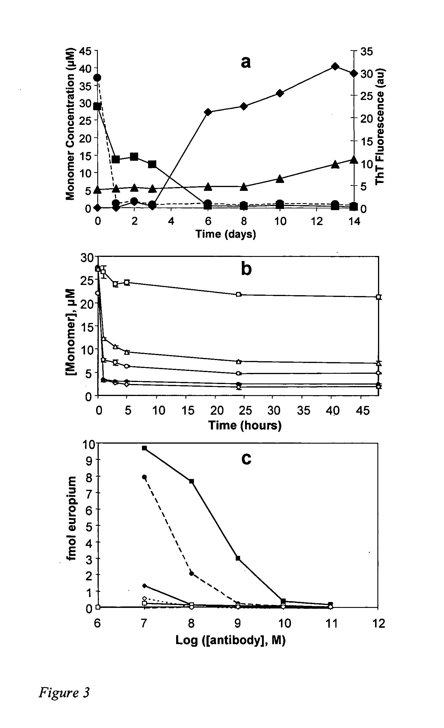
[0012] Described herein is chemical biology approach to structural analysis of Aβ protofibrils. Library screening yielded several molecules that stimulate Aβ aggregation. One of these compounds, calmidazolium chloride (CLC), rapidly and efficiently converts Aβ(1-40) monomers into clusters of protofibrils, e.g., stabilized Aβ protofibrillar aggregates. As monitored by electron microscopy, these stabilized Aβ protofibrillar aggregates persist for days when incubated in phosphate buffered saline (PBS) at 37° C., with a slow transition to fibrillar structures apparent only after several weeks. Like normal protofibrils, the stabilized Aβ protofibrillar aggregates exhibit a low thioflavin T response. Like Aβ fibrils, the stabilized Aβ protofibrillar aggregates bind the anti-amyloid antibody WO1. The stabilized Aβ protofibrillar aggregates exhibit the same protection from hydrogen-deuterium exchange as do protofibrils isolated from a spontaneous Aβ fibril formation reaction: about 12 of the 39 Aβ(1-40) backbone amide protons are protected from exchange in the protofibril, compared to about twice that number in amyloid fibrils. Scanning proline mutagenesis analysis shows that the Aβ molecule in these stabilized Aβ protofibrillar aggregates exhibits the same flexible N- and C-termini as do mature amyloid fibrils. The major difference in Aβ conformation between fibrils and stabilized Aβ protofibrillar aggregates is added structural definition in the 22-29 segment in the fibril. Besides aiding structural analysis, compounds capable of facilitating oligomer and protofibril formation have therapeutic potential, and / or can be used to screen for therapeutics. In addition, stabilized Aβ protofibrillar aggregates can be used to screen for antibodies useful for both research and therapeutics.

Problems solved by technology

Isolated Aβ protofibrils incubated in buffer tend to dissociate (7), suggesting a fundamental instability incompatible with many biophysical techniques.
More detailed characterization of such structures, however, is made difficult by their metastable, transient nature.
ADDLs have not been complexed with a small molecule, and their long range stability is unknown.

Method used


Image

  • Stabilized A-beta protofibrillar aggregates
  • Stabilized A-beta protofibrillar aggregates
  • Stabilized A-beta protofibrillar aggregates

Examples


example 1

Materials and Methods

[0091] Below are examples of specific embodiments for carrying out the present invention. The examples are offered for illustrative purposes only, and are not intended to limit the scope of the present invention in any way. Efforts have been made to ensure accuracy with respect to numbers used (e.g., amounts, temperatures, etc.), but some experimental error and deviation should, of course, be allowed for.

[0092] The practice of the present invention will employ, unless otherwise indicated, conventional methods of protein chemistry, biochemistry, recombinant DNA techniques and pharmacology, within the skill of the art. Such techniques are explained fully in the literature. See, e.g., T. E. Creighton, Proteins: Structures and Molecular Properties (W. H. Freeman and Company, 1993); A. L. Lehninger, Biochemistry (Worth Publishers, Inc., current addition); Sambrook, et al., Molecular Cloning: A Laboratory Manual (2nd Edition, 1989); Methods In Enzymology (S. Colowic...

example 2

Library Screening

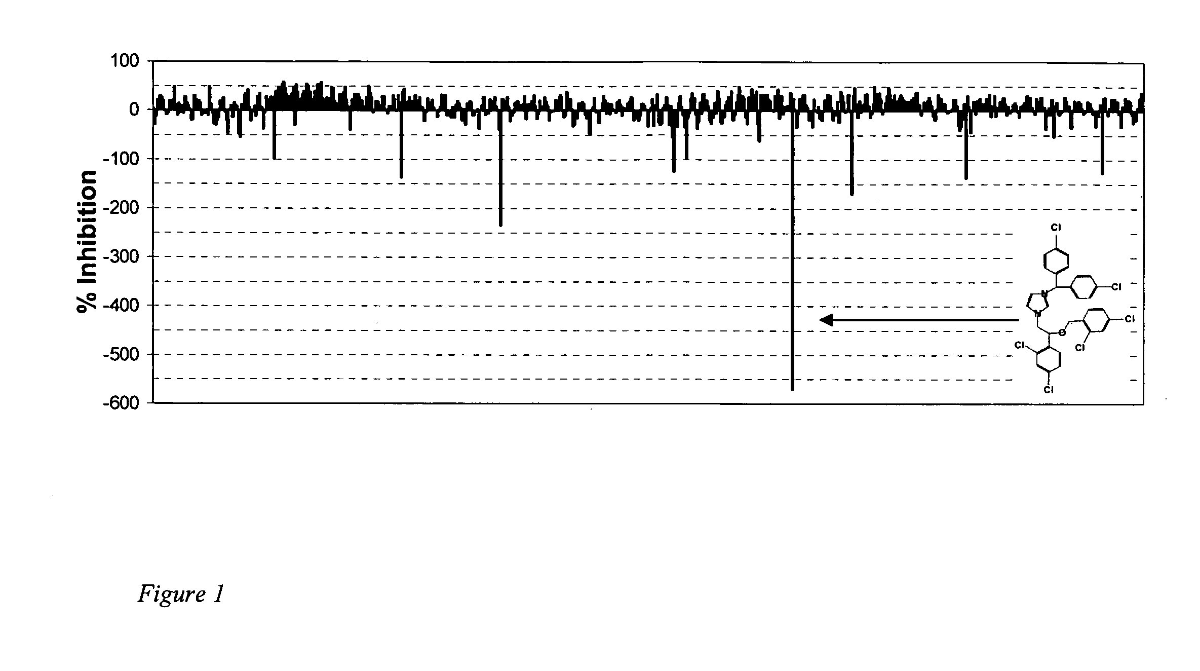
[0103] We screened a small library of 640 compounds, the LOPAC library (Sigma), using a microplate-based Aβ elongation assay. In this assay, plastic wells containing immobilized fibrils are incubated with a biotin-tagged monomer in the presence or absence of potential modifiers (13, 18). We found a number of compounds with potent abilities to enhance deposition of the biotin-tagged Aβ(1-40) (FIG. 1). Seven of the compounds were selected for further analysis:—amiloride HCl, phenamil methane sulfonate, naftopidil dihydrochloride, NPC 15437 dihydrochloride, calmidazolium chloride (R24571), 5-(nonoxyl)-tryptamine hydrogen oxalate, and L-α-methyl DOPA. The compounds have a wide variety of structures and biochemical properties. The structures of these seven compounds are shown in FIG. 9, along with the percent stimulation observed in the experiments.

[0104] The most potent of these compounds is calmidazolium chloride (CLC, FIG. 1 insert), which at 100 μM stimulates the d...

example 3

Characterization of CLC Stabilized Protofibrillar Aggregates

[0106] Although clearly distinct structures, Aβ mature fibrils exhibit a number of similarities to protofibrils and CLC-Aβ aggregates, including a fundamental fibrillar character in the EM, a shared epitope for the antibody WO1, and common possession of a subset of unusually stable H-bonds. Both mature amyloid fibrils and CLC aggregates appear to be assembled from very similar conformations of the peptide, as revealed by scanning proline mutagenesis, which can reveal sites of restrictive peptide backbone geometry within fibrils (15, 22).

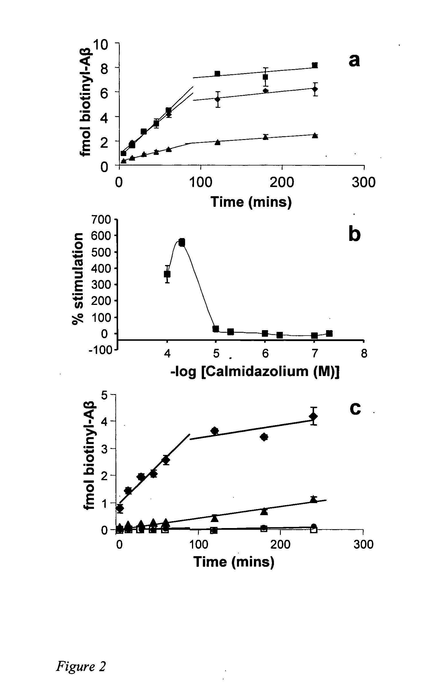
[0107] We characterized the activity of CLC in the microplate assay in more detail. The time course of Aβ deposition in the presence of CLC yields a pattern similar to the reaction without added compound (FIG. 2a), suggesting a simple acceleration of the normal mechanism. However, when the assay is repeated on a microplate lacking Aβ fibrils, the dramatic deposition of biotinylated Aβ in t...


PUM

PropertyMeasurementUnit
Structureaaaaaaaaaa
Immunogenicityaaaaaaaaaa

Abstract

Described and claimed herein are methods for producing chemically stabilized Aβ protofibrillar aggregates, and compositions made therefrom. Compositions produced are useful in facilitating detailed studies of Aβ protofibril structure, fibril formation, and progression of Aβ related diseases, e.g., Alzheimers disease. In addition, chemically stabilized protofibrillar structures can be used as tools to generate and / or screen for antibodies specific for protofibrils. Antibodies specific for protofibrils can be used as diagnostic tools or as therapeutics in the diagnosis or treatment of, e.g., Alzheimer's disease. Finally, chemically stabilized protofibrillar structures can be used in the preparation of therapeutic or prophylactic vaccines.

Description

CROSS REFERENCE TO RELATED APPLICATIONS [0001] This application claims the benefit of U.S. Provisional Application No. 60 / 617,114, filed Oct. 7, 2004 and U.S. Provisional Application No. 60 / 674,101, filed Apr. 22, 2005, the entire disclosures of which are hereby incorporated by reference in their entirety for all purposes.STATEMENT REGARDING FEDERALLY SPONSORED RESEARCH OR DEVELOPMENT [0002] The U.S. Government has certain rights in this invention pursuant to Grant Nos. R01AG18416 and R01 AG18927 awarded by the National Institutes of Health.BACKGROUND OF THE INVENTION [0003] 1. Field of the Invention [0004] The invention relates to methods for producing stabilized Aβ protofibrillar aggregates using stabilizing compounds, e.g., small molecules such as CLC; the stabilized Aβ protofibrillar aggregates produced by the methods; and use of the methods and aggregates for screening for stabilizing compound and specific binding reagents, e.g., antibodies. The invention is useful for the stud...

Claims


Application Information

Patent Timeline
IPC IPC(8): A61K38/17A61K31/4164
CPCA61K31/4164A61K38/1709
Inventor WETZEL, RONALD B.
Owner UNIV OF TENNESSEE RES FOUND
Learn More