

Methods for measuring the rates of replication and death of microbial infectious agents in an infected
a technology of infectious agents and microbial replication, which is applied in the field of methods for determining the rate of replication and death of infectious microbial agents, can solve the problems of inability to reliably, relevantly, and usefully culture ex vivo infectious organisms, and achieves the effects of accelerating the death of infectious organisms, and preventing the growth of infectious organisms
- Summary
- Abstract
- Description
- Claims
- Application Information
AI Technical Summary
Benefits of technology
Problems solved by technology
Method used
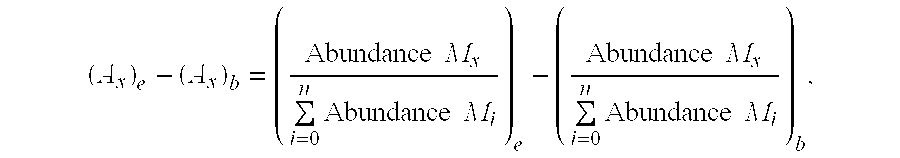
Image


Examples
example 1
[0111] In an illustrative embodiment of the methods of the invention, an isotopically-labeled amino acid such as leucine (e.g., [13C]-leucine, [2H]-leucine, [14C]-leucine) is administered by constant intravenous infusion to a human being who is known to be infected with the human immunodeficiency virus and who is under treatment with an anti-retroviral agent. Blood is removed from the subject during and after completion of the infusion of labeled leucine and the human immunodeficiency virus is isolated from the blood plasma by ultracentrifugation. Gel electrophoresis is then performed to isolate specific proteins contained in the virus (e.g., the Gag protein products, P24, P17, and P7). These proteins are then hydrolyzed in acid to free leucine, which is analyzed by mass spectrometry. The rate of rise of isotopically-labeled leucine in the leucine isolated from the Gag protein products during the infusion of isotopically-labeled leucine reveals the synthesis or replication rate of v...
example 2
[0112] Isotopically-labeled water is administered orally to a human being who is known to be infected with the human immunodeficiency virus and who is under treatment with an anti-retroviral agent. In one such embodiment, this is 2H2O (at a dose of 50 ml, for example) given orally to drink with morning and evening meals for 42 days (6 weeks). A blood or urine aliquot (10 ml) is collected from the individual at a defined time point or points (e.g., on the final day of the 2H2O protocol (day 42)).
[0113] Blood is removed from the subject during and after completion of isotope labeled water administration and the human immunodeficiency virus is isolated from the blood plasma by ultracentrifugation. Gel electrophoresis is then performed to isolate specific proteins contained in the virus (e.g., the Gag protein products, P24, P17, and P7). These proteins are then hydrolyzed in acid to free amino acids, which are analyzed by mass spectrometry.
[0114] The rate of rise of isotopically-label...
example 3
[0115] Isotopically-labeled water is administered orally to a human being who is known to be infected with Mycobacterium tuberculosis and who is under treatment with an antibiotic or immune stimulant. In one such embodiment, this is 2H2O (at a dose of 50 ml, for example) given orally to drink with morning and evening meals for 42 days (6 weeks). A blood or urine aliquot (10 ml) is collected from the individual at a defined time point or points (e.g., on the final day of the 2H2O protocol (day 42).
[0116] A sputum sample or lung biopsy is taken from the subject during and after completion of administration of labeled water and the Mycobacterium tuberculosis is isolated by techniques known in the art. Alternatively, M. tuberculosis DNA is isolated from the sample taken from the host organism without isolating the M. tuberculosis from the sample. The total DNA is then isolated. The DNA is hydrolyzed into nucleosides, which are analyzed by mass spectrometry. Alternatively, isolated nucl...
PUM
| Property | Measurement | Unit |
|---|---|---|
| Force | aaaaa | aaaaa |
| Electric charge | aaaaa | aaaaa |
| Electrical conductance | aaaaa | aaaaa |
Abstract
Description
Claims
Application Information