Covalently attached collagen VI for cell attachment and proliferation

a collagen vi and cell technology, applied in the field of useable surfaces, can solve problems such as creating further unwanted complications

Inactive Publication Date: 2006-05-18
BECTON DICKINSON & CO
View PDF5 Cites 12 Cited by
  • Summary
  • Abstract
  • Description
  • Claims
  • Application Information

AI Technical Summary

Problems solved by technology

The sera conventionally used for cell culture, which includes undefined mixtures of proteins that vary from lot to lot of serum, can create further unwanted complications.

Method used


Image

  • Covalently attached collagen VI for cell attachment and proliferation
  • Covalently attached collagen VI for cell attachment and proliferation
  • Covalently attached collagen VI for cell attachment and proliferation

Examples


example i

Attachment and Proliferation of HEP G2 Human Hepatoma Cells In Serum-Free Medium

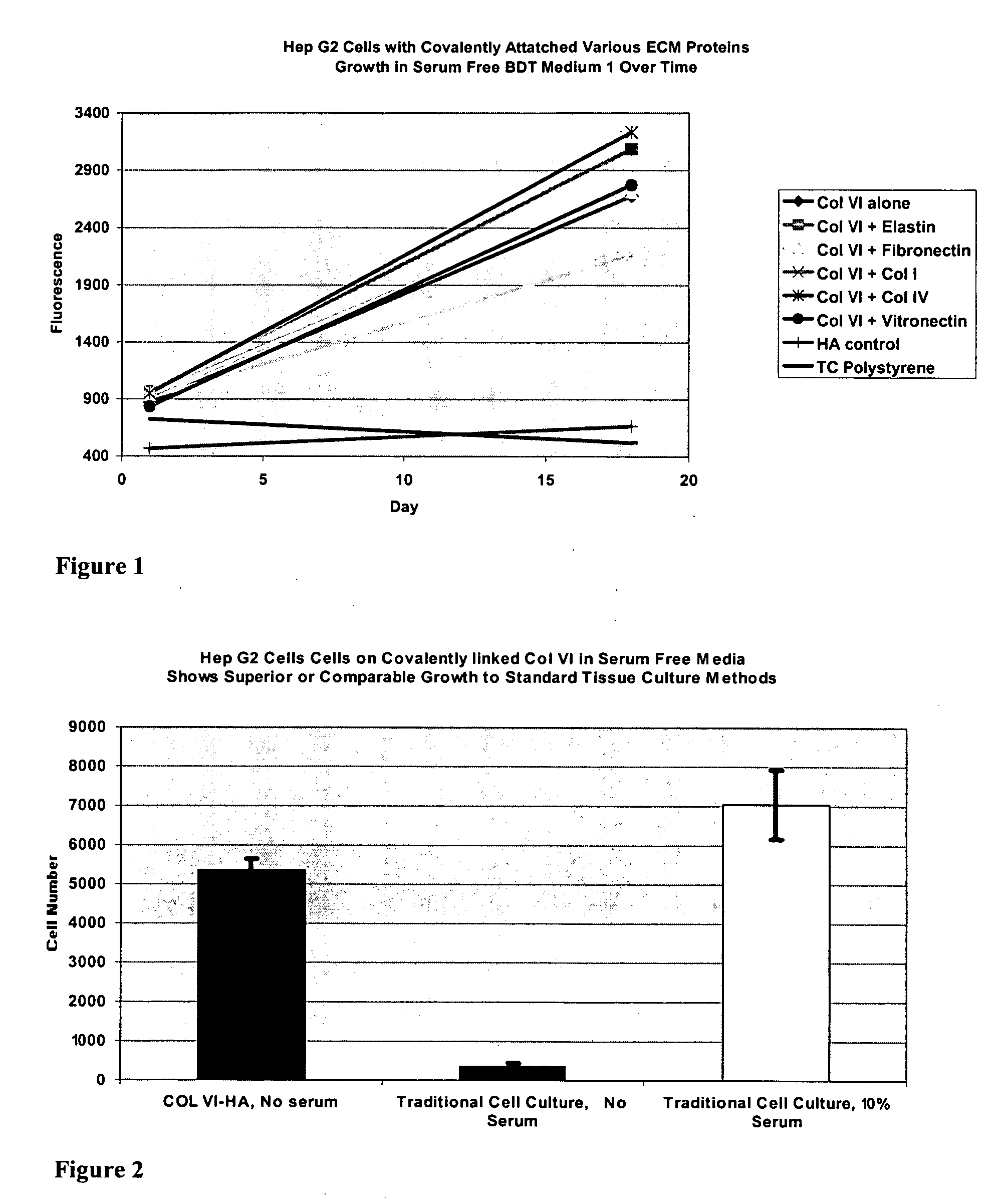
[0070] HepG2 human hepatoma cells were grown in BD Medium #1, a serum free chemically defined medium, on surfaces comprising hyaluronic acid (HA) to which was covalently attached collagen VI, either alone or in combination with other covalently attached extracellular matrix (ECM) proteins. (The components of BD Medium #1 are summarized in Table 2.) The ECM combinations tested were: collagen VI alone, or collagen VI in combination with either elastin, fibronectin, collagen I, collagen IV or vitronectin. In control samples, cells were seeded in BD Medium # 1 onto standard tissue culture treated polystyrene.

[0071] The cells were seeded in wells of 96-well microplates at an initial density of 104 cells / well, incubated in a CO2 incubator at 37° C, and stained at the time points indicated in the figure, using propidium iodide. Fluorescence was measured with a BMG Polarstar fluorometer at excitation of 544 nm...

example ii

Comparison of Proliferation of HEP G2 Human Hepatoma Cells in Serum-Free Medium to Commercial Media

[0072] HepG2 human hepatoma cells were grown as described in Example I, except the only ECM covalently bound to the HA surface was collagen VI. Proliferation of the cells on this collagen VI-surface in BD Medium #1 was compared to proliferation under the standard tissue culture conditions, either with or without serum. The cell number after 5 days of culture is shown graphically in FIG. 2.

[0073] The cells were stained with propidium iodide. Fluorescence microscopy images were obtained on an HT Imager (Discovery-1, Universal Imaging Corporation, a subsidiary of Molecular Devices, Downington, Pa.) at excitation of 535 nm and emission of 700 nm. Cell numbers were determined from these fluorescence microscopy images using UIC Metamorph™ analysis software. FIG. 2 shows that collagen VI combined with serum free BD Medium #1 promoted the proliferation of Hep G2 cells to the same extent as t...

example iii

Attachment and Proliferation of Rat Epithelial Stem Cells in Serum-Free Medium

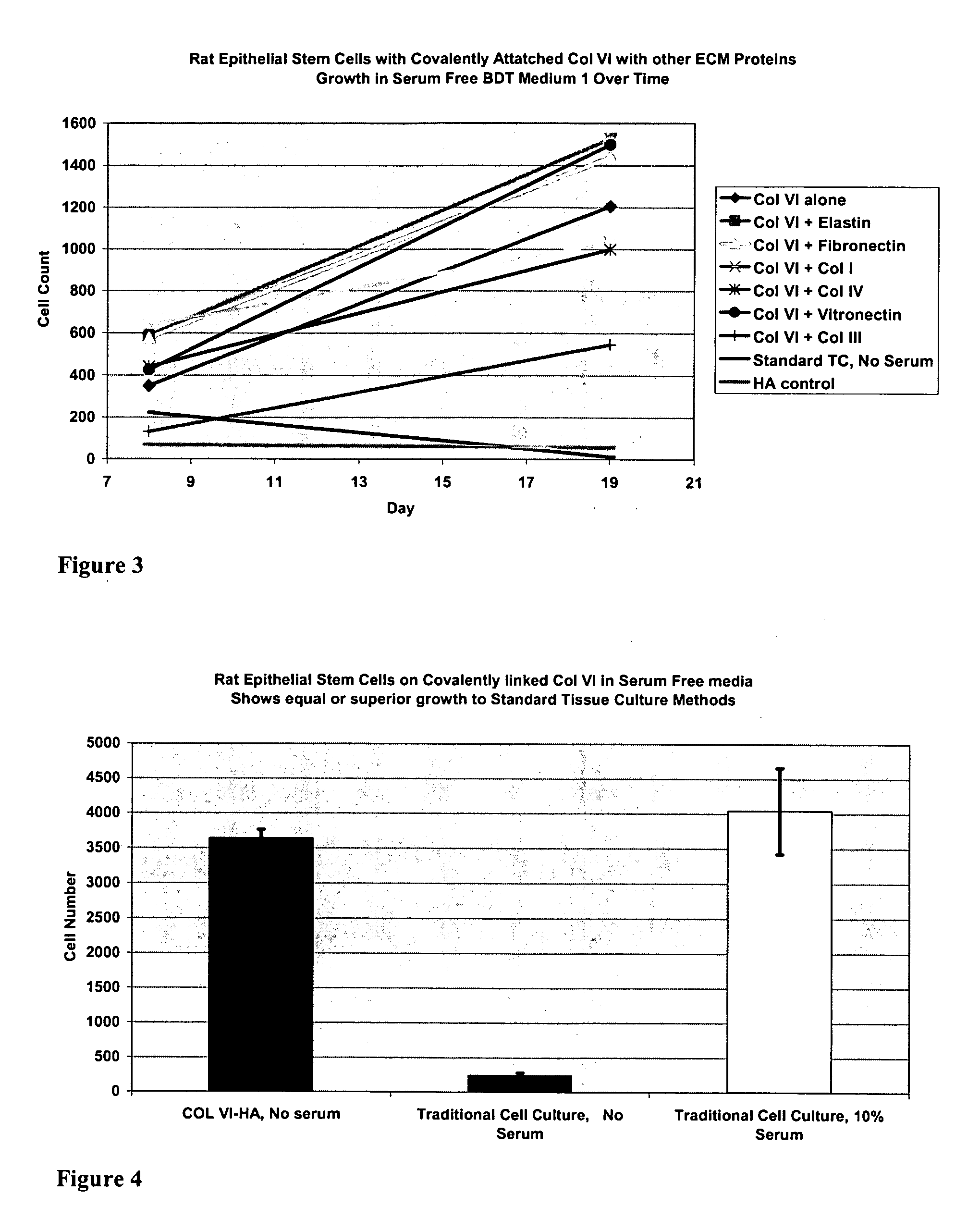
[0074] Rat epithelial stem cells (passage 6) were grown in BD Medium #1 on surfaces comprising hyaluronic acid (HA) to which was covalently attached collagen VI alone, or in combination with either elastin, fibronectin, collagen I, collagen IV vitronectin, or collagen III. Control samples were (1) cultured under “standard tissue culture conditions,” which comprised seeding cells onto tissue culture PS plates using commercial medium (DMEM / F12 mixed 1:1), or (2) cultured on a hyaluronic acid (HA) surface with no extracellular matrix protein present in BD Medium #1. The cells were stained with propidium iodide and analyzed as described in Example II. The proliferation over time was assayed.

[0075] As shown in FIG. 3, the number of cells on the collagen VI surfaces increased between day 8 and day 19. The proliferation in BD Medium #1 on the surfaces comprising collagen VI was superior to proliferation in comm...


PUM

PropertyMeasurementUnit
cell adhesion resistantaaaaaaaaaa
adhesion resistantaaaaaaaaaa
conformational statesaaaaaaaaaa

Abstract

Surfaces useful for cell culture comprise a support to which is bound a CAR material, and, bound to the CAR material, collagen VI or a biologically active fragment or variant thereof and, optionally, one or more other ECM proteins (or fragments or variants thereof) such as elastin, fibronectin, vitronectin, tenascin, laminin, entactin, aggrecan, decorin, collagen I, collagen III, and collagen IV. Also, optionally present on the surface is one or more polycationic polymers, such as poly-D-lysine or poly-D-ornithine. This surface is used in cell culture to promote cell attachment, survival, and / or proliferation of a number of different cell types such as (a) liver cells (e.g., HepG2 tumor cells, and a newly discovered line of rat liver epithelial stem cells) (b) osteoblasts, such as the murine cell line MC3T3 cell line and (c) primary bone marrow cells. Kits comprising the surfaces and additional reagents are also disclosed.

Description

CROSS-REFERENCE TO RELATED APPLICATIONS [0001] This application is a divisional of U.S. application Ser. No. 10 / 660,781, filed Sep. 12, 2003, which is hereby incorporated in its entirety by reference herein.BACKGROUND OF THE INVENTION [0002] 1. Field of the Invention [0003] This invention relates generally to useful surfaces for culturing cells in vitro, and to methods for using those surfaces. [0004] 2. Description of the Background Art [0005] Typically, for cell culture, cells are dispersed in a culture medium supplemented with serum, and the culture medium is then dispensed into a vessel that is made of a synthetic cell culture substrate such as tissue culture-grade polystyrene (PS). Under these conditions, non-specific protein adsorption to the PS surface rapidly occurs, generating a protein layer comprised of many different serum proteins in a spectrum of conformational states ranging from almost native to highly denatured. In stationary cultures, the cells subsequently settle ...

Claims


Application Information

Patent Timeline
Patent Type & Authority Applications(United States)
IPC IPC(8): C12N5/08C12N5/00C12N5/02G01N33/543
CPCC12N5/0068C12N2500/99C12N2533/52C12N2533/54C12N2533/80G01N33/54393C12N2500/90
Inventor GUARINO, RICHARD DAVIDROWLEY, JONATHAN A.LIEBMANN-VINSON, ANDREAHEMPERLY, JOHN JACOBHEIDARAN, MOHAMMAD A.PRESNELL, SHARON COLLINS
Owner BECTON DICKINSON & CO
Why Eureka
Try Eureka