Methods and apparatus for rapid crystallization of biomolecules

a biomolecule and crystallization technology, applied in the field of biomolecule crystallization methods and apparatuses, can solve the problems of slow crystallization process, tedious process, and method rarely used in protein crystallization, and the other methods known in the ar

Inactive Publication Date: 2006-06-29
BUKSHPAN SHMUEL
View PDF38 Cites 15 Cited by
  • Summary
  • Abstract
  • Description
  • Claims
  • Application Information

AI Technical Summary

Benefits of technology

[0058] Another particular advantage of the method of the present invention is that protein crystals may be obtained from a protein solution that is not highly purified, since the method of the invention comprises isoelectric focusing which is directed to protein separation and resolution from impurities.

Problems solved by technology
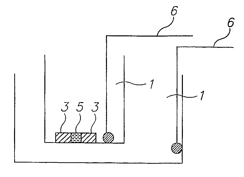
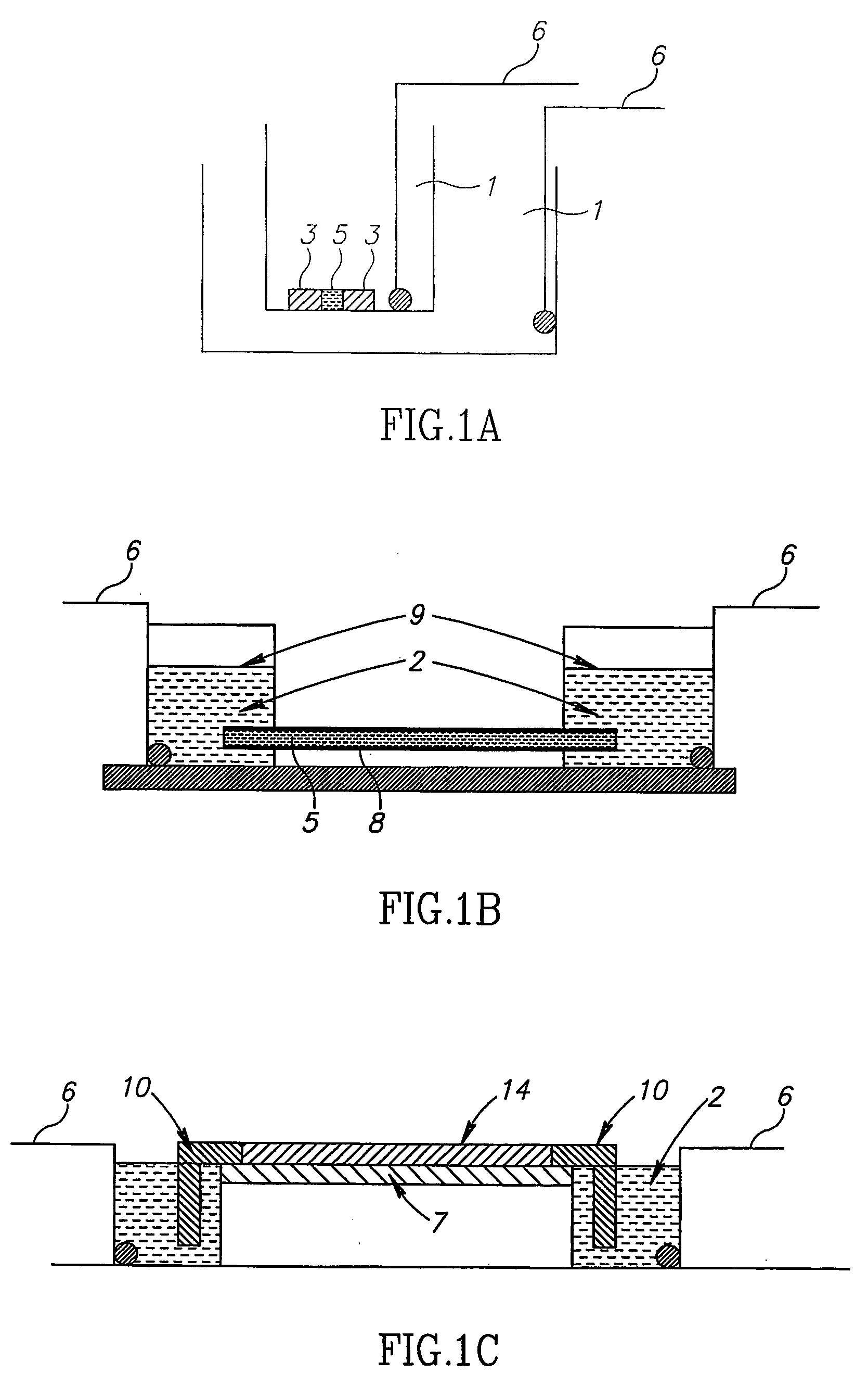
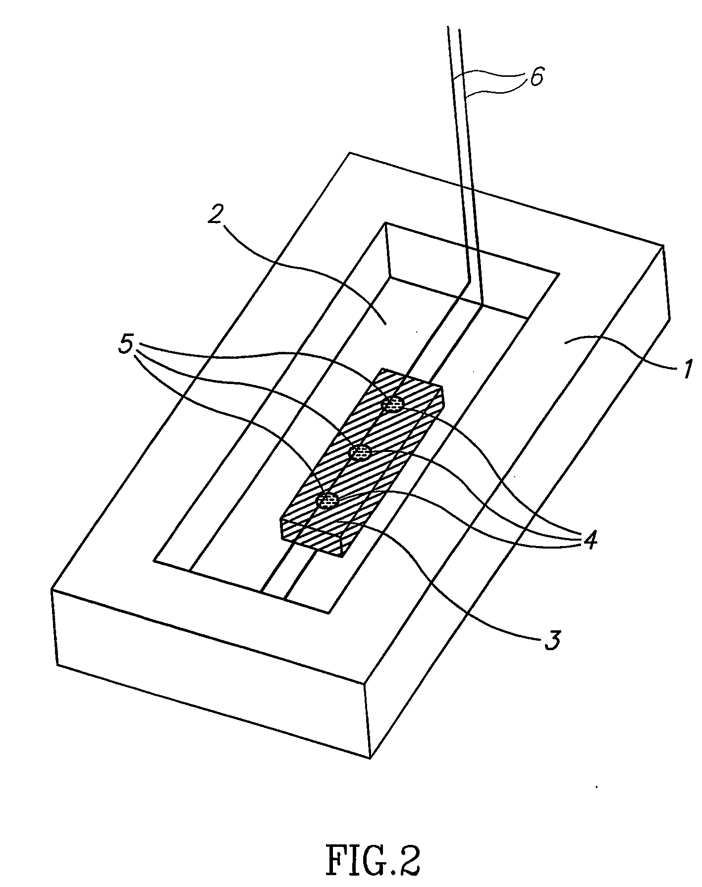
Crystal formation is a slow, and tedious process typically based on preparation of a saturated protein solution following nucleation, at optimal conditions.
It was shown that using this method, appropriate crystals were obtained (Sauter et al., Proteins: Structure, Function and Genetics 48:146-150, 2002), however, this method is rarely employed for protein crystal growth and is as slow as the other methods known in the art.

Method used


Image

  • Methods and apparatus for rapid crystallization of biomolecules
  • Methods and apparatus for rapid crystallization of biomolecules
  • Methods and apparatus for rapid crystallization of biomolecules

Examples


example 1

Crystallizing a Component of the p53 Protein

[0238] Three protein samples were supplied: [0239] (a) p53 human DBD [pI (theoretical)=8.83][0240] (b) β-transcription factor from C Elegans [pI(theoretical)=5.98][0241] (c) human β-transcription factor -[pI(theoretical)=5.5]

[0242] All three samples were analyzed by an initial IEF on commercial IPG strips pH 3-10 (Amersham) to determine the pH of the specific protein (FIG. 4). As can be seen in FIG. 4, each one of the three “purified” protein solutions was found to be composed of a large number of different protein bands.

[0243] For crystallization according to the method of the invention, a band corresponding to a certain pI of a specific protein was selected. Crystallization was carried out using a homemade IPG strip with pH values in the vicinity of the theoretical pI and an apparatus as schematically described in FIG. 4C. A few crystals were rapidly formed. The crystals were extracted from the IPG gel and the scattering pattern obtain...

example 2

Feasibility Demonstration of Crystallization of an IEF Separated Protein by Using an IPG Zoom Strip

[0244] Since our crystallization technique requires only microgram quantities of proteins, separation by IEF can serve as a source of material to obtain crystals. Such an experiment was performed on one of the bands separated from the p53 sample as seen in FIG. 5.

[0245] The following procedure was employed: from the IPG strip we selected and cut out the band at pH˜9.6, for crystallization we chose to utilize the system presented in FIG. 4C. For that purpose a short IPG zoom strip was prepared in the range of 9.5-9.75 in steps of 0.05. The strip was prepared by standard density gradient method. The protein band cut out from the IEF experiment was glued on with polyacrylamide close to the edge of the IPG strip. The strip was placed on top of the electrophoresis chamber shown in FIG. 4C. A standard IEF procedure resulted in focusing and crystallization of the separated fraction generati...

example 3

Myorlobin Crystallization

[0246] Horse Heart Myoglobin (75 μg) was dissolved in 1.5 ml of water and introduced into the crystallization apparatus. This solution served as the running buffer (pH=7.00) for the IEF procedure. Immobiline™ buffer (Amersham) was polymerized into 10% polyacrylamide gel cylindrical cavities of 1 mm diameter and 1 mm depth (pH=6.80). An IEF separation was performed under the following conditions: 1200V, 30 min and distance between electrodes of 0.5 cm.

[0247] IEF was performed under intensive shaking of the apparatus, at 4° C., in order to facilitate protein migration within the buffer solution. An efficiency calibration which was conducted for the IEF procedure, indicated that the amount of protein, which was accumulated in the gel cavity, was about 50% of the total protein in the solution.

[0248] Inspection of the gel cavity under a microscope, revealed a highly developed crystalline structure in the gel (FIG. 7A).


PUM

PropertyMeasurementUnit
concentrationsaaaaaaaaaa
temperatureaaaaaaaaaa
electric field intensityaaaaaaaaaa

Abstract

The present invention relates to methods and apparatus for promoting rapid formation of biomolecule crystals from a solution of biomolecules, preferably proteins, wherein the protein solution undergoes rapid concentration according to its isoelectric point in an electric field. Protein crystallization according to the methods of the present invention takes place within a period of hours or less.

Description

FIELD OF THE INVENTION [0001] The present invention relates to methods and apparatus for promoting rapid formation of biomolecule crystals from a solution of biomolecules, preferably proteins, wherein the protein solution undergoes rapid concentration according to its isoelectric point in an electric field. Protein crystallization according to the methods of the present invention takes place within a period of hours or less. BACKGROUND OF THE INVENTION [0002] Protein crystallization has three major applications: (1) structural biology and drug design, (2) bioseparations, and (3) purification. Crystal formation is a slow, and tedious process typically based on preparation of a saturated protein solution following nucleation, at optimal conditions. Optimal conditions for protein crystallization include optimal protein concentration in the saturated solution, pH and temperature among others whereas these conditions are determined by extensive trial and error experiments. [0003] Convent...

Claims


Application Information

Patent Timeline
Patent Type & Authority Applications(United States)
IPC IPC(8): C30B7/00C30B21/02C30B28/06C07K1/00C07K1/30C30BC30B29/54G01N1/10
CPCC07K1/306C30B7/00C30B29/58
Inventor BUKSHPAN, SHMUEL
Owner BUKSHPAN SHMUEL
Why Eureka
Try Eureka