Device for aggregating, imaging and analyzing thrombi and a method of use

a technology for aggregating, imaging and analyzing thrombosis, applied in image data processing, biochemistry apparatus, biochemistry apparatus and processes, etc., can solve the problem of not being able to differentiate between anti-adhesive and anti-aggregatory treatment, and the method is less informative of the underlying biology and pharmacological respons

Inactive Publication Date: 2006-09-21
MILLENNIUM PHARMA INC
View PDF19 Cites 58 Cited by
  • Summary
  • Abstract
  • Description
  • Claims
  • Application Information

AI Technical Summary

Problems solved by technology

This device requires the use of anti-coagulated blood; however, all anti-coagulants affect thrombotic process and therefore can cause misreading of the anti-thrombotic properties of anti-platelet drugs.
The platelet rich plasma is further known to activate platelets and makes the method less informative of underlying biology and pharmacological response.
An assay of samples of anti-coagulated whole blood produces results in less than thirty minutes following blood puncture, however, there can be drawbacks to this analyzer.
It measures time for occlusion under high shear rates, but differentiation cannot be made between an anti-adhesive and anti-aggregatory treatment.
Nor does this system allow for a precise study of the level of inhibition achieved by anti-thrombotic drugs, the kinetics of thrombosis and the antithrombotic profiles of therapeutic agents and their combination.
The flow device in this patent is complex, requires assembly, and requires the use of a screw to seal the plates.
This device and method, however, is imprecise because of the inability to perform a dose response curve with anti-thrombotic agents, for example.
The cross-sectional dimension of the capillary tube are a limitation on the assay because the tubes, as presently configured, require a minimum volume of blood sample in order to run an assay.

Method used


Image

  • Device for aggregating, imaging and analyzing thrombi and a method of use
  • Device for aggregating, imaging and analyzing thrombi and a method of use
  • Device for aggregating, imaging and analyzing thrombi and a method of use

Examples


example 1

A Method to Detect the Kinetics of Thrombosis; Choice of the Anticoagulant

[0146] Antithrombotic activity of antiplatelet agents is artificially improved by the use of anticoagulants (see Andre et al. (2003) Circulation 108, 2697-2703). Several anticoagulants have been studied for their effects on the antithrombotic activity of a proprietary direct P2Y12 antagonist in the perfusion chamber assay. Whole blood was perfused over type III collagen-coated capillaries for 4 minutes at 1000 / sec. At the end of the experiment, thrombotic deposits were rinsed, fixed and stained with toluidine blue for measurement of thrombus size. Factor Xa inhibitors (and direct thrombin inhibitors like hirudin) have the least impact on the antithrombotic activity of P2Y12 antagonist. It is expected that Corn Trypsin Inhibitor (which shut down contact activation pathway of coagulation) will provide similar profile. Citrate and PPACK artificially increased the antithrombotic effects of P2Y12 antagonist (FIG. ...

example 2

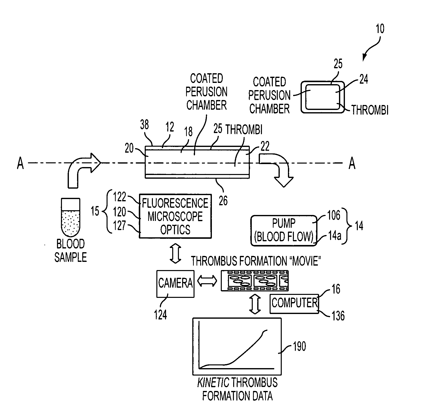
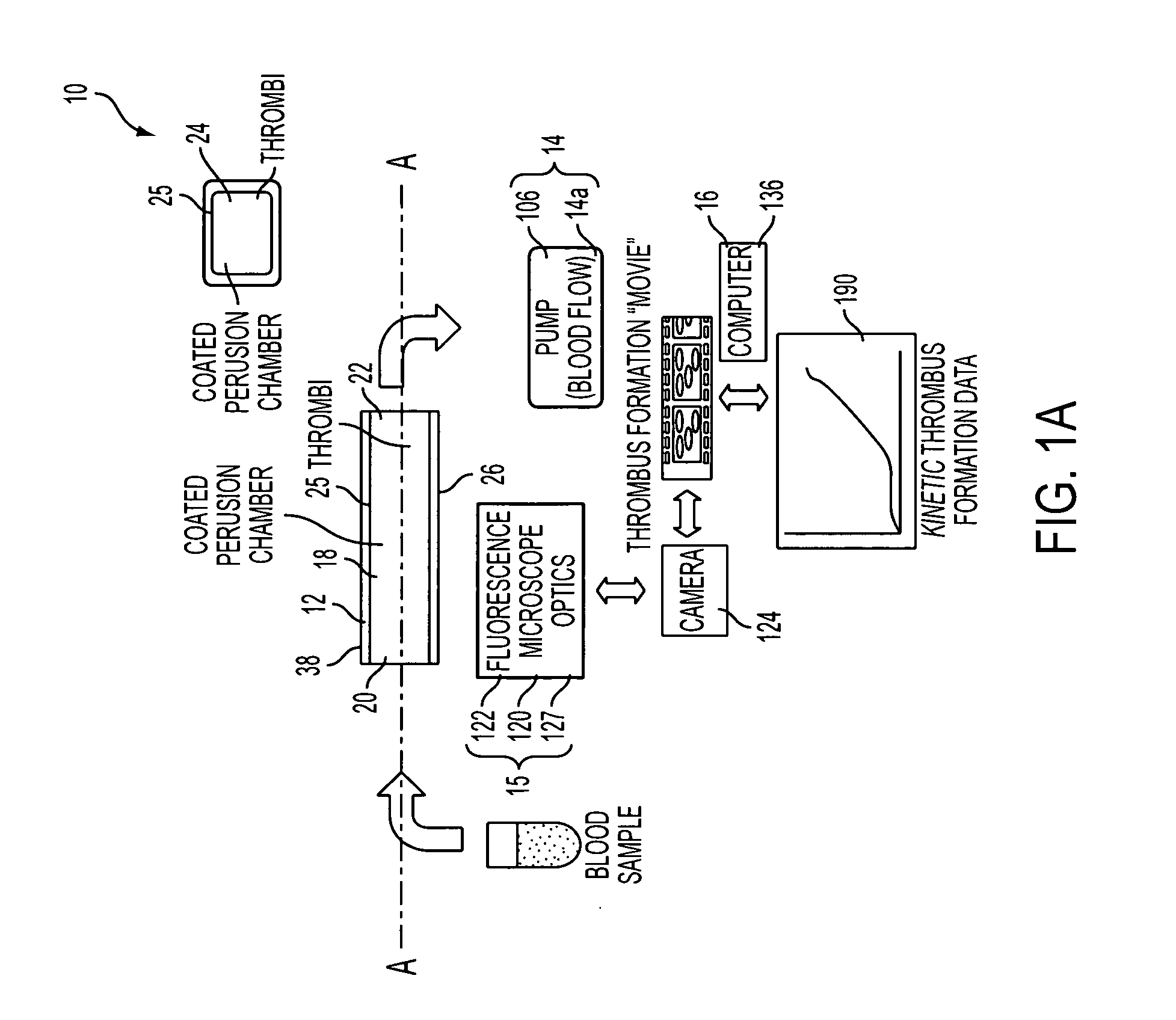
A Method to Monitor in Real Time the Kinetics of Thrombosis

[0147] The methodology and device described herein allows the monitoring in real time of the deposition of fluorescently labeled platelets into a transparent perfusion chamber (FIG. 11). The thrombosis profiler consists of a custom built epifluorescence microscope to monitor thrombus formation and a syringe pump to establish the desired flow and wall shear rate in the capillary perfusion chamber. A thermostatic sample compartment maintains the blood sample at a temperature of 37° C. Platelets are labeled by adding an aliquot of Rhodamine 6G (final concentration 1.25 μg / ml) to whole blood. The dye is excited with light from a high-power light emitting diode with a spectral maximum at 530 nm and a spectral half width of 35 nm (Luxeon V, Lumileds Lighting, San Jose, Calif.). Excitation and emission light are filtered with a set of fluorescence filters (31002, Chroma Technologies, Rockingham, Vt.). A microscope objective images...

example 3

A Method to Detect the Effect of Shear Rates on the Kinetics of Thrombosis

[0149] Whole blood is collected using a butterfly needle (avoid the use of vacutainer which activates platelets via high shear). Factor Xa inhibitor anticoagulated whole blood was collected from one donor. Six experiments were successively performed at increasing shear rates (from 125 / sec to 2000 / sec). The increase in shear rates leads to an exponential increase in platelet deposition when whole blood is perfused through a human type III collagen coated perfusion chamber (FIG. 13). FIG. 14 indicates the variability in thrombotic profiles between perfusion chambers for the same blood donor. Whole blood (anticoagulated with a factor Xa inhibitor) from one blood donor is perfused for 5 min through a collagen-coated capillary perfusion chamber at 1000 / sec 15, 30, 45, 60, 75 and 90 minutes after blood has been collected. Four individual donors were studied. Experiments demonstrated reproducibility in the kinetics ...


PUM

PropertyMeasurementUnit
heightaaaaaaaaaa
volumeaaaaaaaaaa
widthaaaaaaaaaa

Abstract

An instrument for capturing an image of thrombus formation, blood coagulation, recruitment of circulating inflammatory or tumour cells in a blood sample. The instrument comprises a member defining a channel therethrough, a fluid handling assembly that permits the blood sample to move through the channel at a flow rate, and an imaging assembly including a microscopy device. The imaging assembly is disposed relative to the channel so as to capture light rays defining the image of thrombus formation in the channel.

Description

[0001] This application claims the benefit of U.S. Provisional Application Ser. No. 60 / 635,659 filed Dec. 14, 2004, which is incorporated by reference herein in its entirety.FIELD OF INVENTION [0002] The invention relates generally to a device and method for producing and analyzing blood deposits to obtain a blood deposit profile. More particularly, to a device and system for analyzing the kinetics of thrombosis (platelet adhesion, thrombus growth, stability and reversal), blood coagulation and biological behavior of blood sample constituents (leukocytes and circulating tumor cells. The assays and analytical tools embodied in the systems enable novel and clinically relevant information for use in characterizing modifiers of constituent responses as affected by genetic, experimental and / or pharmacological modulation and or variation. DESCRIPTION OF RELATED ART [0003] Evaluation of the thrombotic process in humans has been achieved using different approaches. One way is the use of a p...

Claims


Application Information

Patent Timeline
Patent Type & Authority Applications(United States)
IPC IPC(8): C12Q1/56G06F19/00G06K9/00C12M1/34
CPCB01L3/5027C12Q1/56G01N1/31G01N33/86G01N2015/0084G01N2015/0092G06T7/0012G06T7/602G06T7/62
Inventor ANDRE, PATRICKLUEDEMANN, HANSPHILLIPS, DAVIDSHAPURIAN, GOLNAZMUIR, CRAIG
Owner MILLENNIUM PHARMA INC
Why Eureka
Try Eureka