Polyurethanes for osteoimplants

a polyurethane and osteoimplant technology, applied in the field of polyurethanes for osteoimplants, can solve the problems of fractures and other orthopedic injuries that take a long time to heal, bone is unable to support physiologic loading, and the bone density around the implant site is decreased, so as to achieve the effect of increasing the surface concentration

Inactive Publication Date: 2006-09-28
WARSAW ORTHOPEDIC INC
View PDF38 Cites 144 Cited by
  • Summary
  • Abstract
  • Description
  • Claims
  • Application Information

AI Technical Summary

Problems solved by technology

While bone wounds can regenerate without the formation of scar tissue, fractures and other orthopedic injuries take a long time to heal, during which the bone is unable to support physiologic loading.
Use of metal implants may result in decreased bone density around the implant site due to stress shielding.
Furthermore, metal implants are permanent and unable to participate in physiological remodeling.
In general, however, it is a limitation of allograft bone that larger allografts do not completely remodel, and residual allograft bone may persist at the graft site for many years or indefinitely, potentially acting as a stress riser and a possible fracture site.
The use of bone grafts is further limited by the availability of tissue with the appropriate shape and size, as well as the desired mechanical strength and degradation rate.

Method used


Image

  • Polyurethanes for osteoimplants
  • Polyurethanes for osteoimplants

Examples


example # 1

Example #1

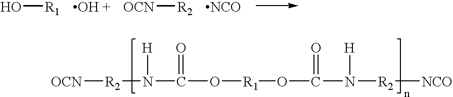
[0067] To determine the compressive strength of a composite implant made of approximately 66.6% bone and 33.3% castor bean polyurethane resin; 20 grams of bovine bone powder (particle size 120 μm-500 μm) were combined with a two part polyurethane (Doctors Research Group, Plymouth Conn. and described in “Vegetal Polyurethane Resin Implant Cranioplasty. Experimental Studies in Rabbits” by Luiz Fernando Francisco, Sao Jose do Rio Preto, 1998, which is incorporated herein by reference). Firstly, 6.10 grams of liquid comprising a polyisocyanate terminated molecule “prepolymer” were combined with 3.60 grams of a liquid comprising castor bean oil fatty acid triglyceride “diol”. Next, bone particles were gradually mixed into the polyurethane solution, until the bone appeared well coated. The mixture was then packed by hand into three 5 cc syringes (packed with light hand pressure). The samples were then set aside to polymerize over a 48-hour period at room temperature. After polym...

example # 2

Example #2

[0068] To determine the compressive strength of an implant made of 100% two-part castor bean polyurethane resin, (Doctors Research Group, Plymouth Conn. and described in “Vegetal Polyurethane Resin Implant Cranioplasty. Experimental Studies in Rabbits” by Luiz Fernando Francisco, Sao Jose do Rio Preto, 1998) enough of the prepolymer and diol (as indicated in Example 1) were mixed together to fill a 5 cc syringe. The material was hand packed into the syringe and allowed to polymerize for 18 hours at room temperature (air bubbles were noticed within the sample). After polymerization was complete, the samples were removed from the syringe and cut to length (approx. 13 mm). The results of mechanical static compression tests, using the Bionix MTS 858 (Edin Prarrie Minn.), are shown in column 5 of Table 2. The MPa values listed are only approximate at the point of visible plastic deformation of the implant. Samples did not mechanically fail at 20 MPa, but rather plastically defo...

example # 3

Example #3

[0069] To determine the compressive strength of a composite implant made of approximately 75% bone and 25% castor bean polyurethane resin, 20 grams of bovine bone powder (particle size 120 μm-500 μm) were combined with a 6.82 grams of a two part polyurethane (Doctors Research Group, Plymouth Conn. and described in “Vegetal Polyurethane Resin Implant Cranioplasty. Experimental Studies in Rabbits” by Luiz Fernando Francisco, Sao Jose do Rio Preto, 1998). The mixture was then packed by hand into three 5 cc syringes (packed with light hand pressure). The samples were then set aside to polymerize over a 48-hour period at room temperature. After polymerization was complete, the samples were removed from the syringes and cut to length (approx. 14 mm). Of the 6 samples tested; 4 were tested dry, while two were hydrated in Simulated Body Fluid (SBF) for 24 hours and tested wet. The results of mechanical static compression tests using the Bionix MTS 858 (Edin Prarrie Minn.) are show...


PUM

PropertyMeasurementUnit
time periodaaaaaaaaaa
thickaaaaaaaaaa
widthaaaaaaaaaa

Abstract

Biological-based polyurethanes and methods of making the same. The polyurethanes are formed by reacting a biodegradable polyisocyanate (such as lysine diisocyanate) with an optionally hydroxylated biomolecule to form polyurethane. The polymers formed may be combined with ceramic and / or bone particles to form a composite, which may be used as an osteoimplant.

Description

[0001] This application is a continuation-in-part of U.S. application Ser. No. 10 / 771,736, filed Feb. 4, 2004, which claims the benefit of U.S. Provisional Application No. 60 / 444,759, filed Feb. 4, 2003, the entire contents of both of which are incorporated herein by reference in its entirety.BACKGROUND OF THE INVENTION [0002] Vertebrate bone is a composite material composed of hydroxyapatite, collagen, and a variety of noncollagenous proteins, as well as embedded and adherent cells. Vertebrate bone can be processed into an implantable biomaterial, such as an allograft, for example, by removing the cells, leaving behind the mineral and extracellular matrix. The processed bone biomaterial can have a variety of properties, depending upon the specific processes and treatments applied to it, and may incorporate characteristics of other biomaterials with which it is combined. For example, bone-derived biomaterials may be processed into load-bearing mineralized grafts that support and int...

Claims


Application Information

Patent Timeline
Patent Type & Authority Applications(United States)
IPC IPC(8): A61K31/785C08G63/91C08G63/48C08G18/10C08G18/38C08G18/64C08G18/73C08G18/77C08G18/79
CPCA61F2/28A61F2002/2817A61K31/785A61L27/18A61L27/46A61L27/48C08G18/10C08G18/3882C08G18/6484C08G18/73C08G18/771C08G18/798C08G2230/00C08L75/04C08G18/36A61F2310/00359
Inventor KNAACK, DAVIDWINTERBOTTOM, JOHNKAES, DAVIDBOYCE, TODDSHIMP, LARRY
Owner WARSAW ORTHOPEDIC INC
Why Eureka
Try Eureka