Real time ultrasound monitoring of the motion of internal structures during respiration for control of therapy delivery

a technology of internal structure and real-time ultrasound, applied in the field of radiation therapy precision targeting, can solve the problems of loss of accuracy, movement can be particularly problematic, loss of accuracy,

Inactive Publication Date: 2006-10-26
CIVCO MEDICAL INSTR CO
View PDF12 Cites 59 Cited by
  • Summary
  • Abstract
  • Description
  • Claims
  • Application Information

AI Technical Summary

Problems solved by technology

However, much of the accuracy attainable with these products is lost when treating any tumor or lesion in a body region that moves with respiration.
Such movement can be especially problematic in a patient who is breathing voluntarily and not intubated with an endotracheal tube where volume input may be controlled.
Normal respiration is a complex mixture of diaphragmatic and chest wall movements that may vary from breath to breath, and the result is that even a similar breath may not result in a similar position of internal organs that move with respiration.
These variables have made accurate targeting of tumors that move with respiration a persistent problem.
As a consequence, in typical prior art treatment methods, the loss of accuracy has been addressed simply by enlarging the treatment field to include the entire excursion area of the moving target throughout respiration.
The additional morbidity and reduced treatment effectiveness using this method have been accepted in the past as being unavoidable.
Because of the inconsistency of normal respiration, fundamental inaccuracies inherent in breath measurement, and the significant expense and / or the invasiveness required by these different “gating” methods, none of these approaches has achieved universal acceptance.
The optimized therapy that is regularly achieved for static organs generally is not being achieved in the cases where respiratory movement affects the treatment field.
However, there is no suggestion to use U / S images to directly “gate” or time the treatment delivery.
Moving the entire treatment table to move the patient between medical devices, rather than moving just the patient, reduces the chance that internal organs within the patient will shift during movement.

Method used


Image

  • Real time ultrasound monitoring of the motion of internal structures during respiration for control of therapy delivery
  • Real time ultrasound monitoring of the motion of internal structures during respiration for control of therapy delivery
  • Real time ultrasound monitoring of the motion of internal structures during respiration for control of therapy delivery

Examples


example 1

[0064] In an exemplary use of the method of the present invention, a lung lesion is to be treated with radiation therapy, particularly ERT. As described previously, the lung moves during the respiratory cycle, and thus it is desirable to irradiate the patient only to the extent that the radiation is directed at the lesion itself. The lung cannot be readily imaged using ultrasound, and thus this hollow organ cannot be imaged with ultrasound. In this case, the liver may be chosen as the surrogate organ and a suitable surrogate AF within an easily visualized area of the liver may be chosen, while the lung contains the target lesion. Because lung lesion movement may be correlated to movement of a surrogate AF within the liver as described above, the radiation beam may be triggered based on movement of the surrogate AF with respect to a reference image. Because the target lesion is in a known anatomic position when the surrogate AF is in its “snapshot” position, the beam may be timed and...

example 2

[0065] In another exemplary use of the method of the present invention, a lesion of the liver is to be treated with radiation therapy, particularly ERT. As described previously, the liver moves during the respiratory cycle, and thus it is desirable to irradiate the patient only to the extent that the radiation is directed at the lesion itself. The liver may be readily imaged using ultrasound, simultaneously with radiation treatment of the liver, so long as the ultrasound transducer does not interfere with the field of the radiation treatment. No surrogate organ is needed. Because liver lesion movement may be tracked, the radiation beam may be triggered based on movement of the liver with respect to an AF within the liver chosen from a reference 2-D ultrasound image.

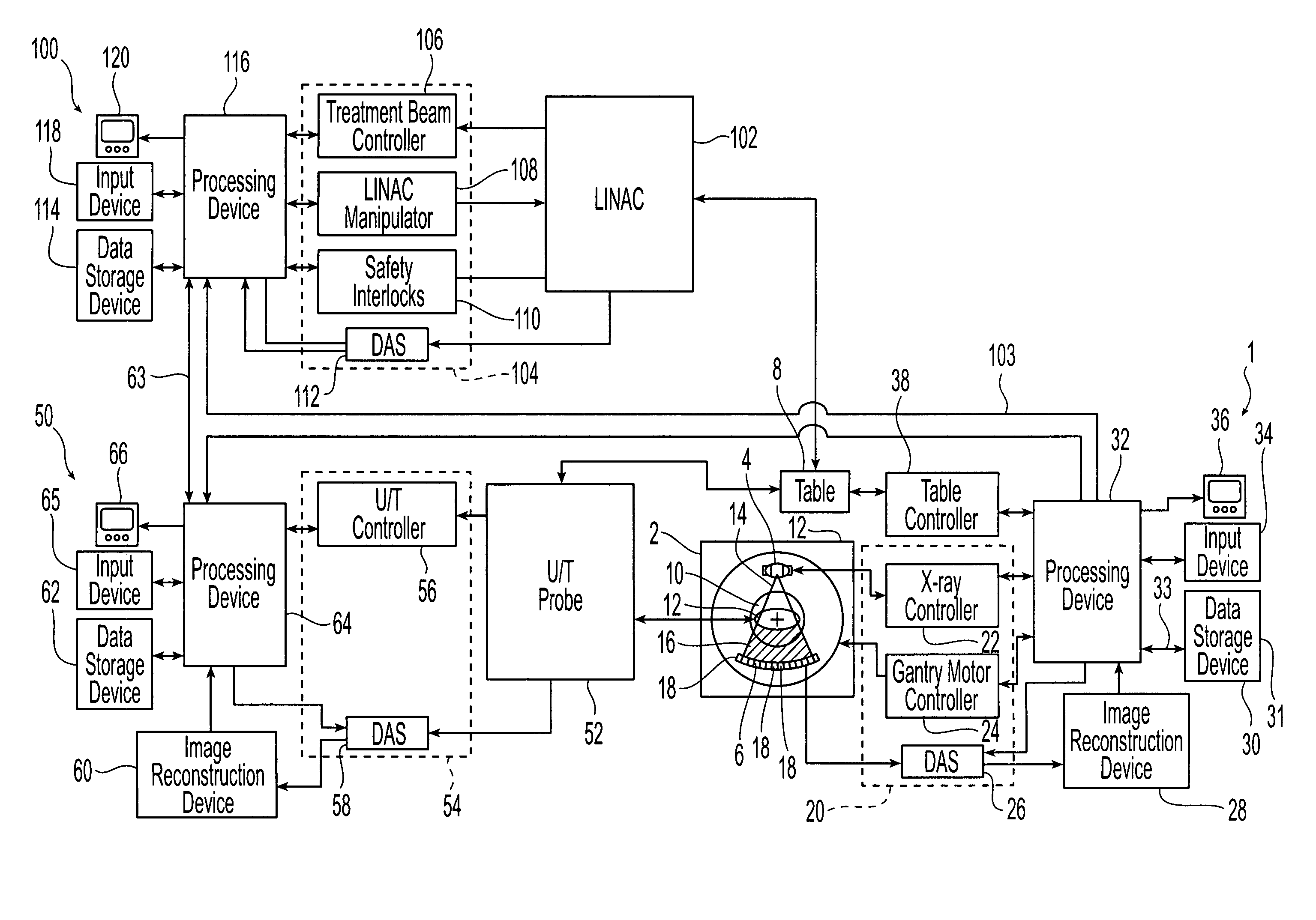
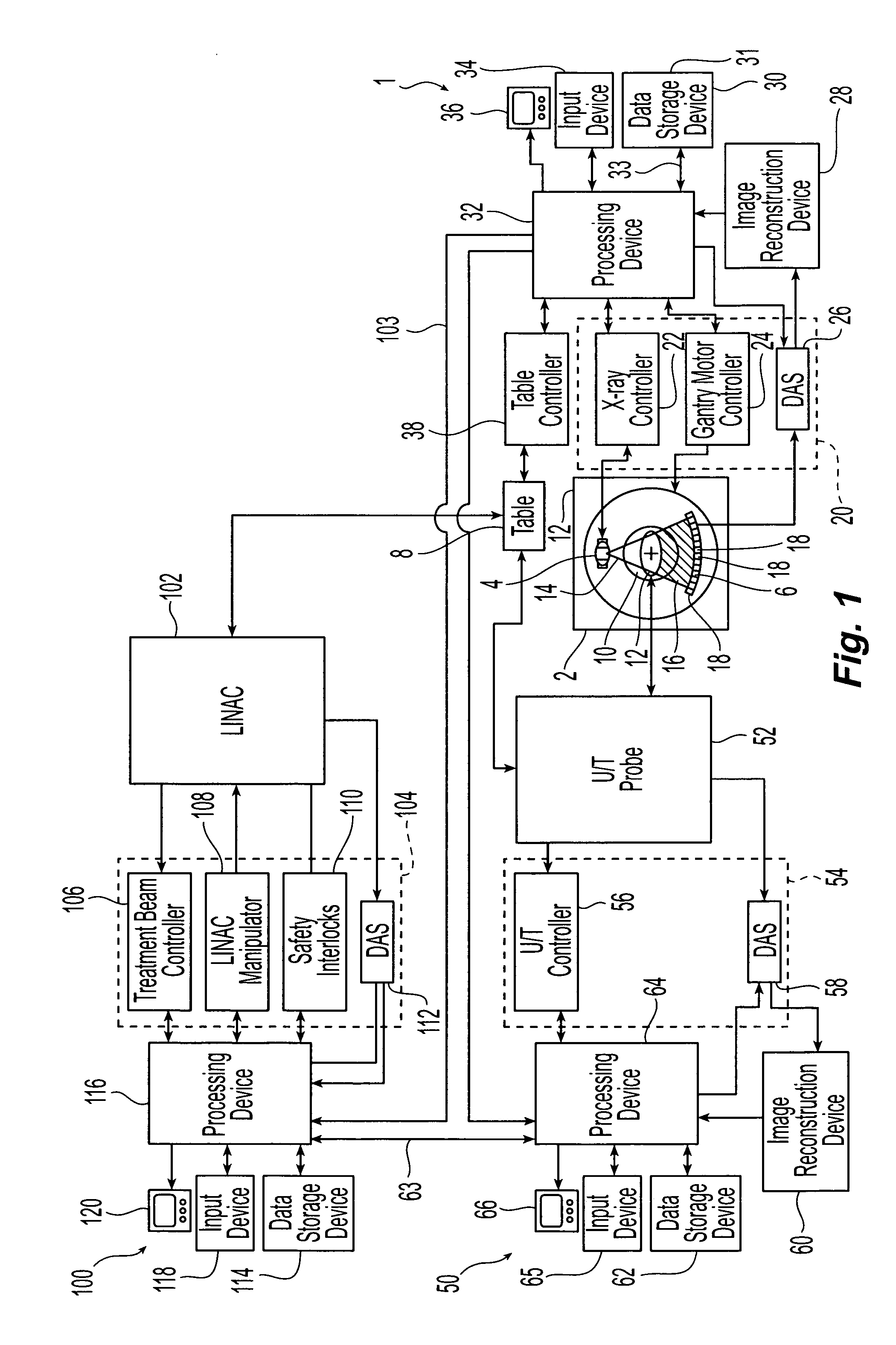
[0066] A representative computer system is now described in conjunction with which the embodiments of the present invention may be implemented. The computer system may be a personal computer, workstation, or a larger sys...


PUM


Abstract

A method of targeting therapy such as radiation treatment to a patient includes: identifying a target lesion inside the patient using an image obtained from an imaging modality selected from the group consisting of computed axial tomography, magnetic resonance tomography, positron emission tomography, and ultrasound; identifying an anatomical feature inside the patient on a static ultrasound image; registering the image of the target lesion with the static ultrasound image; and tracking movement of the anatomical feature during respiration in real time using ultrasound so that therapy delivery to the target lesion is triggered based on (1) movement of the anatomical feature and (2) the registered images.

Description

CROSS-REFERENCE TO RELATED APPLICATIONS [0001] The benefits of Provisional Application No. 60 / 629,403 filed Nov. 22, 2004 are claimed under 35 U.S.C. § 119(e), and the entire contents of this application are expressly incorporated herein by reference thereto.FIELD OF THE INVENTION [0002] The invention relates to the application of continuous ultrasound imaging during the delivery of therapy to eliminate the errors induced by respiratory movement. More particularly, the invention relates to precision targeting of radiation therapy. BACKGROUND OF THE INVENTION [0003] In the field of radiation oncology, tumors of all types are treated with ionizing radiation that disrupts the basic chemistry of tumor cells in order to cause cell death. The radiation may be delivered by either an invasive procedure that uses an implanted or temporary internal source, or non-invasively by an external source such as a linear accelerator. During treatment, a sufficient dose of ionizing radiation preferably...

Claims


Application Information

Patent Timeline
Patent Type & Authority Applications(United States)
IPC IPC(8): A61B8/00A61H1/00
CPCA61B5/7285A61B6/541A61B8/08A61N5/1037A61B8/565A61N5/1064A61N5/107A61N2005/1058A61N5/1049A61B8/5276
Inventor WHITMORE, WILLET F. IIISCHENCK, ARTHUR J.ONIK, GARY M.
Owner CIVCO MEDICAL INSTR CO
Why Eureka
Try Eureka