Protein subcellular localization assays using split fluorescent proteins

a technology of fluorescent proteins and subcellular localization, which is applied in the field of protein subcellular localization assays using split fluorescent proteins, can solve the problems of misfolding of the fused protein, interference with protein processing and/or intracellular transport, and limited information provided by methods

Active Publication Date: 2006-11-16
TRIAD NAT SECURITY LLC
View PDF7 Cites 24 Cited by
  • Summary
  • Abstract
  • Description
  • Claims
  • Application Information

AI Technical Summary

Benefits of technology

[0013] A distinct advantage of the present invention over tagging using full-length GFP is the absence of background fluorescence prior to complementation. Only if complementation occurs in a particular compartment to which one or more fragments are localized, does that compartment become fluorescent. It is necessary only to measure the fluorescence of the cell to determine whether the specific loc

Problems solved by technology

However, although protein localization methods using GFP fused to a protein of interest have been useful, these methods provide limited information and are compromised by problems inherent in using a relatively large terminally fused reporter protein tag.
In particular, GFP and related fluorescent proteins may cause misfolding of the fused protein, and may interfere with protein processing and/or intracellular transport.
Misfolding of the reporter can result in the generation of insoluble aggregates of the fusion, which may be unable to freely move through intracellular processing and transport systems within the cell.
In general, the use of GFP fusions in extracytoplasmic compartments has been limited because of export deficiencies.
As an example, efforts to obtain functional GFP following export through the sec-dependent pathway failed because of improper folding of the protein; the secreted fraction of GFP was not fluorescent (Feilmeier, Iseminger et al.
Accordingly, it is difficult to obtain a true picture of a test protein's trafficking or distribution within a cell using terminal GFP fusion tags, and systems capable of visualizing subcellular compartmentalization through a wider lens and over time are n

Method used


Image

  • Protein subcellular localization assays using split fluorescent proteins
  • Protein subcellular localization assays using split fluorescent proteins
  • Protein subcellular localization assays using split fluorescent proteins

Examples


example 2

Complementation of Split GFP in Specialized Compartments

Cloning of Localization Sequences in Fusion with SR-s11 and GFP1-10

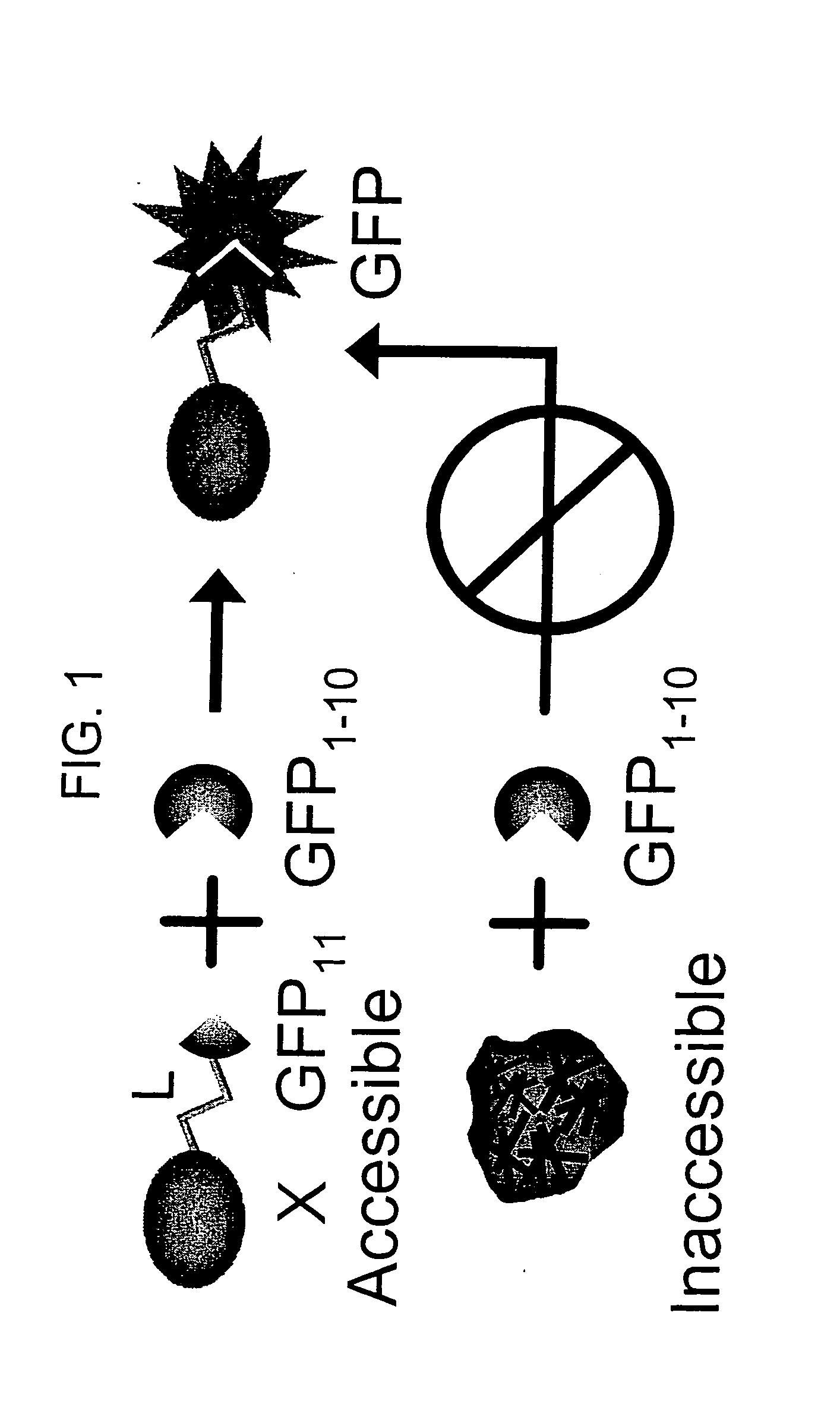
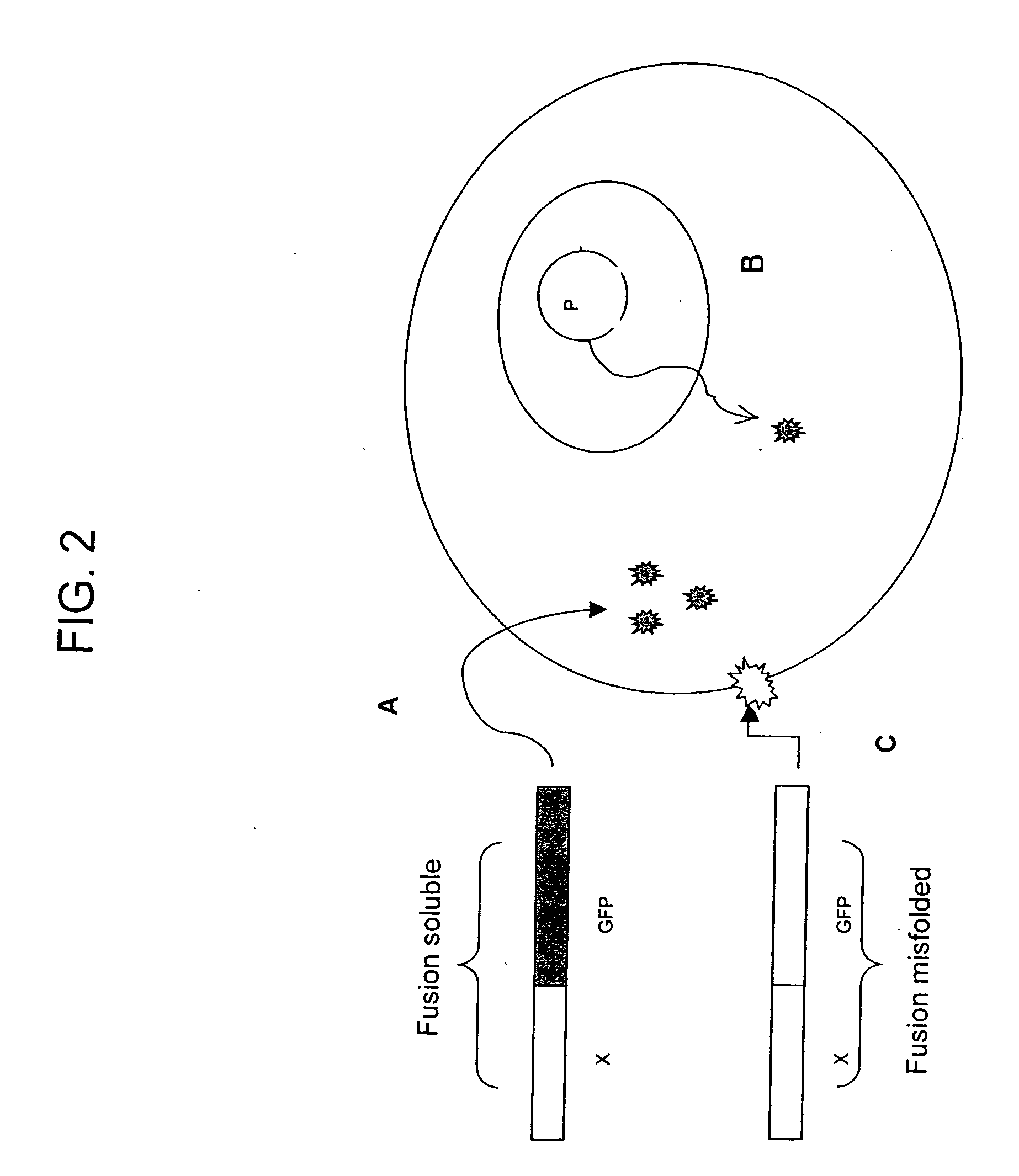
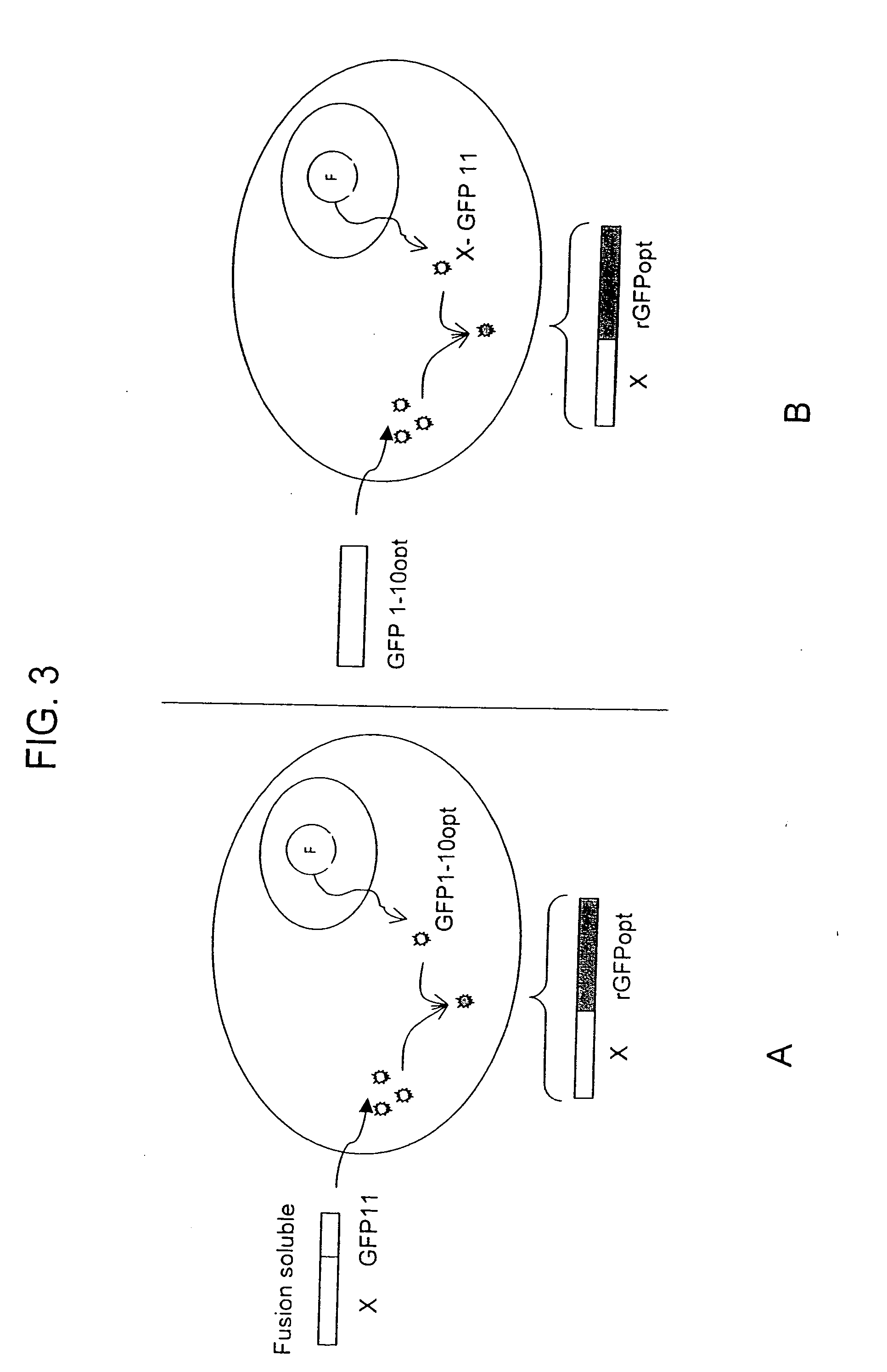
[0113] If both fragments contains a sequence that targets the gene of interest in a specialized compartment, then the two tagged split-GFP fragments will be targeted to this particular location and will be able to complement in the compartment.

[0114] Various subcellular localization signal sequences or tags are known and / or commercially available. These tags are used to direct split-fluorescent protein fragments to particular cellular components or outside of the cell. Mammalian localization sequences capable of targeting proteins to the nucleus, cytoplasm, plasma membrane, endoplasmic reticulum, golgi apparatus, actin and tubulin filaments, endosomes, peroxisomes and mitochondria are known.

[0115] Detailed protocols for localization in the nucleus, mitochondria and endoplasmic reticulum follow.

[0116] Localization in mitochondria: The mitochondrial targetin...


PUM

PropertyMeasurementUnit
Volumeaaaaaaaaaa
Volumeaaaaaaaaaa
Volumeaaaaaaaaaa

Abstract

The invention provides protein subcellular localization assays using split fluorescent protein systems. The assays are conducted in living cells, do not require fixation and washing steps inherent in existing immunostaining and related techniques, and permit rapid, non-invasive, direct visualization of protein localization in living cells. The split fluorescent protein systems used in the practice of the invention generally comprise two or more self-complementing fragments of a fluorescent protein, such as GFP, wherein one or more of the fragments correspond to one or more beta-strand microdomains and are used to “tag” proteins of interest, and a complementary “assay” fragment of the fluorescent protein. Either or both of the fragments may be functionalized with a subcellular targeting sequence enabling it to be expressed in or directed to a particular subcellular compartment (i.e., the nucleus).

Description

RELATED APPLICATIONS [0001] This patent application claims the benefit of the filing date of U.S. Provisional patent application No. 60 / 633,488.STATEMENT AS TO RIGHTS TO INVENTIONS MADE UNDER FEDERALLY SPONSORED RESEARCH OR DEVELOPMENT [0002] This invention was made with government support under Contract No. W-7405-ENG-36 awarded by the United States Department of Energy to The Regents of The University of California. The government has certain rights in this invention.BACKGROUND OF THE INVENTION [0003] Understanding the intracellular transport and localization of proteins is important because many processes in biology, including transcription, translation, and metabolic or signal transduction pathways, are mediated by proteins and non-covalently-associated multiprotein complexes localized to particular cellular compartments Reed, L. J. (1974) Multienzyme Complexes. Acc. Chem. Res. 7, 40-46. Proteins and protein complexes are the workhorses of the chemical machinery in living system...

Claims


Application Information

Patent Timeline
IPC IPC(8): G01N33/567G01N33/53
CPCG01N33/5005G01N33/5041G01N33/6842G01N33/582G01N33/6803G01N33/542
Inventor WALDO, GEOFFREYCABANTOUS, STEPHANIE
Owner TRIAD NAT SECURITY LLC
Why Eureka
Try Eureka