
Cell-based luminogenic and nonluminogenic proteasome assays

a luminogenic and non-luminogenic technology, applied in the field of cell-based luminogenic and non-luminogenic proteasome assays, can solve the problems of apoptosis of cells, high cost of many currently available synthetic substrates, and inability to detect luminogenic proteins,

Inactive Publication Date: 2007-03-01
PROMEGA
View PDF28 Cites 35 Cited by
  • Summary
  • Abstract
  • Description
  • Claims
  • Application Information

AI Technical Summary

Benefits of technology

[0016] Therefore, in one embodiment, a combined luminogenic / nonluminogenic assay format of the present invention allows multiplexing of assays for one or more peptides or polypeptides, e.g., enzymes, one or more molecules which are bound by and / or altered by the peptide(s) or polypeptide(s), e.g., a peptide or polypeptide substrate for at least one enzyme, and / or one or more cofactors for an enzyme-mediated reaction, or other molecules or conditions, or a combination thereof. In one embodiment, the invention provides a method to detect the activity of a protease associated with proteasomes (a first enzyme-mediated reaction) and the presence or amount of a second molecule for a second enzyme-mediated reaction, and optionally other molecules including molecules for other enzyme-mediated reactions. The method includes contacting a cellular sample with a reaction mixture for the first enzyme-mediated reaction and the second enzyme-mediated reaction. In one embodiment, a cell membrane permeabilization reagent in an amount which does not substantially disrupt intracellular membrane bound organelles or compartments is added to the cells before the cells are contacted with the reaction mixture. In another embodiment, a cell membrane permeabilization reagent in an amount which does not substantially disrupt intracellular membrane bound organelles or compartments is added to the reaction mixture before the reaction mixture is contacted with the cells. In one embodiment, luminescence is employed to detect proteasome activity and fluorescence or colorimetry is employed to detect a molecule for a second enzyme-mediated reaction. The activity of proteasomes and the presence or amount of the second molecule are then detected.
[0022] The assay also has use as a drug discovery tool. Thus, the presence or amount of a modulator, for instance, an inhibitor, of a protease associated with proteasomes may be detected using an assay of the invention, e.g., a fluorogenic or luminogenic assay. The invention thus provides a method in which one or more agents are contacted with a reaction mixture comprising cells, a cell membrane permeabilization reagent in an amount which does not substantially disrupt intracellular membrane bound organelles or compartment in the cells, and a luminogenic or fluorogenic substrate for a protease associated with proteasomes, so as to yield a mixture. Also provided is a method in which one or more agents are contacted with cells, and that mixture contacted with a reaction mixture comprising a cell membrane permeabilization reagent in an amount which does not substantially disrupt intracellular membrane bound organelles or compartment in cells, and a luminogenic or fluorogenic substrate for a protease associated with proteasomes. Luminescence or fluorescence in the mixture is compared to luminescence or fluorescence in a corresponding mixture which lacks the one or more agents.BRIEF DESCRIPTION OF THE DRAWINGS

Problems solved by technology

Proteases, however, are not easy to assay with their naturally occurring substrates.
Moreover, many currently available synthetic substrates are expensive, insensitive, and nonselective.
Inhibition of the proteasome pathway results in apoptotic cell death.

Method used


Image

  • Cell-based luminogenic and nonluminogenic proteasome assays
  • Cell-based luminogenic and nonluminogenic proteasome assays
  • Cell-based luminogenic and nonluminogenic proteasome assays

Examples


example i

Protease Retention and Release Cell Viability Multiplex Assays

[0075] Live cell and dead cell assays are widely used to monitor the change in cellular viability in response to specific chemical, biological or physical treatments. Viability and cytotoxicity assays are generally converse and measure different biomarkers. Methods for assessment of general changes in cell viability by cytotoxicity have historically related to changes in outer membrane permeability. Classical methods of detecting compromised membrane structure include trypan blue exclusion, nucleic acid staining, and lactate dehydrogenase release (Riss et al., 2004; Myers et al., 1998). Assays for the assessment of cell function or proliferation include tritiated thymidine incorporation, ATP content, tetrazolium dye conversion or fluorescein diacetate (Cook et al., 1989). The assumption is that intact cell membranes do not allow bulky charged molecules or peptides to enter from the extracellular space into the cytosol. C...

example ii

Cell-Based Assays for Proteasome Activities

Materials and Methods

Plate Preparation

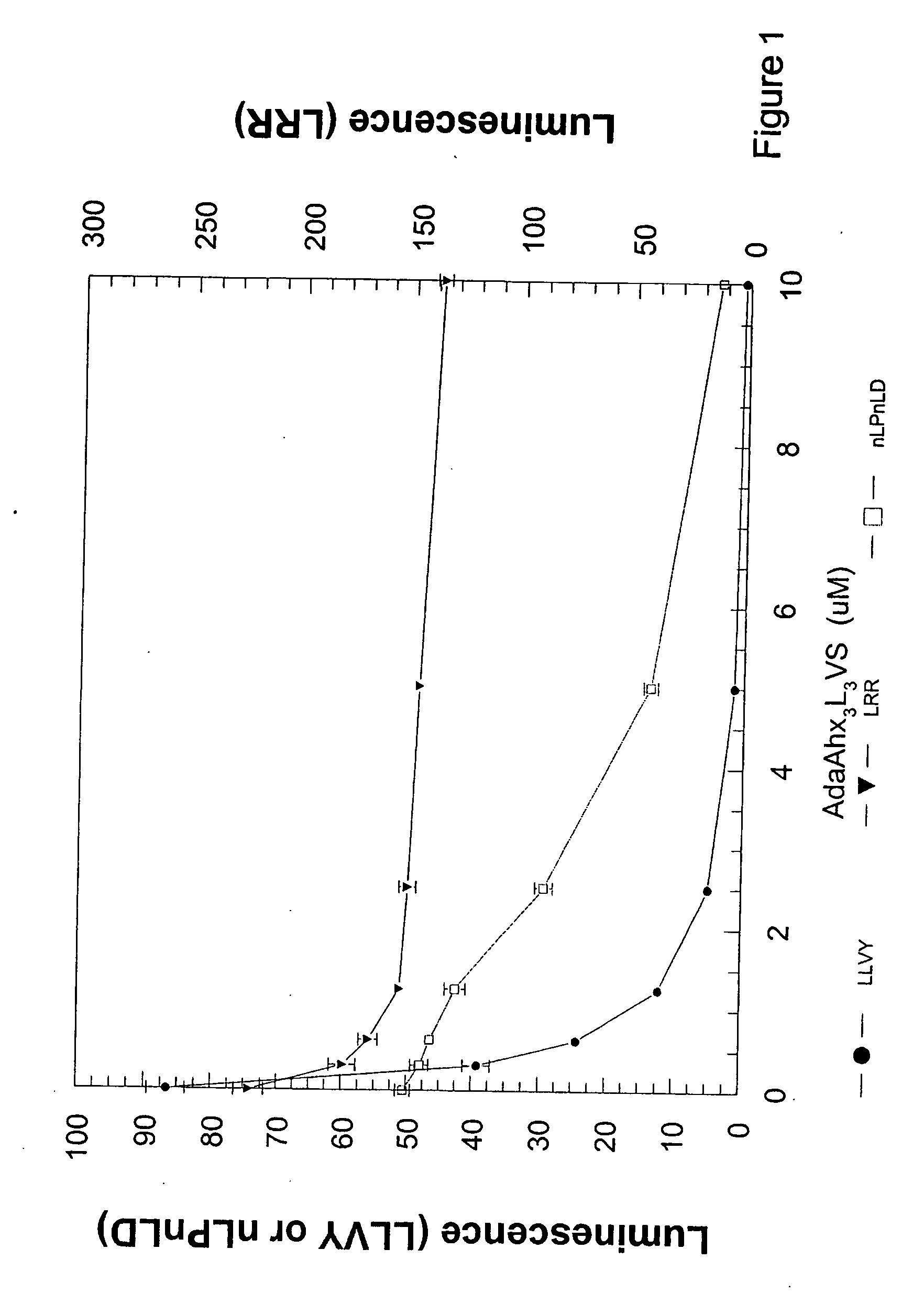
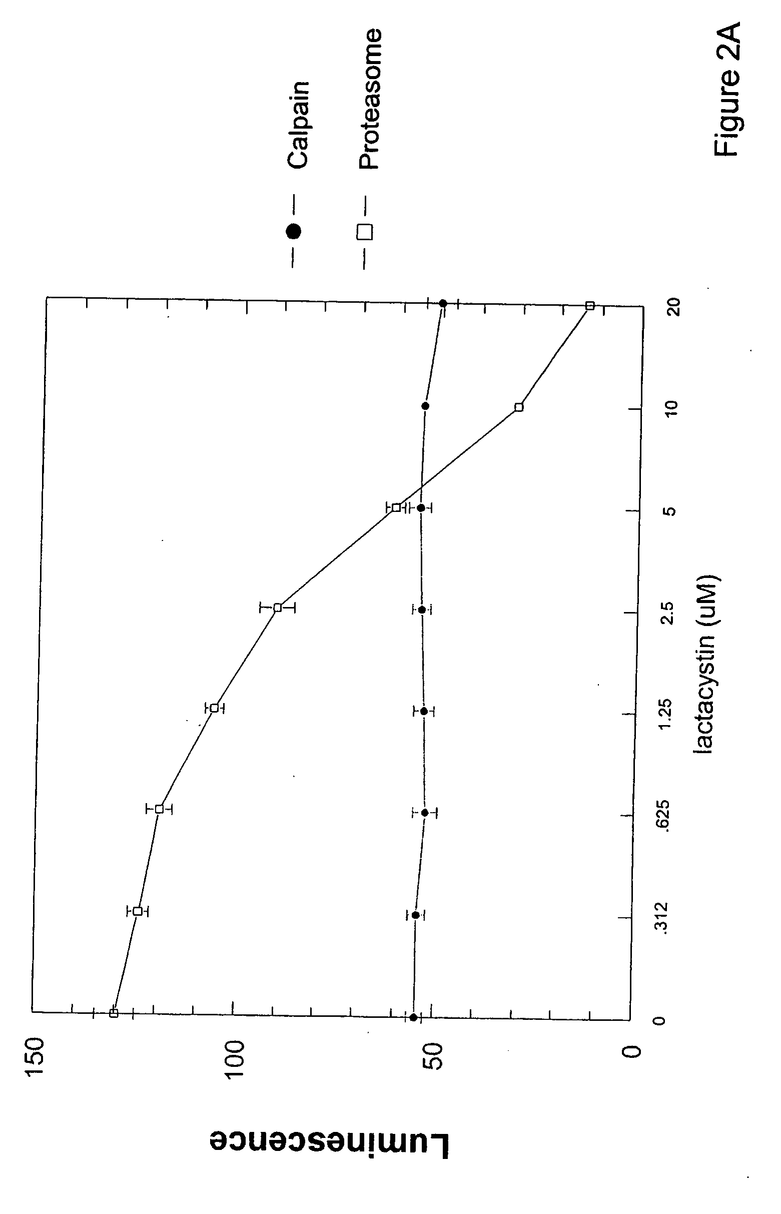
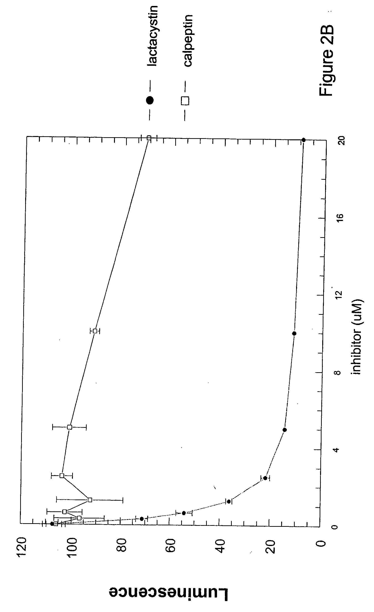
[0113] NCI-H226 cells (ATCC #CRL-5826) were maintained as an attached line using RPMI 1640 (Sigma #R-8005) with 10% fetal bovine serum (Hyclone # SH30070) and passaged as needed. Jurkat, HL-60 and U937 suspension cell lines were likewise maintained and passaged. To prepare cells for a cell-based proteasome assay, adherent cells were harvested from the parent flask by removing medium, rinsing the flask with D-PBS and incubated with trypsin-EDTA (Sigma T-4040) for 3 to 4 minutes at 37° C. The trypsin reaction was stopped by adding complete medium containing serum, and cells were then centrifuged 4 minutes at 200×G. The cell pellet was suspended in fresh medium, cells counted by trypan blue exclusion and adjusted to 1.11×10⁵ cells / ml. A 96-well clear bottom / white walled plate (Costar# 3610) was obtained and 90 μl / well of cell suspension or medium alone was dispensed. Plate was cultured in a humidified...


PUM


Abstract

A method to detect proteasome activity in permeabilized cells, and optionally in a multiplex assay to detect presence or amount of at least one molecule for a different enzyme-mediated reaction, is provided.

Description

CROSS-REFERENCE TO RELATED APPLICATIONS [0001] This application claims benefit under 35 U.S.C. 119(e) of U.S. Provisional Application Ser. No. 60 / 713,906, filed Sep. 1, 2005, which is incorporated herein by reference in its entirety.BACKGROUND [0002] Luminescence is produced in certain organisms as a result of a luciferase-mediated oxidation reaction. Luciferase genes from a wide variety of vastly different species, particularly the luciferase genes of Photinus pyralis and Photuris pennsylvanica (fireflies of North America), Pyrophorus plagiophthalamus (the Jamaican click beetle), Renilla reniformis (the sea pansy), and several bacteria (e.g., Xenorhabdus luminescens and Vibrio spp), are extremely popular luminescence reporter genes. Firefly luciferase is also a popular reporter for determining ATP concentrations, and, in that role, is widely used to detect biomass. Luminescence is also produced by other enzymes when those enzymes are mixed with certain synthetic substrates, for ins...

Claims


Application Information

Patent Timeline
IPC IPC(8): C12Q1/66
CPCC12Q1/37C12Q1/66G01N2500/02G01N2333/96466G01N2333/96433
Inventor MORAVEC, RICHARD A.RISS, TERRY L.
Owner PROMEGA
Learn More