
Methods for direct visualization of active synapses

a synapse and active technology, applied in the field of can solve the problems of difficult caveats and elusive direct visualization of active synapses in complex neuronal networks, and achieve the effects of improving the transport of fragments, modulating neuronal transport, and easy manipulation of composition

Inactive Publication Date: 2007-04-26
INST PASTEUR +1
View PDF16 Cites 3 Cited by
  • Summary
  • Abstract
  • Description
  • Claims
  • Application Information

AI Technical Summary

Benefits of technology

This patent describes a method for visualizing an active synapse in the brain using a biomarker called tetanus toxin. The method involves exposing cells in the brain to a biomarker and then visualizing it using a special technique. This method can be used to screen molecules that can modulate synapse activity, which is important for brain function. It can also be used to diagnose neurodegenerative diseases by detecting the accumulation of the biomarker in dendritic spines. The patent also describes a method for delivering desired compositions into the brain using tetanus toxin. Overall, this patent provides a useful tool for studying brain function and disease diagnosis.

Problems solved by technology

However, direct visualization of active synapses in complex neuronal networks has been elusive to date because of the lack of biological markers being specifically addressed and / or accumulated in these neuronal structures.
Nevertheless, the non-proteic nature of these molecules and the transient labeling that they provide represent difficult caveats when trying to study long-term synaptic activity and remodeling.
Moreover, staining of more physiological relevant networks such as those presented in brain slices or intact animals with styryl dyes has been proven to be arduous, requiring the reduction of nonspecific background fluorescence while preserving the specific fluorescent signal by adding sulforhodamine or other fluorescent quenching reagents (Pyle, 1999).

Method used


Image

  • Methods for direct visualization of active synapses
  • Methods for direct visualization of active synapses
  • Methods for direct visualization of active synapses

Examples


example 1

Plasmid Constructions

[0130] (A) TTC Cloning:

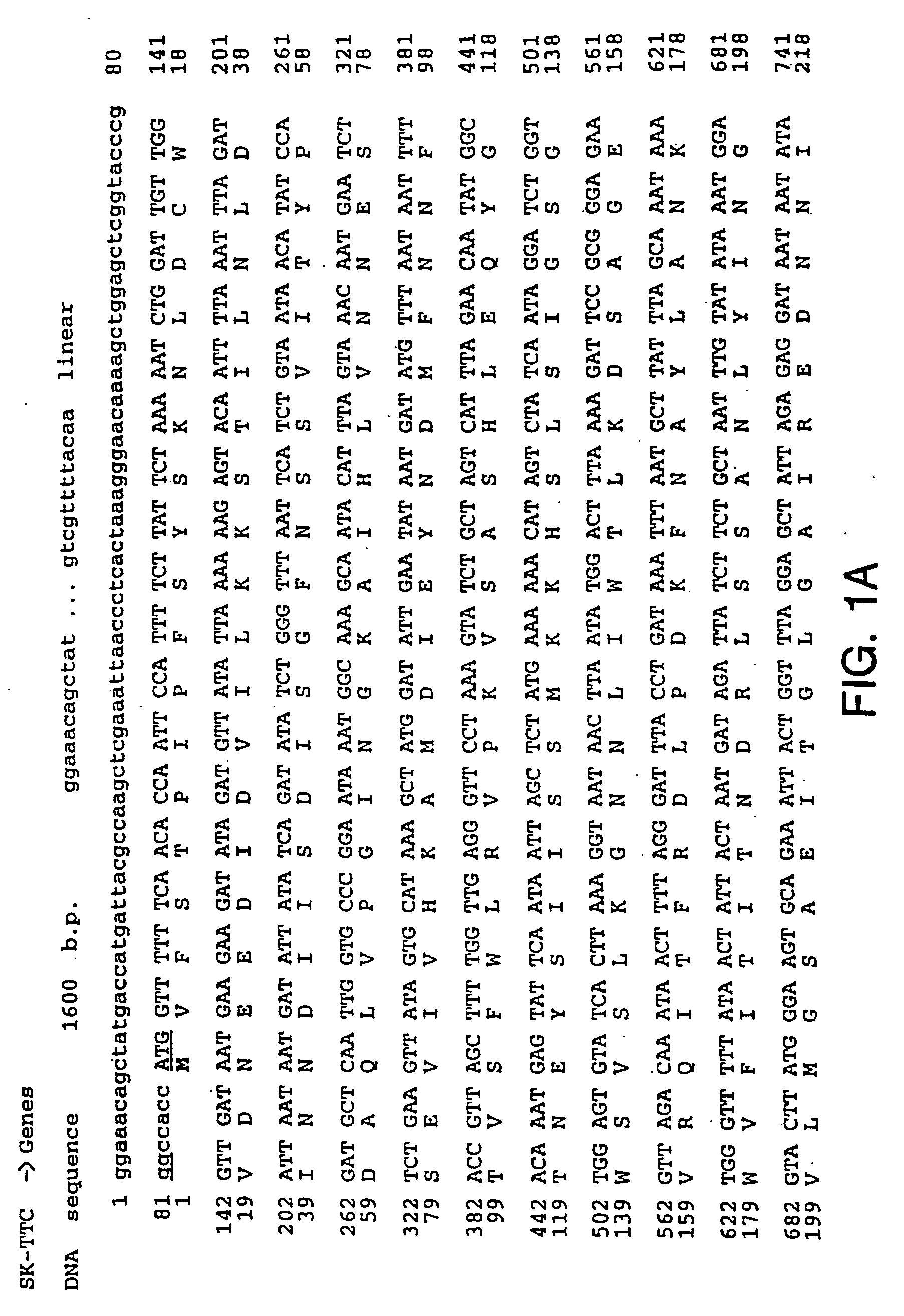
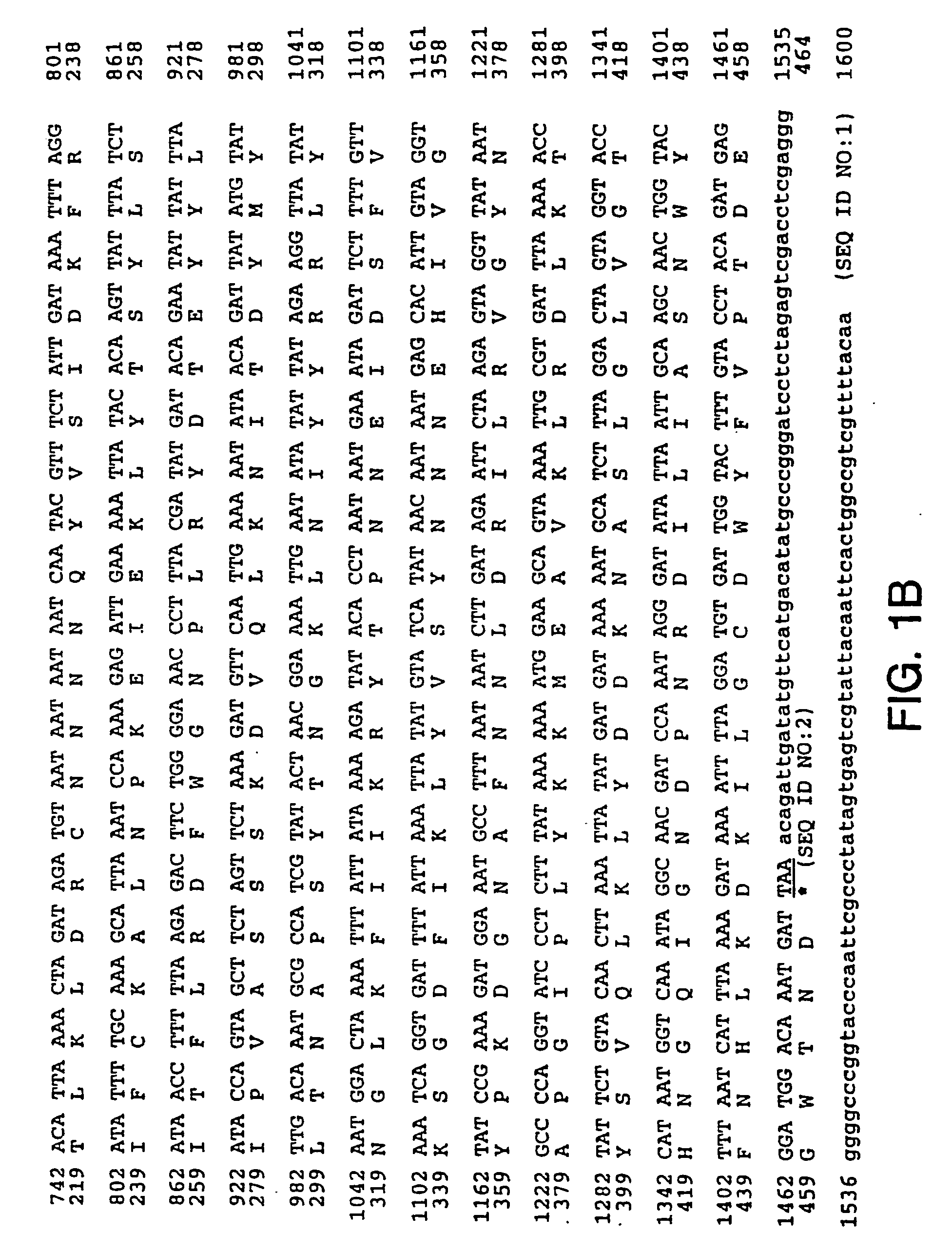
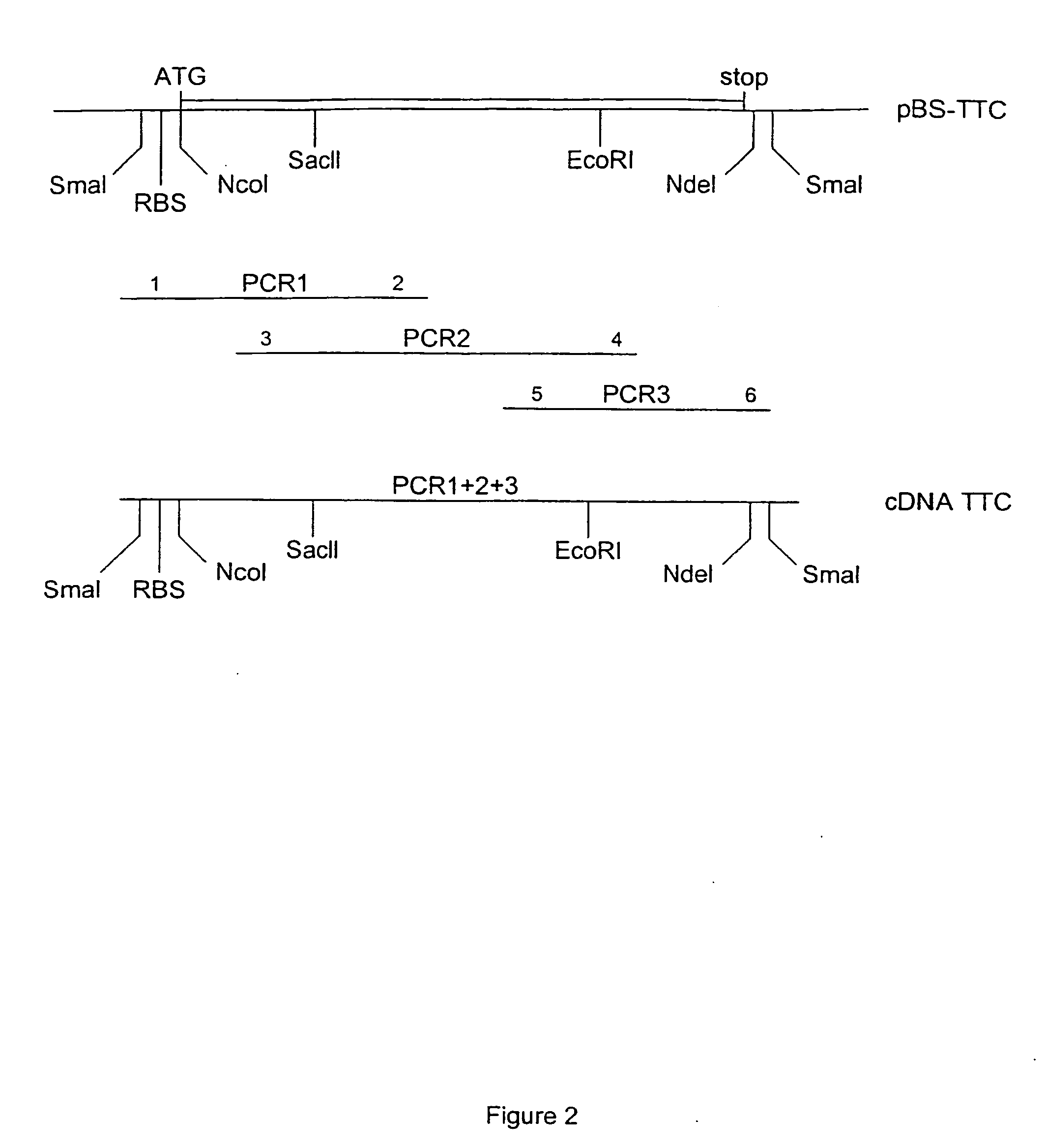
[0131] Full length TTC DNA was generated from the genomic DNA from the Clostridium Tetani strain (a gift from Dr. M. Popoff, Institut Pasteur) using PCR. Three overlapping fragments were synthesized: PCR1 of 465 bp (primer 1: 5′-CCC CCC GGG CCA CCA TGG TTT TTT CAA CAC CAA TTC CAT TTT CTT ATT C-3′ and primer 2: 5′-CTA AAC CAG TAA TTT CTG-3′), PCR2 of 648 bp (primer 3: 5′-AAT TAT GGA CTT TAA AAG ATT CCG C-3′ and primer 4: 5′-GGC ATT ATA ACC TAC TCT TAG AAT-3′) and PCR3 of 338 bp (primer 5: 5′-AAT GCC TTT AAT AAT CTT GAT AGA AAT-3′ and prirner 6: 5′-CCC CCC GGG CAT ATG TCA TGA ACA TAT CAA TCT GTT TAA TC-3′). The three fragments were sequentially introduced into pBluescript KS+ (Stratagene) to give pBS:TTC plasmid. The upstream primer 1 also contains an optimized eukaryotic Ribosome Binding Site (RBS) and translational initiation signals. Our TTC fragment (462 amino acids) represents the amino acids 854-1315 of tetanus holotoxin, i.e. the ca...

example 2

Purification of the Hybrid Protein

[0139] The E. coli strain SR3315 (a gift from Dr. A. Pugsley, Institut Pasteur) transfected with pGEX:lacz-TTC was used for protein production. An overnight bacterial culture was diluted 1:100 in LB medium containing 100 μg / ml ampicillin, and grown for several hours at 32° C. until an OD of 0.5 was reached. Induction from the Ptac promoter was achieved by the addition of I mM IPTG and 1 mM MgCl2 and a further 2 hrs incubation. The induced bacteria were pelleted by centrifugation for 20 min at 3000 rpm, washed with PBS and resuspended in lysis buffer containing 0.1M Tris pH 7.8, 0.1M NaCl, 20% glycerol, 10 mM EDTA, 0.1% Triton-X100, 4 mM DTT, 1 mg / ml lysosyme, and a mixture of anti-proteases (100 μg / ml Pefablok, 1 μg / ml leupeptin, 1 μg / ml pepstatin, 1 mM benzamidine). After cell disruption in a French Press, total bacterial lysate was centrifuged for 10 min at 30000 rpm. The resulting supernatant was incubated overnight at 4° C. with the affinity ma...

example 3

Binding and Internalization of Recombinant Protein in Differentiated 1009 Cells

[0141] The 1009 cell line was derived from a spontaneous testicular teratocarcinoma arising in a recombi nant inbred mouse strain (129×B6) (17). The 1009 cells were grown in Dulbecco's modified Eagle's medium (DMEM) containing 10% fetal calf serum and passaged at subconfluence. In vitro differentiation with retinoic acid and cAMP was performed as described (18). Eight days after retinoic acid treatment, cells were used for the internalization experiments with either the hybrid protein or β-gal.

[0142] Binding and internalization of the β-Gal-TTC fusion were assessed using a modified protocol (16). Differentiated 1009 cells were incubated for 2 hrs at 37° C. with 5 μg / ml of β-Gal-TTC or β-Gal protein diluted in binding buffer (0.25% sucrose, 20 mM Tris acetate 1 mM CaCl2, 1 mM MgCl2, 0.25% bovine serum albumin, in PBS). The cells were then incubated with 1 μg / ml Pronase E (Sigma) in PBS for 10 min at 37° ...


PUM

PropertyMeasurementUnit
diameteraaaaaaaaaa
sizeaaaaaaaaaa
ODaaaaaaaaaa

Abstract

A method for visualizing an active synapse wherein said method comprises: (a) exposing cells forming the active synapse to a biomarker comprising at least fragment C of tetanus toxin and a reporter protein; and (b) visualizing the biomarker; wherein the accumulation of the biomarker into dendritic spines of the cells allows visualization of an active synapse. Also, a method for screening molecules capable of modulating synapse activity is provided. A kit useful for the early diagnosis of neurodegenerative disease comprises a biomarker comprising at least fragment C of tetanus toxin and a reporter protein.

Description

BACKGROUND OF THE INVENTION [0001] This invention relates to the use of part of tetanus toxin for delivering a composition to the central nervous system of a human or animal. This invention also relates to a hybrid fragment of tetanus toxin, a polynucleotide that hybridizes with natural tetanus toxin, and a composition containing the tetanus toxin fragment as an active molecule. Further, this invention relates to a vector comprising a promoter and a nucleic acid sequence encoding the tetanus toxin fragment. In addition, this invention relates to methods of using the tetanus toxin fragment. [0002] Tetanus toxin is produced by Clostridium tetani as an inactive, single, polypeptide chain of 150 kD composed of three 50 kD domains connected by protease-sensitive loops. The toxin is activated upon selective proteolytic cleavage, which generates two disulfide-linked chains: L (light, 50 kD) and H (heavy, 100 kD) [Montecucco C. and Schiavo G. Q. Rev. Biophys., (1995), 28: 423-472]. [0003] E...

Claims


Application Information

Patent Timeline
Patent Type & Authority Applications(United States)
IPC IPC(8): A61K49/00
CPCA61K49/0041A61K49/0047A61K49/0056G01N33/5058G01N33/5091G01N2333/33G01N2800/28
Inventor VAZQUEZ-MARTINEZ, RAFAELBRULET, PHILIPPE
Owner INST PASTEUR
Learn More