

Assessment of human papilloma virus-related disease
a human papilloma virus and disease technology, applied in the field of cytological and molecular assays, can solve the problems of inconvenient diagnosis and tracking of hpv infection, inconvenient use, and inability to detect hpv infection in conventional viral detection assays, including serologic assays and cell culture growth,
- Summary
- Abstract
- Description
- Claims
- Application Information
AI Technical Summary
Benefits of technology
Problems solved by technology
Method used
Image

Examples
example 1
General Methods for Nucleic Acid Analysis
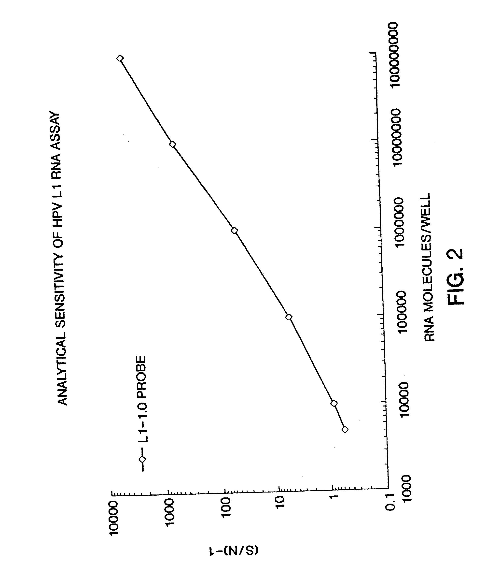
[0069] The assay for nucleic acids follows in general principle the method for detecting HIV RNA by the Digene Hybrid Capture HIV Test, described in WO 93 / 10263 by Digene. Briefly, following lysis, 50 μl of probe mix (containing DNA biotinylated probe) was added to each well. The plate was sealed and incubated at 65° C. for 2 hours for hybridization to occur. After hybridization, samples were transferred to a strepavidin-coated microplate, and 25 μL of anti-hybrid antibody was added to each well. The plate was agitated at 1100 RPM, for 1 hour, at room temperature. Wells were washed 6× times with 65° C. wash buffer, followed by one wash using distilled water. 100 μl of a chemiluminescent substrate was added to each well and the plate was incubated at room temperature for 30 minutes. The plate was then read in the DML 2000 luminometer. The data was then expressed as signal-to-noise. Using a calibration curve, the chemiluminescent signal genera...
example 2
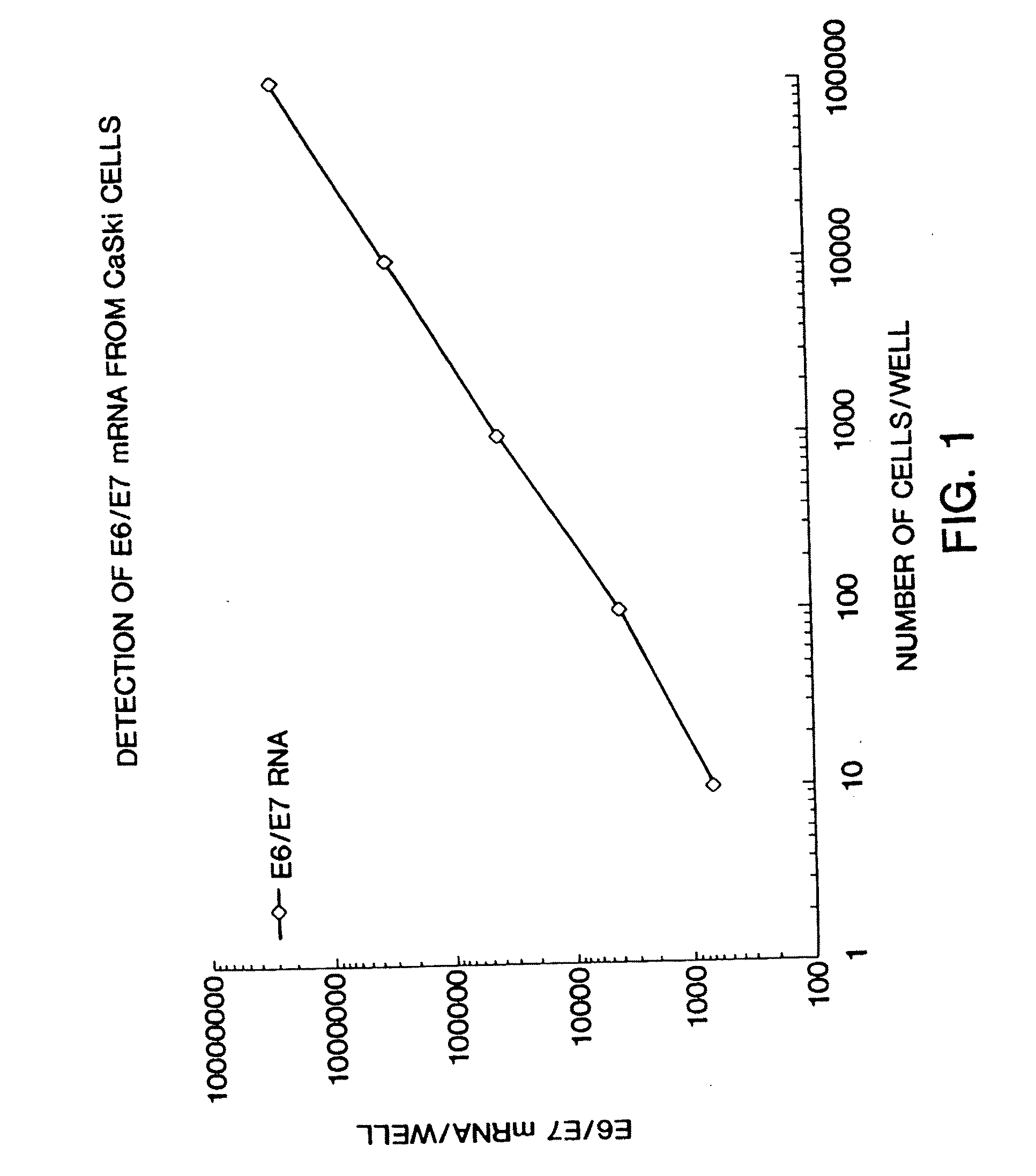
Quantitation of HPV
[0070] This example illustrates the measurement of HPV E6 / E7 expression for use in the disclosed method. A method for detecting and quantitating HPV mRNA, including E6 / E7 and mRNA has been developed. This example measures expression in CaSki cells, but the method is generally applicable to other cell lines and clinical specimens. CaSki cells contain an integrated high-risk HPV-16 genome (about 600 copies / cell). CaSki cells were maintained in subconfluence in RMPI 1640 media containing 10% FBS and 10 mM sodium pyruvate. For this example, CaSki cells were grown to confluence and were removed from the dishes by treatment with 0.1% trypsin-0.5 mM EDTA. Using trypan blue, viable cells were counted under microscopy. Cells were seeded, in 10 μl volumes, at final concentrations of 10, 102, 103, 104, and 105 cells / well in a polystyrene, tissue culture treated, 96 well plate. Each dilution was performed and seeded in triplicate.
[0071] The cells were lysed with Proteinase ...
example 3
Quantitation of HPV mRNA Using Preservative Collection Medium
[0072] CaSki cell line was trypsinized by incubating with 0.25% Trypsin-EDTA for 5 minutes at 37° C. Cells were pelleted from the suspension by centrifugation at 800 rpm for 3 minutes in Sorvall RT 6000 centrifuge. Cell pellet was resuspended in 500 μl of 1×PBS and counted under microscope Trypan Blue solution. Cells were diluted to 50 and 500 cells / μl in 1×PBS. 10 μl of each cell concentration, including zero point (10 μl of 1×PBS) were spiked in 3 ml of PreservCyt reagent. 100 μl of Sample Conversion Buffer were added into each tub to help visualize the cell pellet. All samples were mixed well and were spun down at 3800 rpm for 15 minutes in Sorvall RT 6000 centrifuge. Supernatants were discarded and tubes were drained by inversion on the Kimtowels for 2 to 5 minutes on the bench. All pellets were resuspended with 50 μl of the lysis reagent (50 units of Proteinase K) and mixture was transferred into the plate coated wit...
PUM
 Abstract
Description
Claims
Application Information