Bioengineered Intervertebral Discs and Methods for Their Preparation

a technology of intervertebral discs and bioengineered discs, which is applied in the field of bioengineered intervertebral discs, can solve the problems of limited mobility, low back pain, and less synthesized, and achieve the effects of increasing density and gags, reducing the time of in vitro cell manipulation, and high density

Active Publication Date: 2007-12-20
THE UNIVERSITY OF HONG KONG
View PDF10 Cites 17 Cited by
  • Summary
  • Abstract
  • Description
  • Claims
  • Application Information

AI Technical Summary

Benefits of technology

[0012] A bioengineered IVD for disc replacement as a clinically feasible treatment for patients with severely degenerated IVD has been developed. The disc is built of layers of cells such as MSCs that are encapsulated in a material such as collagen or extracellular matrix. The composition can include factors affecting the differentiation if MSCs such as TGF-beta for chondrocytic lineage. The bioengineered IVD is produced by including the self-assembly of a multi-layered structure containing the extracellular matrix and the living cells via MSCs-induced matrix contraction. the MSCs or other living cells can be evenly distributed throughout the bulk pf the ECM using this method. The method produces a bioengineered IVD with extra high density collagen as the outermost lamellae of the multi-layered structure with nucleus, inner and outer annulus, strengthened and stabilized by photochemical crosslinking followed by controlled dehydration. The method dehydrates the outermost ECM lamellae in a controlled manner.
[0014] The method enables the “growth” of IVD-like structures using living cells and natural extracellular matrix in a short period of time, preferably 10 days, so as to minimize the time of in vitro manipulation of the cells. The methods can produce multiple bioengineered IVDs that van be used for replacement of multiple discs at the same time, or for selection of the structure with the best functional characteristics after in vitro testing using a bioreactor such as that from the commercial source. These methods are capable of producing other multi-layered or heterogenous tissue structures including, but not limited to G.I. tract and blood vessels. The methods can also be used for cell encapsulation and delivery therapies for disorders such as, cardiovascular, neurological and musculoskeletal systems.
[0015] The bioengineered IVD is intact, viable and mechanically comparable to native discs and is ready for implantation as a treatment for severely degenerated IVD the needs replacement, The bioengineered IVD is structurally similar to the native disc with heterogeous structure including a distinct nucleus having collagen and glycosaminoglycans (GAGs) at a ratio to that of the native the disc, an inner annulus with clear demarcation separating the nucleus and with multiple lamellae of collagen matrix with increasing density and GAGs with decreasing density together with evenly distributed cells of increasing density; and an outer annulus with several lamellae of high density collagen matrix and cells. The collagen-GAGs core can be produced at different ratios and with water content similar to those in native tissues, the method of production also enhances the retention of GAGs in the collagen-GAGs core. The bioengineered IVD has mechanical properties comparable to that of the native disc.

Problems solved by technology

Disc degeneration is a common clinical problem affecting human population worldwide, causing low back pain and limited mobility.
In early disc degeneration, disc cells, in particular the NP cells, become less capable of synthesizing ECM, the gelatinous NP becomes more fibrous and therefore reduces the water content of the disc.
At advanced stages, structural collapse and eventual loss of disc function in resisting loading result.
There is no satisfactory clinical treatment for advanced disc degeneration.
Spinal fusion does not solve the problem but does relieve pain.
However, the fused spine loses motility and is non-functional.
However, these prostheses cannot integrate with the surrounding tissues and allow new tissue formation.
These methods are for early stage degeneration where the disc is still functional and retains its structural integrity, not in advanced degenerative cases where structural and functional replacement is needed.
Unfortunately, penetration of the cells into preformed scaffolds is usually limited to the surface (Seguin, et al., Spine, 29(12):1299-306 (2004)).
Large size ensures better penetration but compromises the mechanical properties.
Efforts such as agitation during seeding and creating channels in the scaffolds have improved the amount of cells reaching the half thickness of the scaffolds to around 38% (Rose, et al., Biomaterials, 25(24):5507-14 (2004)) but the high speed agitation detrimentally affects cell viability and wide channels detrimentally affect the scaffold properties.
Another limitation of the cell-seeding approach is that the distribution of cells in the scaffolds is not homogenous, which may affect the quality of the engineered tissue structure.
As a result, the above mentioned approaches may not have sufficient amount of extracellular matrix deposition to provide the necessary mechanical support for intervertebral disc.
Finally, the cell-seeding approach using preformed scaffolds can not be used for production of tissue with heterogeneous structures, consisting of different cell type and intensity, and with different ECM type and intensity, such as invertebral discs.
However, these synthetic materials have limitations in biocompatibilities.

Method used


Image

  • Bioengineered Intervertebral Discs and Methods for Their Preparation
  • Bioengineered Intervertebral Discs and Methods for Their Preparation
  • Bioengineered Intervertebral Discs and Methods for Their Preparation

Examples


example 1

Encapsulation of MSCs in Collagen Microshperes

[0076] Materials and Methods

[0077] Rattail sollagen solution type I was neutralized and diluted into 0.5 mg / ml in the presence of different concentrations (2×10⁴, 1×10⁵ and 5×10⁵ cells / ml) of human bone marrow derived MSCs in DMEM medium with 10% FBS. The solution was kept at 4° C. in an ice bath before use. A small volume of 5 μl of mixture was dropped onto a non-adhesive substratum using a dispenser. The hMSC containing microdroplets were allowed to reconstitute into microspheres encapsulating the cells by incubating in a 37° C. incubator for 2 hours and were then collected into a DMEM medium containing bath with non-adherent substratum. The collected microcapsules were incubated in 37° C. for three dimensional cultures and the morphological change and the dimensional change of the shperes were recorded for 48 hours before they were ready to be plated onto traditional culture dishes.

[0078] Results

[0079] Microspheres at day 0 showed...

example 2

Three Dimensional Culture of MSCs in Collagen Microcapsules, Outgrowth from the Attached Microcapsules and Replating of the Microcapsules

[0080] Materials and Methods

[0081] hMSCs encapsulated collagen microshperes were plated onto 10 mm diameter culture flask at different densities (50, 125 and 250 microshperes per dish) and incubated for 1-2 hours with 1-2 ml of full medium covering the surface of the dish. Afterwards, the dish was gently filled with 8-9 ml more medium. The microspheres were cultured for 3 days and the morphology of the cells migrating out from the microspheres assessed.

[0082] Results

[0083] Attachment of the microcapsules onto culture flask in necessary for cell outgrowth from the microspheres and depends on the cell density and plating density of the cell-encapsulating collagen microsphere showed similar morphology with spindle and elongates shaped. The hMSC-encapsulated collagen microspheres wer released from the culture dish by flushing with full medium and r...

example 3

Production of MSCs Seeded Collagen Matrix, MSCs Induced Collagen Matrix Contraction; Analysis of the Controlling Parameters of the Extent of Contraction

[0086] Materials and Methods

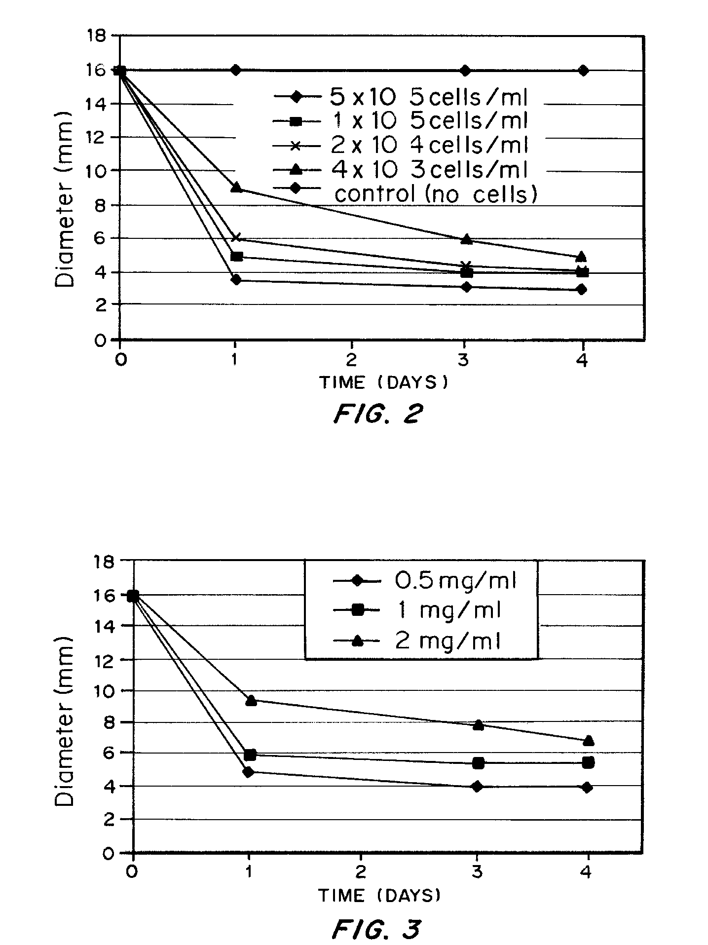
[0087] Rattail collagen type I solution was neutralized and diluted into concentrations at 0.5, 1 and 2 mg / ml i the presence of rabbit bone marrow derived MSCs at a concentration of 4×10³, 2×10⁴, 1×10⁵, 5×10⁵ cells / ml. The collagen MSC mixtures were thoroughly mixed and degassed if necessary and were cast in 4-well plate and incubated for 30 minutes at 37° C. in an incubator the solidified structure was detached from the culture plate by using a sterile pipette tip or a syringe needle without damaging the structure and was transferred into a non-adherent culture dish such as a bacterial culture dish. The dimension of the structure and the morphology of MSCs inside the structure were recorded against time as the index for volume contraction of the structures.

[0088] Results

[0089] Contraction of the colla...


PUM

PropertyMeasurementUnit
compressive modulusaaaaaaaaaa
temperaturesaaaaaaaaaa
densitiesaaaaaaaaaa

Abstract

A bioengineered IVD for disc replacement has been developed that has mechanical and structural support characteristics similar to those of native IVD. Extracellular matrix (ECM) provides support to living cell components and interacts with the living cellular components during the fabrication process without introducing toxicity. The composition can be produced from both natural or synthetic source but preferably natural and induced to self-assemble or reconstitute into its solid form under conditions that are mild enough to support cellular survival and growth. The cells induce a volume change of the structures, leading to changes in dimension, ECM density, cell density mechanical property and stability, etc. The extent of the change i volume of the composition can be precisely controlled by factors such as the density of the ECM, the density of the living cells, the timing for interaction and the serum concentration. Increased structural support is provided by crosslinking.

Description

CROSS-REFERENCE TO RELATED APPLICATIONS [0001] This application claims priority to U.S. Ser. No. 60 / 796.088 filed Apr. 28, 2006.FIELD OF INVENTION [0002] The present invention relates generally to bioengineered intervertebral discs. More specifically, it relates to methods and compositions of producing intervertebral disc-like structures with living cells and natural extracellular matrix, and with mechanical properties comparable to native discs, for replacing degenerated discs in particular severe cases, and to the resulting bioengineered intervertebral discs. BACKGROUND OF THE INVENTION [0003] The intervertebral disc (IVD) separates the vertebrae of the spine and functions to resist loading that the spine is subjected to during daily life, it has a unique structure with an inner water-rich gel-like nucleus pulposus (NP) with random organization of extracellular matrix (ECM); an outer fibrous annulus fibrous (AF) with well-organized collagen sheets; and thin cartilaginous end-plate...

Claims


Application Information

Patent Timeline
Patent Type & Authority Applications(United States)
IPC IPC(8): A61F2/28A61K38/39A61P19/00C12N5/06C12N5/08C12N5/0775
CPCA61F2/3094C12N5/0663A61F2002/2817A61F2002/30006A61F2002/30016A61F2002/30153A61F2002/30677A61F2002/30971A61F2002/4445A61F2002/445A61F2230/0019A61F2250/0015A61F2250/0018A61F2250/0019A61F2310/00365A61K2035/128A61L27/3633A61L27/3834A61L27/3852A61L27/3856A61L2430/38C12N2533/54A61F2/442A61P19/00
Inventor CHAN PUI, BARBARACHEUNG, KENNETH MAN-CHEECHAN, DANNYCHAN, GODFREY CHI-FUNGHUI, TING YAN
Owner THE UNIVERSITY OF HONG KONG
Why Eureka
Try Eureka