
Visualization apparatus for transseptal access

a technology of visualization apparatus and transseptal access, which is applied in the field of medical devices, can solve the problems of difficult visual identification, cramped working area created by inflatable balloons, and interference with fine positioning of imaging systems, and achieves the effect of easy visual identification and difficult visual identification

Active Publication Date: 2007-12-20
INTUITIVE SURGICAL OPERATIONS INC
View PDF103 Cites 265 Cited by
  • Summary
  • Abstract
  • Description
  • Claims
  • Application Information

AI Technical Summary

Benefits of technology

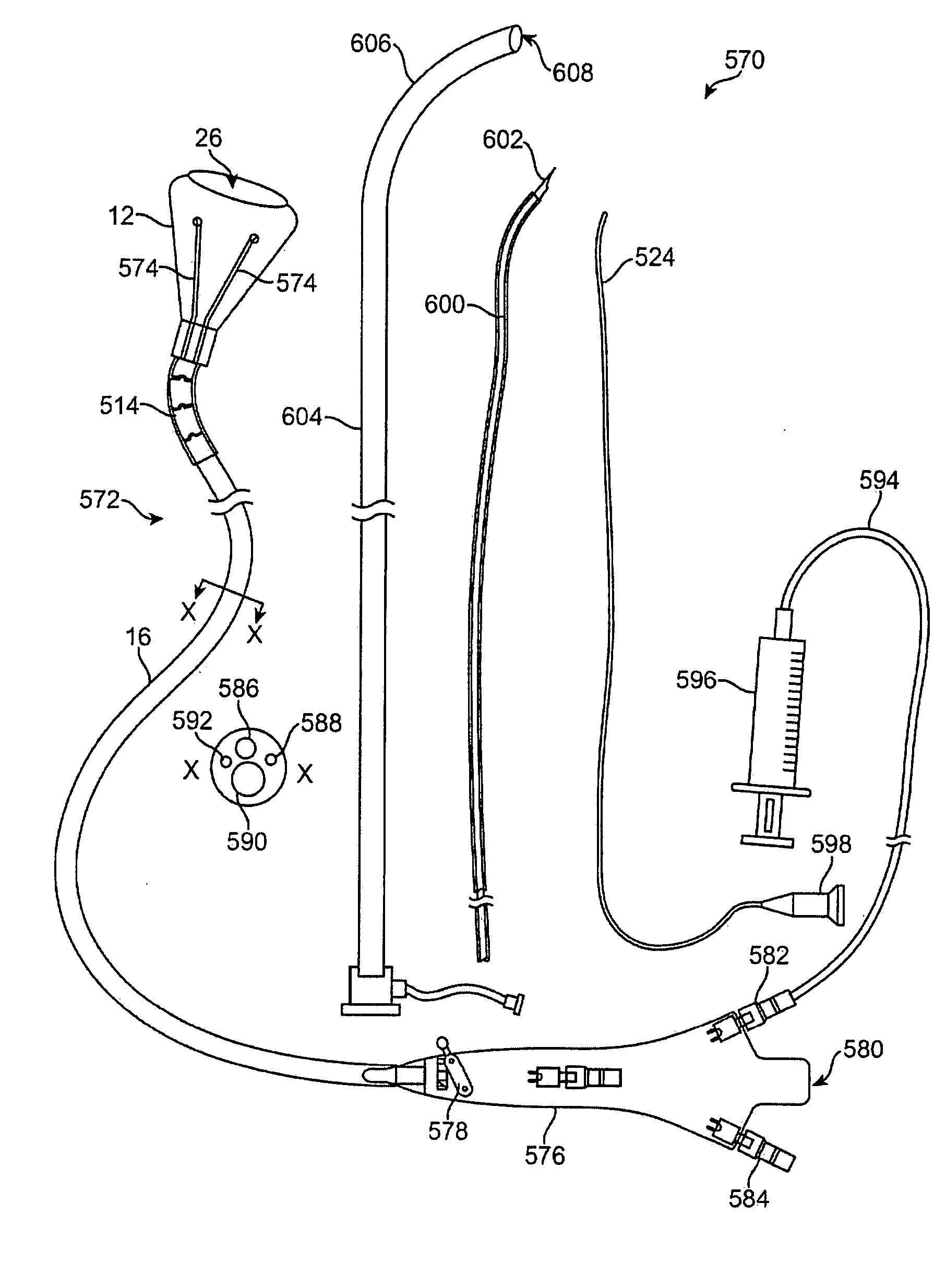
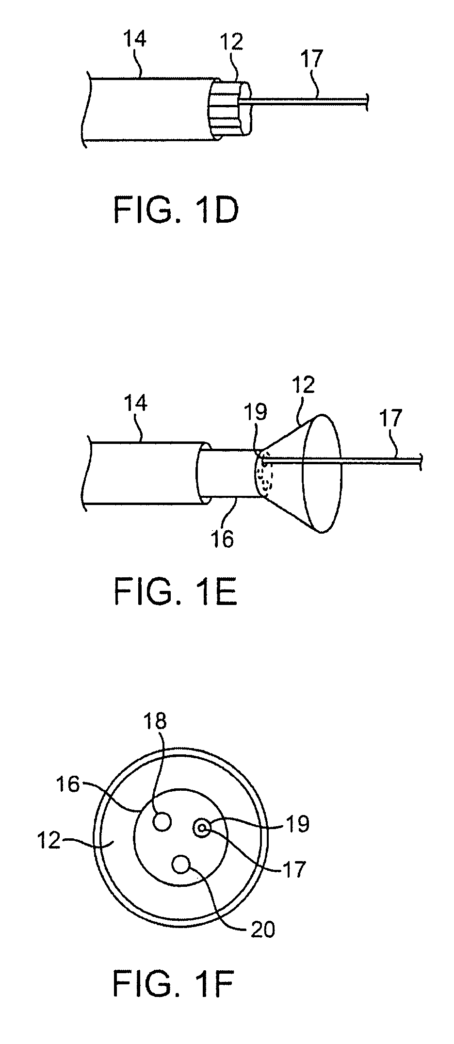
[0022] One variation of the assembly may utilize an off-axis imaging element such as a CCD or CMOS imager, mounted along the hood interior as well as a piercing needle, which may further define one or more tissue engaging features, e.g., helical screw or threads, proximal to the piercing tip for facilitating tissue engagement. When deployed, the imaging hood may be advanced via the deployment catheter distally of the sheath whereupon the hood may be expanded into its deployed configuration. Once a targeted region of tissue, such as the atrial septum, has been engaged the needle may be advanced to pierce through the tissue wall and the guidewire may be advanced through a lumen defined within the needle into the atrial chamber.
[0024] Another variation of a method for obtaining transseptal access may utilize a tissue grasper to engage the tissue allowing the penetrating needle to cross the atrial septum by pulling proximally on the tissue with the tissue graspers and distally pushing the needle across the tissue layer. The guidewire may be concentrically positioned within the needle and subsequently advanced into the left atrium. By pulling proximally on the tissue towards the interior of hood, tenting of the tissue into the left atrium may be avoided when pushing distally with the needle upon the tissue wall. Moreover, by pulling proximally on the tissue, accidental puncturing of surrounding anatomical structures may be potentially avoided when passing the needle through the tissue by preventing or inhibiting tissue tenting.
[0031] Generally, the tissue visualization system described herein may provide for several advantages in intravascular transseptal access. For instance, direct visualization of the underlying tissue such as the fossa ovalis may be obtained as well as visualization of other anatomical landmarks such as the coronary sinus. Visual confirmation of the coronary sinus location, for instance, may be utilized as an orientation indicator to locate and / or confirm a location of the fossa ovalis given the proximity of the fossa ovalis and the coronary sinus to one another. This may be particularly useful in providing a method for identifying an anatomical feature, such as the coronary sinus, that is relatively easy to identify visually to help locate another anatomical feature which is relatively more difficult to identify visually, such as the fossa ovalis. Additionally, in initially accessing the septal wall, the sheath may be curved or pre-bent to point away from the septal wall to allow for the deployment catheter carrying the imaging hood to be curved and steered towards the septal wall particularly when there is limited space in the right atrium for maneuvering the imaging hood.

Problems solved by technology

However, such imaging balloons have many inherent disadvantages.
For instance, such balloons generally require that the balloon be inflated to a relatively large size which may undesirably displace surrounding tissue and interfere with fine positioning of the imaging system against the tissue.
Moreover, the working area created by such inflatable balloons are generally cramped and limited in size.
Furthermore, inflated balloons may be susceptible to pressure changes in the surrounding fluid.
For example, if the environment surrounding the inflated balloon undergoes pressure changes, e.g., during systolic and diastolic pressure cycles in a beating heart, the constant pressure change may affect the inflated balloon volume and its positioning to produce unsteady or undesirable conditions for optimal tissue imaging.
Accordingly, these types of imaging modalities are generally unable to provide desirable images useful for sufficient diagnosis and therapy of the endoluminal structure, due in part to factors such as dynamic forces generated by the natural movement of the heart.
Moreover, anatomic structures within the body can occlude or obstruct the image acquisition process.
Also, the presence and movement of opaque bodily fluids such as blood generally make in vivo imaging of tissue regions within the heart difficult.
However, such imaging modalities fail to provide real-time imaging for intra-operative therapeutic procedures.
However, fluoroscopy fails to provide an accurate image of the tissue quality or surface and also fails to provide for instrumentation for performing tissue manipulation or other therapeutic procedures upon the visualized tissue regions.

Method used


Image

  • Visualization apparatus for transseptal access
  • Visualization apparatus for transseptal access
  • Visualization apparatus for transseptal access

Examples


Embodiment Construction

[0133] A tissue-imaging and manipulation apparatus described below is able to provide real-time images in vivo of tissue regions within a body lumen such as a heart, which is filled with blood flowing dynamically therethrough and is also able to provide intravascular tools and instruments for performing various procedures upon the imaged tissue regions. Such an apparatus may be utilized for many procedures, e.g., facilitating transseptal access to the left atrium, cannulating the coronary sinus, diagnosis of valve regurgitation / stenosis, valvuloplasty, atrial appendage closure, arrhythmogenic focus ablation, among other procedures.

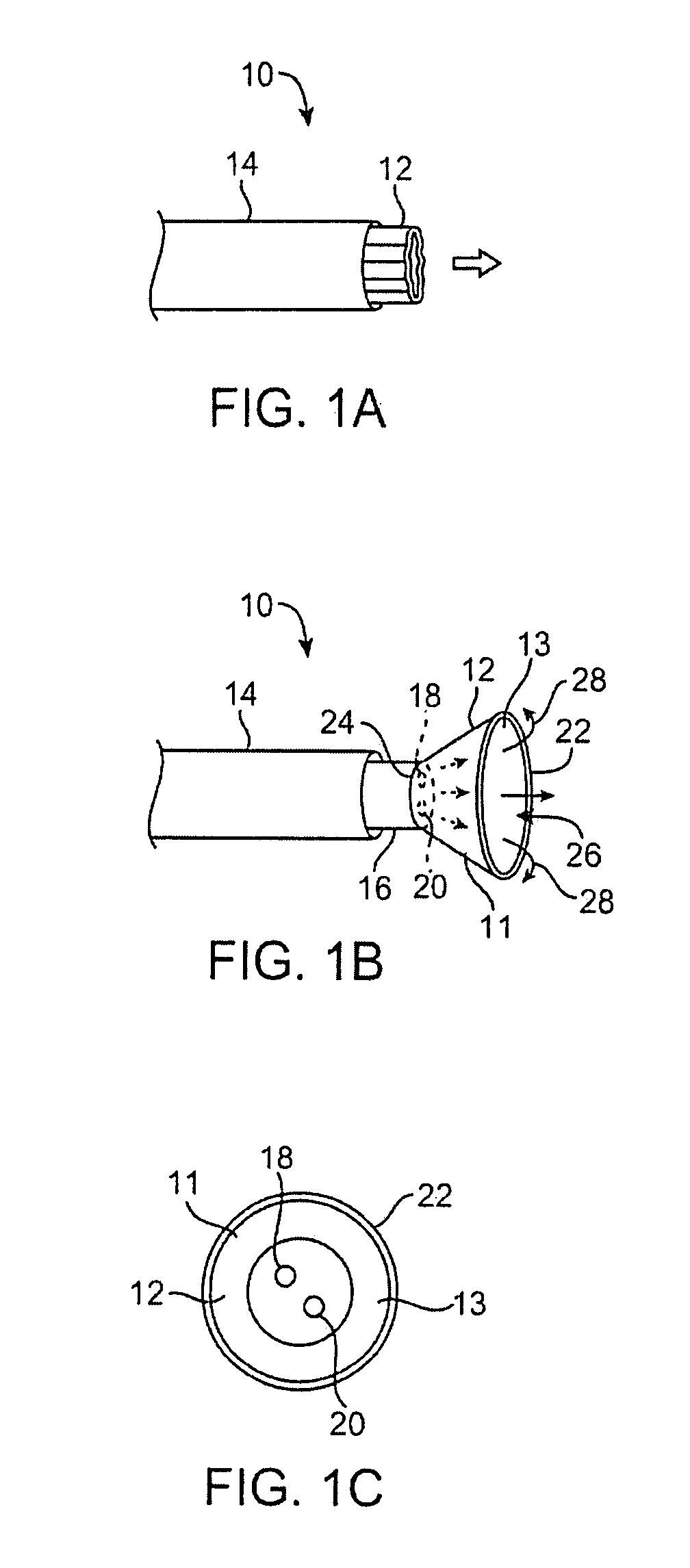
[0134] One variation of a tissue access and imaging apparatus is shown in the detail perspective views of FIGS. 1A to 1C. As shown in FIG. 1A, tissue imaging and manipulation assembly 10 may be delivered intravascularly through the patient's body in a low-profile configuration via a delivery catheter or sheath 14. In the case of treating tissue, such as t...


PUM


Abstract

Visualization apparatus and methods for transseptal access are described herein where intravascular access across a septal wall is facilitated via devices which provide for direct visual viewing of tissue area. Such a system may include a deployment catheter and an attached imaging hood deployable into an expanded configuration. In use, the imaging hood is placed against or adjacent to the tissue to be imaged in a body lumen that is normally filled with an opaque bodily fluid such as blood. A translucent or transparent fluid can be pumped into the imaging hood until the fluid displaces any blood leaving a clear region of tissue to be imaged via an imaging element in the deployment catheter. Any number of therapeutic tools or a guidewire can be passed through the catheter and into the imaging hood for crossing the septal wall and passing the guidewire or instruments therethrough.

Description

CROSS-REFERENCE TO RELATED APPLICATIONS [0001] This application claims the benefit of priority to the following U.S. Prov. Pat. App. Ser. Nos. 60 / 804,801 filed Jun. 14, 2006; 60 / 806,924 filed Jul. 10, 2006; 60 / 806,926 filed Jul. 10, 2006; 60 / 871,415 filed Dec. 21, 2006; 60 / 871,424 filed Dec. 21, 2006; 60 / 888,242 filed Feb. 5, 2007; this is also a continuation-in-part of U.S. patent application Ser. No. 11 / 259,498 filed Oct. 25, 2005, which claims priority to U.S. Prov. Pat. App. Ser. No. 60 / 649,246 filed Feb. 2, 2005; and this is also a continuation-in-part of U.S. patent application Ser. No. 11 / 560,742 filed Nov. 16, 2006, which claims priority to 60 / 737,521 filed Nov. 16, 2005 and is a continuation-in-part of U.S. patent application Ser. No. 11 / 259,498 filed Oct. 25, 2005. Each application is incorporated herein by reference in its entirety.FIELD OF THE INVENTION [0002] The present invention relates generally to medical devices used for visualizing and / or manipulating regions of t...

Claims


Application Information

Patent Timeline
IPC IPC(8): A61B1/12
CPCA61B1/0008A61B1/00082A61B1/00085A61B1/00089A61B1/005A61B8/4472A61B1/018A61B1/04A61B5/02007A61B8/12A61B1/015A61B1/3137
Inventor SAADAT, VAHIDABOLFATHI, AMIRROTHE, CHRIS A.VAN BLADEL, KEVIN H.
Owner INTUITIVE SURGICAL OPERATIONS INC
Learn More