Three-dimensional optical guidance for catheter placement

a three-dimensional optical guidance and catheter technology, applied in the field of optical guidance, can solve the problems of increasing hospital costs, slowing patient care, and insufficient placement of catheters, and achieve the effect of rapid confirmation of placemen

Inactive Publication Date: 2008-02-14
THE TRUSTEES OF THE UNIV OF PENNSYLVANIA
View PDF93 Cites 128 Cited by
  • Summary
  • Abstract
  • Description
  • Claims
  • Application Information

AI Technical Summary

Benefits of technology

[0030] It is also an object to provide a specialized method of this invention, wherein the optically-guided catheter is a central venous catheter, e.g., a Peripherally Inserted Central Catheter (PICC), inserted into a blood vessel leading to the heart of the patient, and wherein the emitted-light is emitted from the distal end of the PICC, said method further comprising moving the light-emitting point in proximity to the patient's heart and observing changes in pattern of emitted light as the light-emitting point approaches the patient's heart, wherein in proximity to the heart, the emitted light fluctuates in intensity synchronously with heart beats, thereby indicating the locat

Problems solved by technology

Post-placement images show that an unacceptably large portion of these catheters are not positioned appropriately using conventional blind placement techniques.
The number of incorrectly positioned catheters slows patient care, increases hospital costs and potentially increases patient risks.
Moreover, in current clinical practice, the final position and often the placement itself, requires the use of either fluoroscopy or x-rays, imaging modalities that result in undesirable exposure of the patient and health care provider to ionizing radiation.
Some medical devices are subject to movement after insertion due to changes in patient position, weakening of the device's securement to the body, rapid infusion of fluids, or removal of guidewires or introducers used during the device insertion process.
Often multiple x-rays are required to locate or confirm the position of an inserted device, subjecting the patient to undesirable levels of ionizing radiation.
This problem increases when handling or movement of the patient necessitates periodic rechecking of tube placement.
Additionally, x-ray equipment can be large and cumbersome to use, and often is not readily available at the patient bedside when a catheter must be inserted, or p

Method used


Image

  • Three-dimensional optical guidance for catheter placement
  • Three-dimensional optical guidance for catheter placement
  • Three-dimensional optical guidance for catheter placement

Examples


example 1

[0177] To demonstrate the effectiveness of the guidance method of the present invention in the alimentary track of a patient, a standard nasogastric feeding tube for an adult human was used. The feeding tube was inserted into the oropharynx of an anesthetized pig. The feeding tube included an optical fiber down the primary lumen of the tube. The tip of the fiber was within 0.5 cm of the tip of the feeding tube. Room lighting was minimized. Using night vision goggles and a camera / monitor system (Gen III intensified CCD camera ITT Industries Night Vision, San Diego, Calif.) insertion of the catheter could be followed very easily from the mouth to the stomach. The point of light emitted from the end of the optical fiber could easily be seen on the monitor as the feeding tube was advanced and placed.

[0178] The system was further tested on a human subject, a 210 lb man. An optical fiber (200 micron diameter core) was inserted into the nasogastric tube until the optical fiber was within ...

example 2

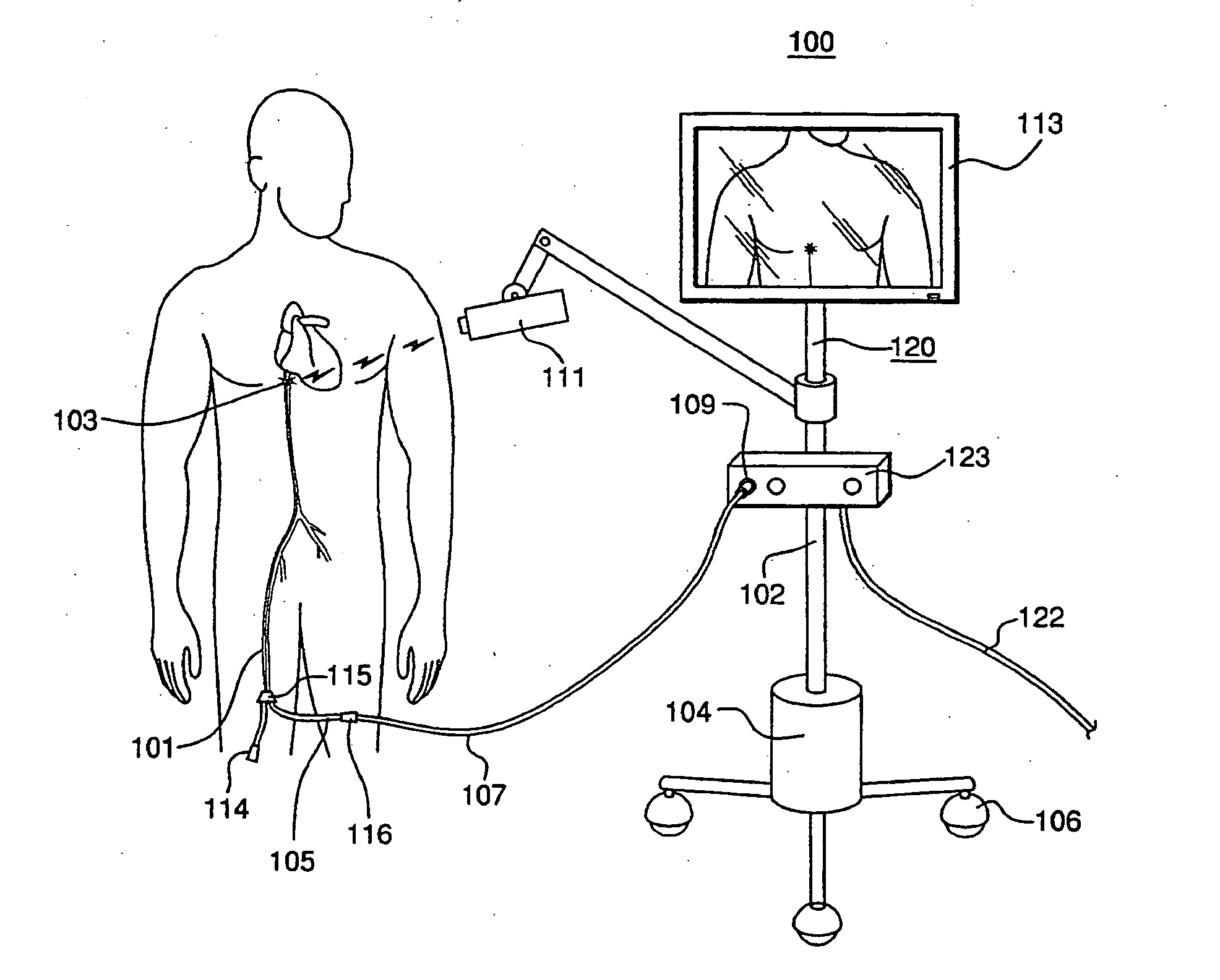
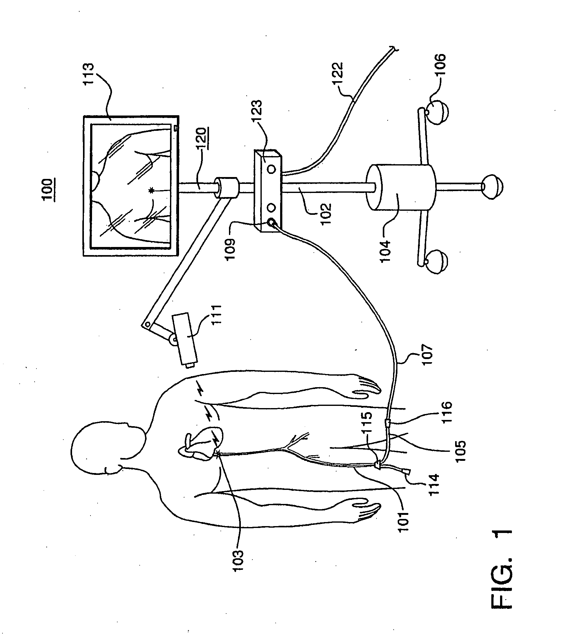
[0182] While demonstrating the effectiveness of the guidance method of the present invention for positioning intravascular catheters, an additional useful feature was noted. When an optical fiber and near infrared light LD system was added, as described above, to a peripherally inserted central venous catheter (PICC) line and placed in accordance with standard PICC practice in a vein leading to the heart, it was observed that as the lighted tip of the catheter neared the heart, the light became modulated by the movement of the beating heart. Moreover, as the lighted tip entered the heart, the light (signal) was greatly attenuated.

[0183] The heart consists of heavy, dense muscle, and the muscle tissue strongly attenuates the near infrared laser light, as compared to the surrounding environment. This is because the heart is suspended in what is mostly open space (lung, chest cavity), which easily transmits near-infrared light. Light emitted from the end of the catheter travels in all...

example 3

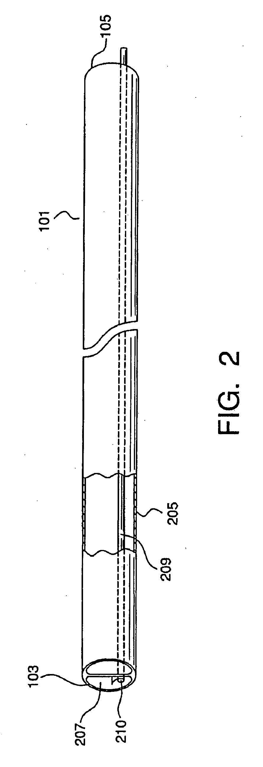
[0186] In another example of the guidance system, a light-guided epidural catheter was inserted into the lower lumbar region of a large pig. Pigs are representative of humans for this invention, as shown in Example 1. The epidural space was accessed in the standard manner by palpation of spinous processes, insertion of an 18 gauge Toughy needle to the depth of the epidural space using the air / fluid technique and a glass syringe. A standard epidural catheter was used, having an optical fiber within its lumen, threaded to the distal tip of the catheter and secured to the catheter (tape was used in this example, but any of the above disclosed methods for securing and / or sealing the optical fiber to the catheter would be effective).

[0187] In ambient light, the epidural catheter was advanced in the subject and the transdermally emitted point of light was captured and followed by the imaging system as it moved from the lower lumbar region to the thoracic region. Using a filtered camera / m...


PUM


Abstract

A system is provided comprising an optically-guided catheter having a proximal end, a distal end, and at least one lumen. A light-emitting means is coupled to the catheter, the catheter is inserted into place in the patient, and light is emitted as a point or points from a selected location, usually the distal tip, of the catheter to which it is coupled. The system further comprises an external detection device that detects the transdermally projected light, emitted by the light-emitting point from within the patient, thereby indicating precise placement of the catheter within the patient. A system and method for three-dimensional visualization using an internally positioned light emitter and an externally positioned detection array are also provided.

Description

CROSS-REFERENCE TO RELATED APPLICATIONS [0001] The present application is a continuation-in-part application claiming priority from (i) a co-pending non-provisional patent application entitled “Optically Guided System for Precise Placement of a Medical Catheter in a Patient,” which was filed on Oct. 4, 2005 and assigned Ser. No. 11 / 242,688, and which claimed priority to a provisional patent application which was filed on Nov. 4, 2004 and assigned Ser. No. 60 / 625,002, and (ii) a co-pending non-provisional patent application entitled “Optical Guidance System for Invasive Catheter Placement,” which was filed on Nov. 2, 2004 and assigned Ser. No. 10 / 482,190, such application having been filed as a national phase application based on PCT / US02 / 19314, filed Jun. 19, 2002, which in turn claimed priority to a provisional patent application which was filed on Jun. 19, 2001 and assigned Ser. No. 60 / 299,299. The present application claims the benefit of each of the aforementioned non-provisiona...

Claims


Application Information

Patent Timeline
IPC IPC(8): A61B5/05
CPCA61B5/06A61B5/061
Inventor WILSON, DAVID F.VINOGRADOV, SERGEI A.APRELEVA, SOFIA
Owner THE TRUSTEES OF THE UNIV OF PENNSYLVANIA
Why Eureka
Try Eureka