
Composite Scaffolds Seeded with Mammalian Cells

a technology of mammalian cells and composite tissue, which is applied in the field of composite tissue scaffolds seeded with mammalian cells, can solve the problems of limited durability, loss of functional tissue, and insufficient restoration of tissue function, and achieves the effect of increasing the production of the desired extracellular matrix and enhancing the retention of mammalian cells

Inactive Publication Date: 2008-04-10
REZANIA ALIREZA +1
View PDF0 Cites 22 Cited by
  • Summary
  • Abstract
  • Description
  • Claims
  • Application Information

AI Technical Summary

Benefits of technology

[0009]The present invention allows for enhanced retention of mammalian cells and increased production of the desired extracellular matrix (ECM) within the composite scaffold.
[0010]In addition, the cell-seeded composite scaffold can act as a vehicle to deliver cell-secreted biological factors. Such biological factors may direct up-regulation or down-regulation of other growth factors, proteins, cytokines or proliferation of other cell types. A number of cells may be seeded on such a composite scaffold before or after implantation into a defect site or site of diseased tissue.

Problems solved by technology

In general, the clinical approaches to repair damaged or diseased musculoskeletal tissue, such as bone, cartilage or muscle, do not substantially restore the original function of the tissue.
Prosthetic joints / devices often have been used to treat such defects with mixed outcomes attributed to loosening, limited durability and loss of functional tissue surrounding the defect.
The current treatments of DM remain inadequate in averting major health complications, such as blindness, kidney failure and ulcers.
Injury to spinal cord can lead to destruction of the white and gray matter in addition to blood vessels.
The CNS, unlike many other tissues, has a limited capacity for self-repair because mature neurons lack the ability to regenerate.
These approaches have resulted in inadequate repair of the CNS following trauma or disease.
So far, conventional materials used in tissue scaffolds, alone or in combination, have proven ineffective to retain seeded cells following implantation.

Method used


Image

  • Composite Scaffolds Seeded with Mammalian Cells
  • Composite Scaffolds Seeded with Mammalian Cells
  • Composite Scaffolds Seeded with Mammalian Cells

Examples


example 1

Forming a Composite Scaffold

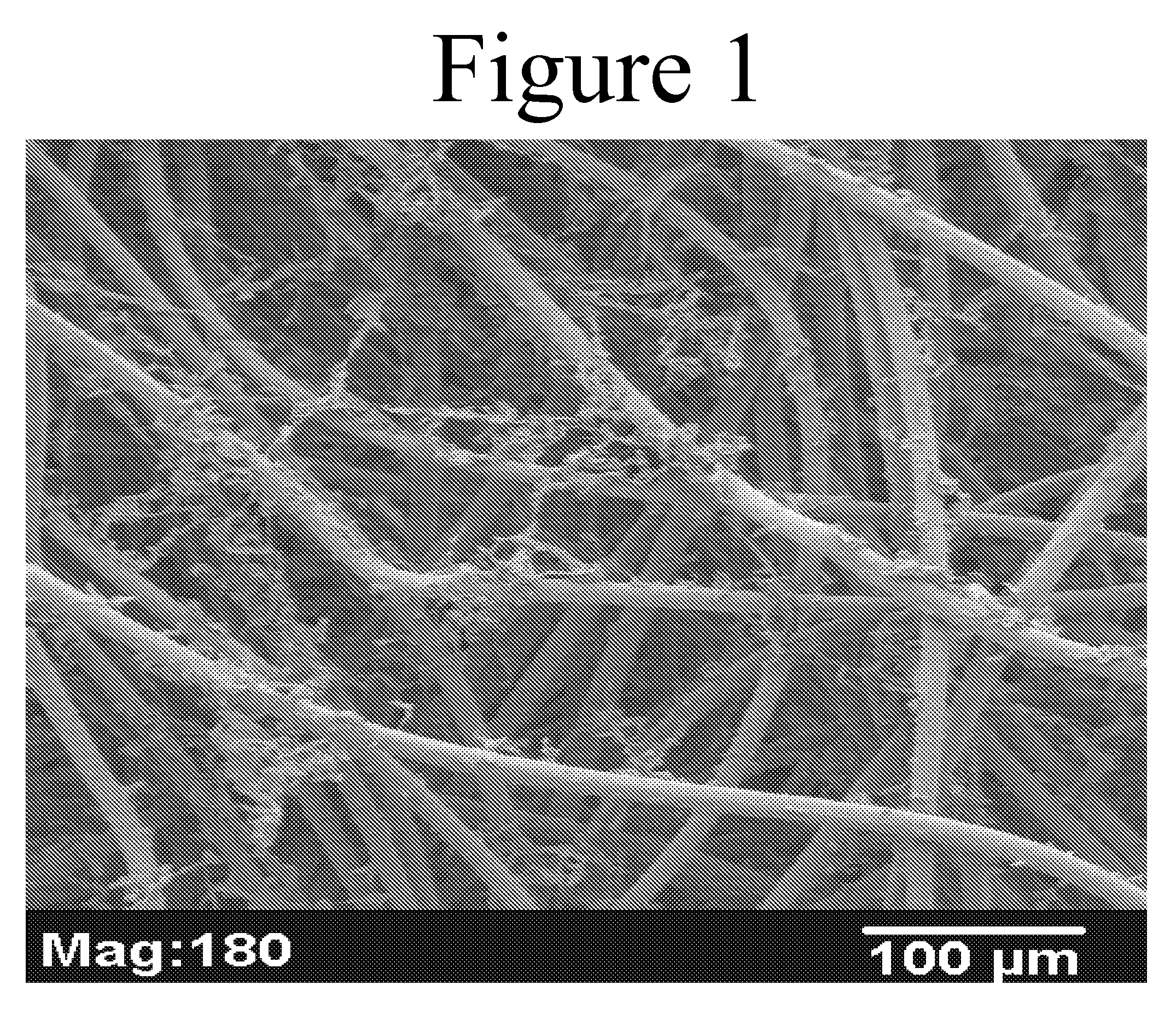
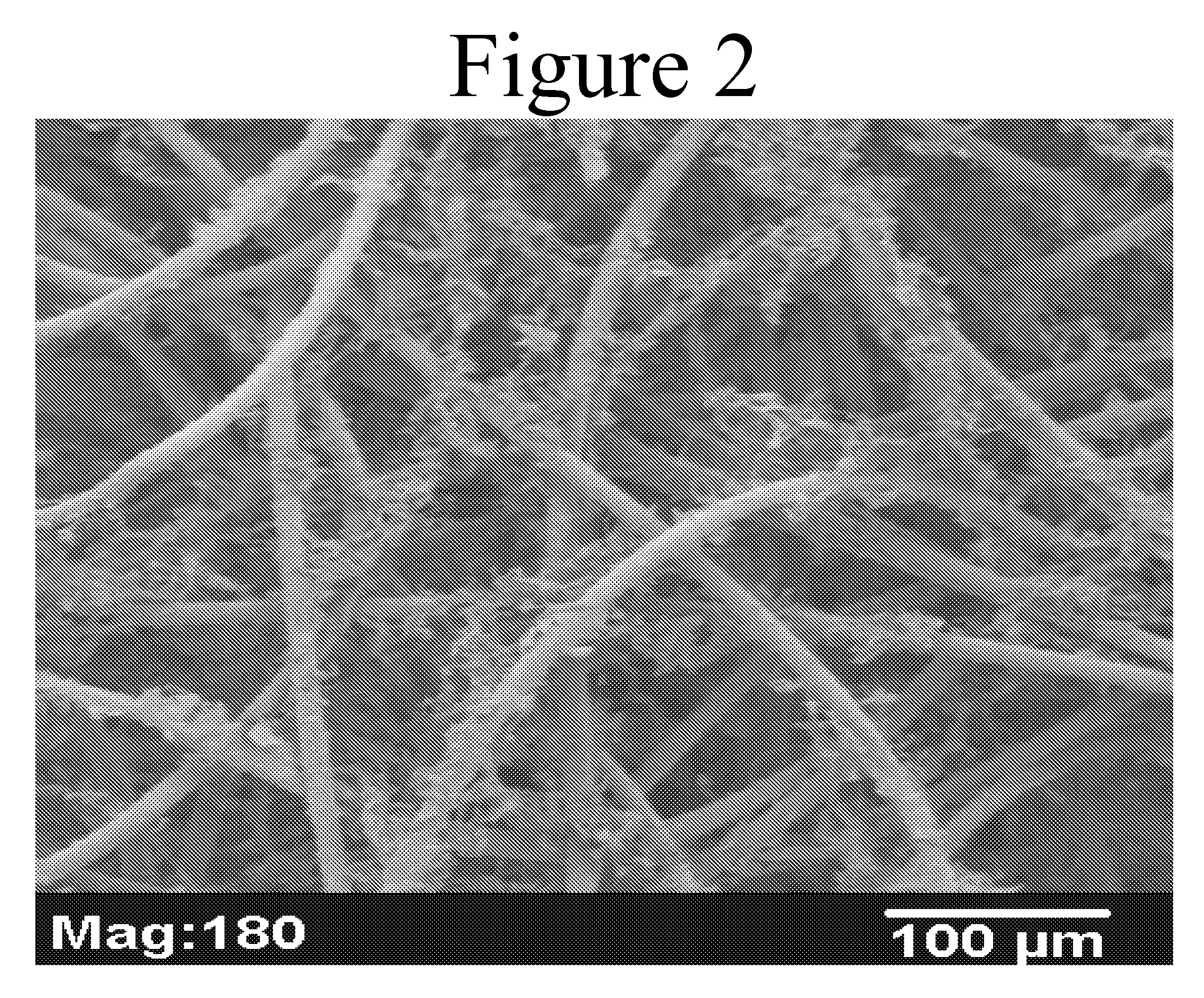
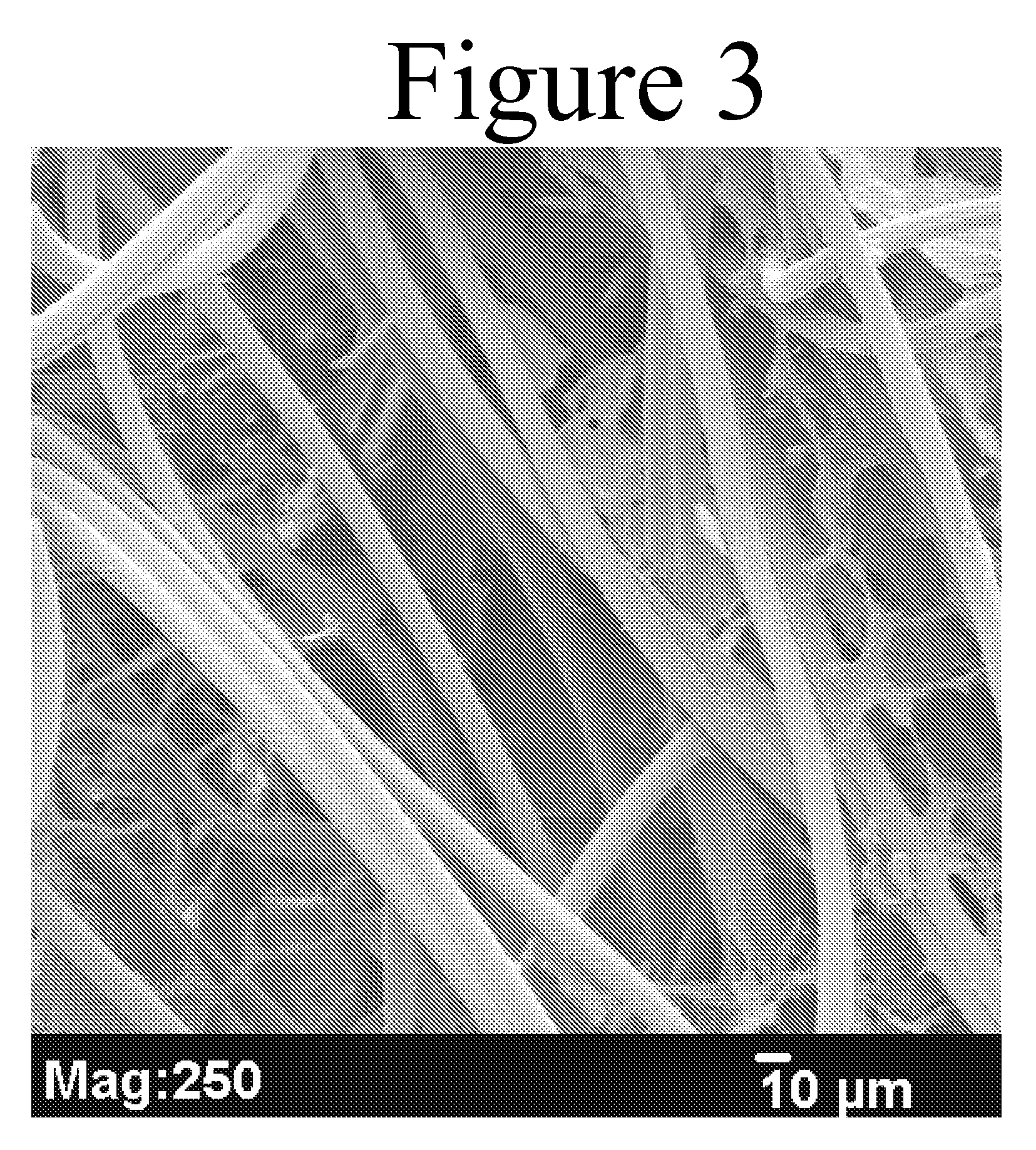
[0070]A needle-punched nonwoven mat (2 mm in thickness) composed of 90 / 10 PGA / PLA fibers was made as described below. A copolymer of PGA / PLA (90 / 10) was melt-extruded into continuous multifilament yarn by conventional methods of making yarn and subsequently oriented in order to increase strength, elongation and energy required to rupture. The yarns comprised filaments of approximately 20 microns in diameter. These yarns were then cut and crimped into uniform 2-inch lengths to form 2-inch staple fiber.

[0071]A dry lay needle-punched nonwoven mat was then prepared utilizing the 90 / 10 PGA / PLA copolymer staple fibers. The staple fibers were opened and carded on standard nonwoven machinery. The resulting mat was in the form of webbed staple fibers. The webbed staple fibers were needle punched to form the dry lay needle-punched, fibrous nonwoven mat.

[0072]The mat was scoured with ethyl acetate for 60 minutes, followed by drying under vacuum.

[0073]A solution of t...

example 2

Forming a Composite Scaffold

[0078]A biodegradable composite scaffold was fabricated following the process of Example 1, except the polymer lyophilized into a foam was a 60 / 40 PLA / PCL copolymer from Birmingham Polymers Inc., Birmingham, Ala., with an I.V. of 1.45 dL / g. The pore size of this composite scaffold was determined using Mercury Porosimetry analysis. The range of pore size was 1-300 μm with a median pore size of 45 μm.

example 3

Forming a Composite Scaffold

[0079]A biodegradable composite scaffold was fabricated following the process of Example 1, except the polymer lyophilized into a foam was a 50:50 blend of 60 / 40 PLA / PCL and 35 / 65 PCL / PGA copolymers from Birmingham Polymers Inc., Birmingham, Ala., with I.V.s of 1.50 dL / g and 1.45 dL / g, respectively.


PUM

PropertyMeasurementUnit
Biological propertiesaaaaaaaaaa
Molar ratioaaaaaaaaaa
Biocompatibilityaaaaaaaaaa

Abstract

Implantable, biocompatible scaffolds containing a biocompatible, porous, polymeric matrix, a biocompatible, porous, fibrous mat encapsulated by and disposed within said polymeric matrix, and a plurality of mammalian cells seeded within said tissue scaffold. The invention also is directed to methods of treating disease or structural defects in a mammal utilizing the scaffolds of the invention.

Description

FIELD OF THE INVENTION[0001]The present invention relates to composite tissue scaffolds seeded with mammalian cells for treating a disease or structural defects in soft or hard tissues.BACKGROUND OF THE INVENTION[0002]There is a clinical need to treat three classes of diseases that afflict many individuals. The first class of disease relates to diseases / damaged musculoskeletal tissues, such as cartilage, bone, meniscus or muscle. In general, the clinical approaches to repair damaged or diseased musculoskeletal tissue, such as bone, cartilage or muscle, do not substantially restore the original function of the tissue. Prosthetic joints / devices often have been used to treat such defects with mixed outcomes attributed to loosening, limited durability and loss of functional tissue surrounding the defect.[0003]The second class of diseases relates to the loss of organ function, such as diabetes mellitus (DM). DM results from destruction of beta cells in the pancreas or from insensitivity ...

Claims


Application Information

Patent Timeline
IPC IPC(8): A61F2/00A61K45/00A61P3/10A61P43/00A61L27/00A61L27/38A61L27/48A61L27/56C12N5/08
CPCA61L27/3847A61L27/3852A61L27/48A61L27/56C08L67/04A61P3/10A61P43/00A61L27/3804
Inventor REZANIA, ALIREZAZIMMERMAN, MARK
Owner REZANIA ALIREZA
Learn More