Neuronal Cell Propagation Using Rotating Wall Vessel

a technology of neuronal cell and rotating wall, which is applied in the field of culturing neurons, can solve the problems of neuronal cell death in select brain areas, loss of intellectual faculties, emotional disturbance, etc., and achieves the effects of reducing doubling rate, increasing susceptibility to apoptosis, and increasing neurite formation

Inactive Publication Date: 2008-11-06
THE ADMINISTRATORS OF THE TULANE EDUCATIONAL FUND
View PDF15 Cites 4 Cited by
  • Summary
  • Abstract
  • Description
  • Claims
  • Application Information

AI Technical Summary

Benefits of technology

[0029]In another embodiment of the present invention is provided a transformed neuronal cell with 3-D phenotype, wherein the 3-D phenotype comprises: reduced doubling rate; increased susceptibility to apoptosis; and increased neurite formation. In a preferred aspect of this embodiment, the 3-D phenotype persists for up to 5 days after said cell is transferred to 2-D culture. In another preferred aspect of this embodiment, the 3-D phenotype further comprises: reduced N-myc expression; reduced HuD expression; reduced Bcl-2 expression; increased Bax expression; and increased Bak expression. In another preferred aspect of this embodiment, the 3-D phenotype further comprising reduced N-myc expression and reduced Bcl-2 expression persists for up to 5 days after said cell is transferred to 2-D culture. In another preferred aspect of this embodiment, the 3-D phenotype further comprising reduced N-myc expression, reduced HuD expression, reduced Bcl-2 expression, increased Bax expression, and increased Bak expression persists for up to 5 days after said cell is transferred to 2-D culture. In a most preferred aspect of this embodiment, the transformed neuronal cell is an SH-SY5Y cell or a PC12 cell.

Problems solved by technology

The neurodegeneration associated with HD causes uncontrolled movements, loss of intellectual faculties, and emotional disturbance.
This expansion produces a mutant form of the Htt protein (mHtt), which results in neuronal cell death in select areas of the brain, and is a terminal illness.
Nevertheless, the benefits of drug therapy often wane after as little as 5 years of treatment, and the drugs themselves produce undesirable side-effects.
In vitro studies of disease pathogenesis in the CNS are often conducted with cultures of primary cells, but when the cells in question are neurons—human neurons, in particular—this becomes problematic because most post-embryonic neurons do not divide.
Thus, the usefulness of neurons in primary culture is severely limited and researchers must employ transformed neuronal cell lines instead (Encinas M, Iglesias M, Liu Y, Wang H, Muhaisen A, Cena V, Gallego C, Comella J X. Sequential treatment of SH-SY5Y cells with retinoic acid and brain-derived neurotrophic factor gives rise to fully differentiated, neurotrophic factor-dependent, human neuron-like cells.
While immortalized cell lines are available for most types of non-neuronal mammalian cells, as well as for many specific disease states, there are very few useful neuronal cell lines available for the study of neurological diseases.
The reason behind the limited availability of neuronal cells is that neuronal cells are particularly difficult to culture.
However, these cell lines are also inherently abnormal since, among other characteristics, they exhibit unregulated cellular division, are known to exhibit an arrested state of cellular differentiation (Abbott A. Cell culture: biology's new dimension.
The inherently abnormal phenotypes of neuronal cell lines complicates the interpretation of experimental results derived from these cells when comparing them to non-transformed cells (i.e., neurons from primary cultures) (Fan L, Iyer J, Zhu S, Frick K K, Wada R K, Eskenazi A E, Berg P E, Ikegaki N, Kennett R H, Frantz C N. Inhibition of N-myc expression and induction of apoptosis by iron chelation in human neuroblastoma cells.
In addition to the limitations introduced by transformed cell lines, traditional monolayer or “2-D” culture systems in Petri dishes are often themselves inadequate to realistically model in vivo conditions (Lelkes P I, Galvan D L, Hayman G T, Goodwin T J, Chatman D Y, Cherian S, Garcia R M, Unsworth B R. Simulated microgravity conditions enhance differentiation of cultured PC12 cells towards the neuroendocrine phenotype.

Method used


Image

  • Neuronal Cell Propagation Using Rotating Wall Vessel
  • Neuronal Cell Propagation Using Rotating Wall Vessel
  • Neuronal Cell Propagation Using Rotating Wall Vessel

Examples


example 1

3-D Culture Changes the Morphology and Proliferation Rate in SY Neuronal Cells

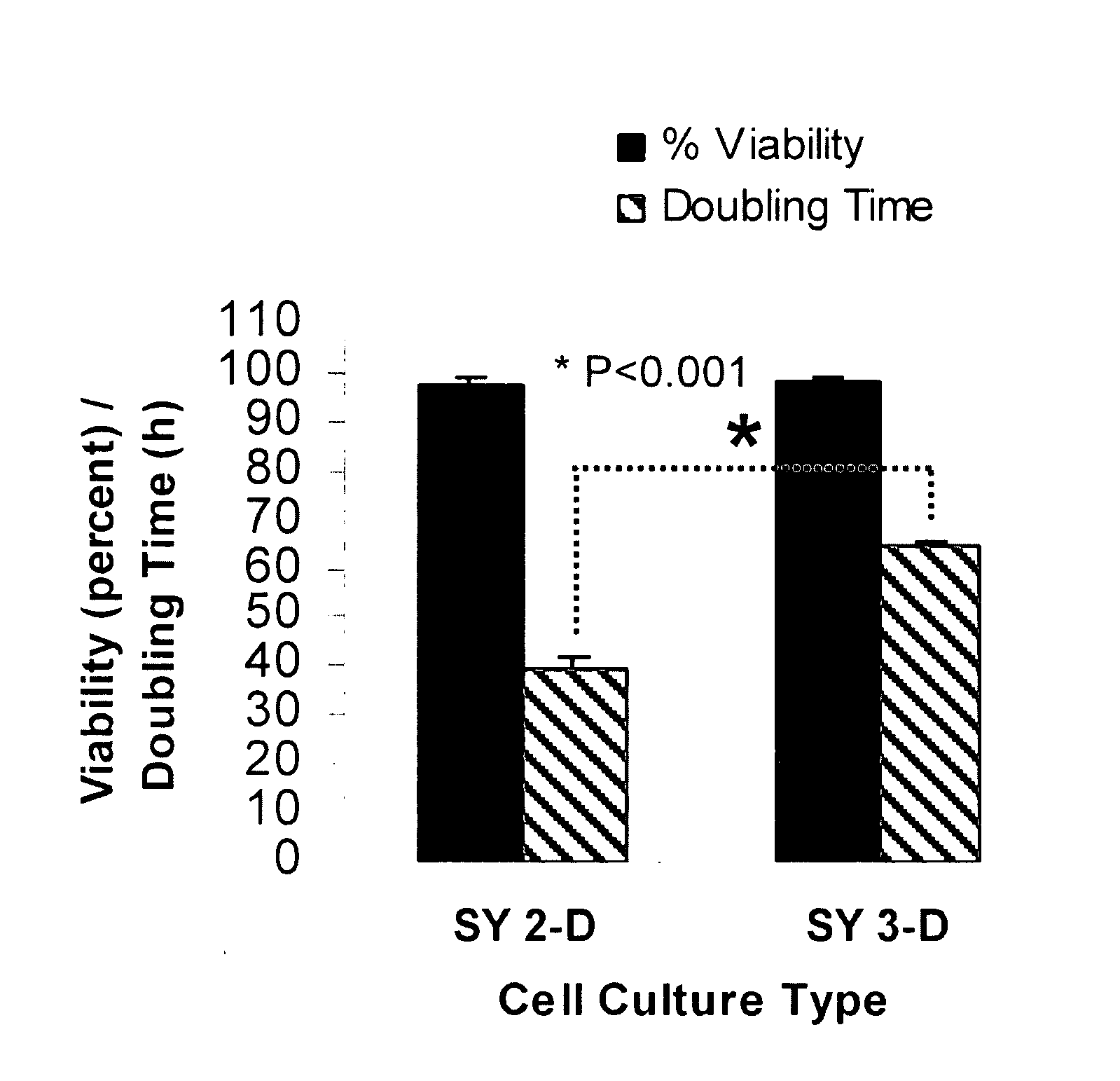
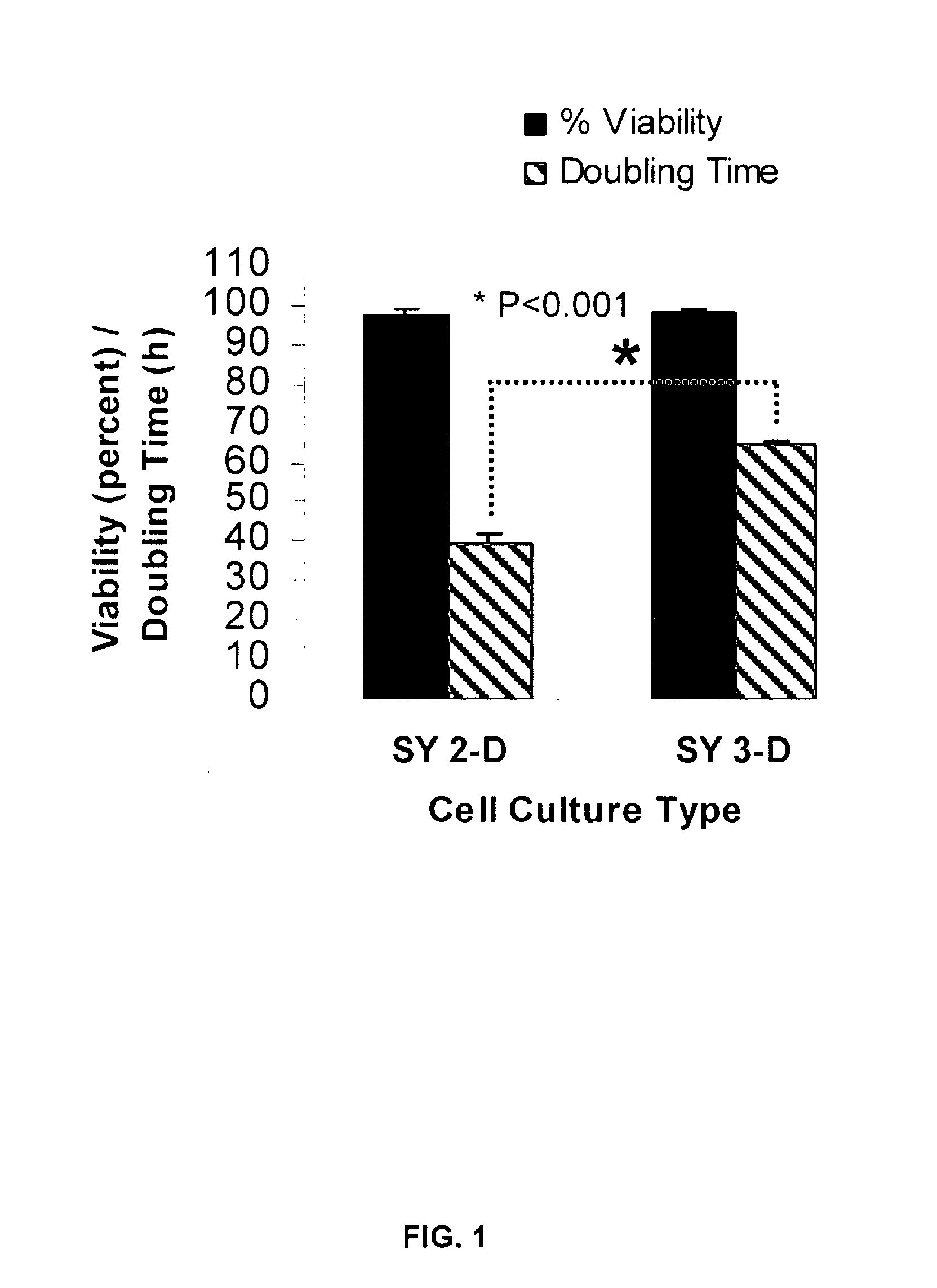
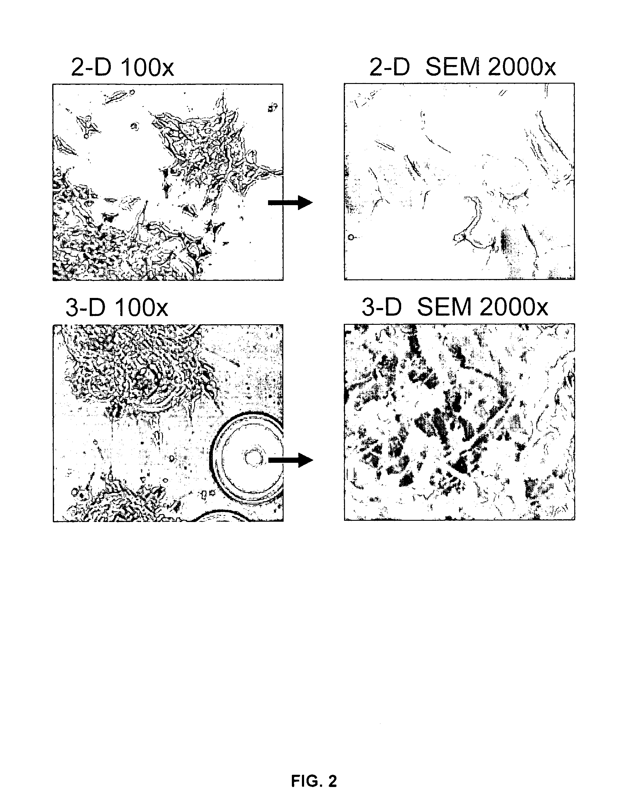
[0078]SY cells cultured for 21 days in the RWV, and then for counting purposes transferred back to 2-D culture flasks for 5 days, revealed a decrease in the cell doubling rate from 40 h to approximately 65 h, with no change in cell viability (FIG. 1). Thus, the 3-D phenotype of SY cells comprises a decrease in the cell doubling rate. Because the carrier beads used in the 3-D culture were coated in collagen, additional SY cells were cultured for 3 weeks and for 4 weeks in 2-D flasks coated with collagen. No detectable difference was observed in the morphology, cell viability or doubling rate of 2-D cells cultured on plastic as compared to collagen. Scanning electron microscopy (SEM) revealed important differences in the morphology of SY cells cultured in 2-D or in 3-D. Specifically, only the 3-D-cultured SY cells acquired a parental, tissue-like conformation with dramatic increases in neurite extension, dir...

example 2

Decreased Expression of N-myc and HuD

[0079]Human neuroblastoma cells are typically characterized by de-differentiation. They have re-entered S-phase of the cell cycle, and are highly resistant to apoptosis (Kang et al., 2006; van Noesel et al., 2003). Amplified expression of the proto-oncogene N-myc has been correlated with cellular de-differentiation and increased resistance to apoptosis, and is believed to have a crucial role in maintenance of the cells' malignant phenotype (Chagnovich and Cohn, 1996; Grandinetti et al., 2006; Smith et al., 2004; van Golen et al., 2003). The RNA binding protein HuD functions in stabilizing N-myc mRNA and may consequently enhance steady-state expression levels of this oncogene (Chagnovich and Cohn, 1996; Grandinetti et al., 2006; Lazarova et al., 1999). Reduced expression of the HuD protein could therefore contribute, through destabilization of N-myc, to an increase in cellular differentiation.

[0080]Western analysis confirmed a culture-dependent sh...

example 3

Apoptosis Resistance is Diminished in 3-D Cultured SY and PC12 Cells

[0081]Cells over-expressing the anti-apoptotic protein Bcl-2 or cells with depleted pro-apoptotic Bax and Bak exhibit resistance to cell death as induced by mitochondrial dysfunction and ER stress (Elyaman W, Terro F, Suen K C, Yardin C, Chang R C, Hugon J. BAD and Bcl-2 regulation are early events linking neuronal endoplasmic reticulum stress to mitochondria-mediated apoptosis. Brain research, 2002; 109: 233-8; Henshall D C, Araki T, Schindler C K, Lan J Q, Tiekoter K I, Taki W, Simon R P. Activation of Bcl-2-associated death protein and counter-response of Akt within cell populations during seizure-induced neuronal death. J Neurosci, 2002; 22: 8458-65; Murakami Y, Aizu-Yokota E, Sonoda Y, Ohta S, Kasahara T. Suppression of endoplasmic reticulum stress-induced caspase activation and cell death by the overexpression of Bcl-xL or Bcl-2. Journal of biochemistry, 2007; 141: 401-10; Scorrano L, Oakes S A, Opferman J T, ...


PUM

PropertyMeasurementUnit
pHaaaaaaaaaa
pressureaaaaaaaaaa
pressureaaaaaaaaaa

Abstract

The present invention provides methods of propagating transformed neurons in a simulated microgravity environment generated by a rotating wall vessel (“3-D culture”) so that the phenotype of the transformed neurons so cultured becomes closer to that of non-transformed neurons (primary neurons) and less like the phenotype of transformed neurons cultured via standard cell culture techniques (“2-D culture”).

Description

CROSS-REFERENCE TO RELATED APPLICATIONS[0001]This Non-Provisional Patent Application, filed under 35 U.S.C. § 111 (a), claims the benefit under 35 U.S.C. § 119(e)(1) of U.S. Provisional Patent Application No. 60 / 915,407, filed under 35 U.S.C. § 111 (b) on 1 May 2007, and which is hereby incorporated by reference in its entirety.STATEMENT REGARDING FEDERALLY SPONSORED RESEARCH[0002]The invention was made with U.S. Government support under grant numbers NS048952 and RR00164 (MTP) awarded by the National Institutes of Health. The United States Government has certain rights in the invention.THE NAMES OF THE PARTIES TO A JOINT RESEARCH AGREEMENT[0003]Not applicable.INCORPORATION-BY-REFERENCE OF MATERIAL SUBMITTED ON COMPACT DISC[0004]Not applicable.BACKGROUND OF THE INVENTION[0005]1. Field of the Invention[0006]The present invention relates to methods of culturing neurons for in vitro laboratory investigations. More particularly, the present invention relates to methods of culturing tran...

Claims


Application Information

Patent Timeline
Patent Type & Authority Applications(United States)
IPC IPC(8): C12N5/06C12N5/08C12N5/0793
CPCC12N5/00C12N5/0619C12N2501/13C12N2525/00
Inventor PHILIPP, MARIO T.NICKERSON, CHERYL A.MYERS, TEREANCE A.
Owner THE ADMINISTRATORS OF THE TULANE EDUCATIONAL FUND
Why Eureka
Try Eureka