
Vascular Tumor Markers

a tumor marker and vascular technology, applied in the field of oncology, can solve the problems of not being able to identify differentially expressed genes, unable to identify differentially expressed proteins on the molecular level, and database entries sometimes contain minor sequencing errors, so as to achieve the effect of treatment, and improving the accuracy of diagnosis

Inactive Publication Date: 2009-01-29
PHILOGEN SRL LLC
View PDF3 Cites 12 Cited by
  • Summary
  • Abstract
  • Description
  • Claims
  • Application Information

AI Technical Summary

Benefits of technology

[0073]It is expected that ligands, preferably antibodies directed/raised against “junction peptides” existing only in the novel periostin isoforms specif

Problems solved by technology

Although differences in gene expression were apparent, it proved difficult to identify the differentially expressed proteins on the molecular level.
Another popular approach has been to raise antibodies to different endothelial structures leading to the identification of new endothelial markers but failed to identify differentially expressed genes, possibly because such proteins are a minor component of the abundant components on the cell surface.
Furthermore, it is common knowledge in the art that database entries sometimes contain minor sequencing errors and may be subject to revisions and changes.
However, Periostin has not been associated with kidney tumors in the prior art.
However, the distribution of isoforms of periostin in brain and liver cDNA libraries showed differences between tumor, fetal and normal adult specimens.
So far, there are no antibodies available which specifically recognize this protein.
Such hypermethylation can result in gene silencing that is clonally propagated through mitosis by the action of DNA-methyltransferase enzymes.

Method used


Image

  • Vascular Tumor Markers
  • Vascular Tumor Markers
  • Vascular Tumor Markers

Examples


example 1

Introduction

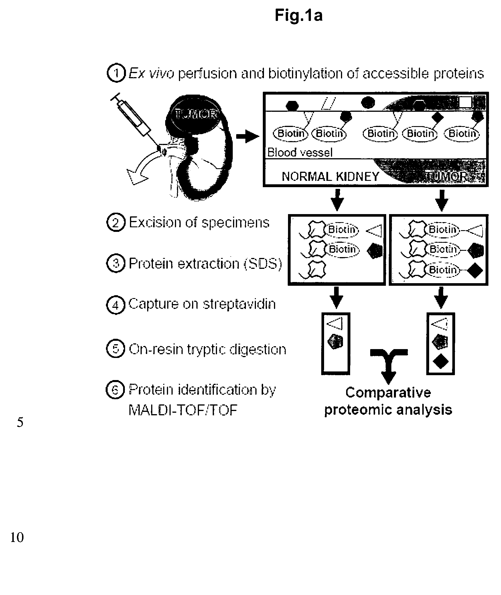
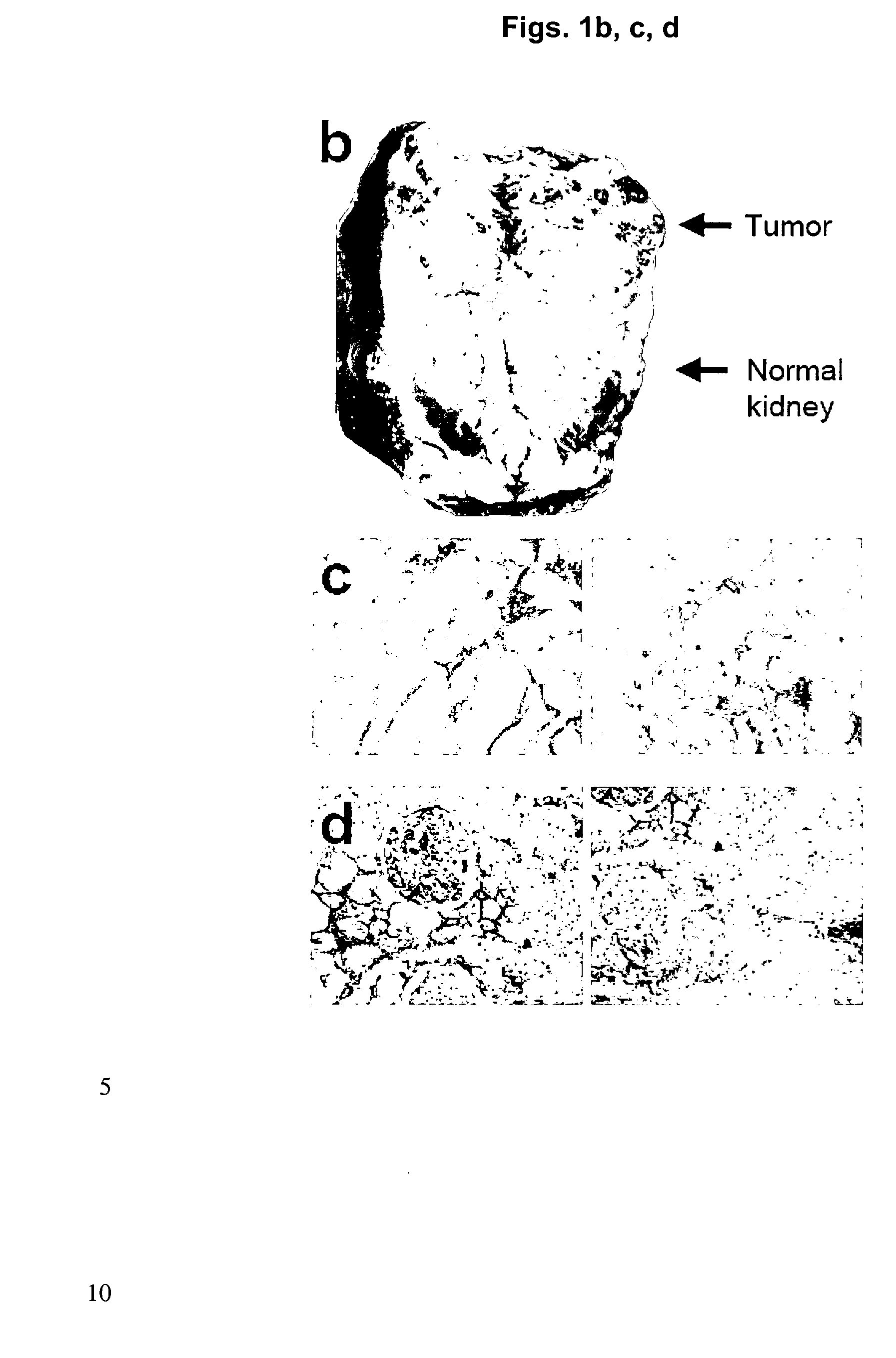
[0184]A chemical proteomic approach based on the ex vivo perfusion and biotinylation of accessible structures within surgically-resected human kidneys with tumor was used to gain information about accessible and abundant antigens which are over-expressed in human cancer. Biotinylated proteins were purified on streptavidin resin and identified using mass spectrometric methodologies, revealing 637 proteins, of which 184 were found in tumor specimens only and 223 in portions of normal kidneys only. Thirty of the accessible tumor-associated antigens identified with this methodology are suitable targets for antibody-based anti-cancer therapies.

[0185]The specific method used for the identification of accessible antigens in normal organs and in tumors is based on the terminal perfusion of tumor-bearing mice with reactive ester derivatives of biotin that was recently published by the present inventors (Rybak et al. 2005 supra). This methodology allows the efficient biotinylation...

example 2

Introduction

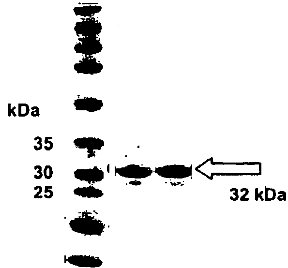
[0204]For assessing periostin's utility as tumor marker recombinant fragments of this protein were cloned and expressed. Human monoclonal antibodies against the recombinant fragments were produced and tested in ELISA and immuno-histochemistry experiments.

Materials and Methods

[0205]Recombinant protein fragments corresponding to the amino acid sequence positions 232-632 (FAS2-FAS4) and 496-632 (FAS4) of periostin (SEQ ID NO: 1) were cloned for use as antigen for biopanning experiments. The fragments were expressed in E. coli strain TG1 using pQE12 vector (Qiagen, Hilden, Germany). Proteins were purified from E. coli lysates using Ni-NTA columns (Qiagen). Antibodies in single chain Fv format (scFv) against the periostin fragments were selected from the ETH-2-Gold phage display library according to the procedure reported in Silacci et al., Proteomics. 2005 June; 5(9):2340-50. ELISA screening for clones expressing scFv antibody binding the antigen and immunohistochemical stai...

example 3

Introduction

[0207]To further study the versican antigen, we have performed an immunohistochemical analysis on kidney tumor sections of different patients.

Materials and Methods

[0208]Immunohistochemical stainings with the monoclonal anti-versican antibody (clone 12C5) on paraformaldehyde-fixed, paraffin-embedded sections of human kidney tumors were performed as described in Example 1.

Results

[0209]Furthermore, a commercial antibody against versican (see also Example 1) was used to evaluate the periostin expression in kidney tumors of different patients (see FIG. 9). Six out of eight patients tested showed positive anti-versican staining in the kidney tumor, either more diffuse in the extracellular matrix or more restricted to areas around tumor blood vessels. These results indicate that versican is expressed at high levels in human kidney tumors of most patients and is likely to be accessible from the vasculature.


PUM

PropertyMeasurementUnit
Fractionaaaaaaaaaa
Sizeaaaaaaaaaa
Sizeaaaaaaaaaa

Abstract

The present invention relates to a method for identifying neovascular structures in mammalian tissue, wherein said neovascular structures are identified by the detection of at least one specific protein in said tissue. It also relates to a method for identifying diseases or conditions associated with neovascularization, methods for targeting and / or imaging neovascular structures and methods for targeting diseases or conditions associated with neovascularization. Furthermore, the present invention is directed to the use of novel and / or known ligands, preferably antibodies, directed against novel and / or known target proteins for identifying tumor cells in mammalian tissue, preferably mammalian kidney tissue, more preferably mammalian vascular kidney tissue. The present invention also relates to novel ligands, preferably antibodies, fusion proteins comprising said ligands or antibodies, pharmaceutical and diagnostic compositions comprising said ligands, antibodies or fusion proteins, diagnostic and therapeutic methods as well as novel proteins and corresponding polynucleotides, vectors and host cells.

Description

[0001]The present invention relates to a method for identifying neovascular structures in mammalian tissue, wherein said neovascular structures are identified by the detection of at least one specific protein in said tissue. It also relates to a method for identifying diseases or conditions associated with neovascularization, methods for targeting and / or imaging neovascular structures and methods for targeting diseases or conditions associated with neovascularization. Furthermore, the present invention is directed to the use of novel and / or known ligands, preferably antibodies, directed against novel and / or known target proteins for identifying tumor cells in mammalian tissue, preferably mammalian kidney tissue, more preferably mammalian vascular kidney tissue. The present invention also relates to novel ligands, preferably antibodies, fusion proteins comprising said ligands or antibodies, pharmaceutical and diagnostic compositions comprising said ligands, antibodies or fusion prote...

Claims


Application Information

Patent Timeline
IPC IPC(8): A61K49/00C07K14/00C12N15/11C12N15/00C12N5/06A61P31/00C07K16/18A61K39/395G01N33/566G01N33/574
CPCG01N33/57438G01N2333/515G01N2500/00G01N2800/7028G01N2800/10G01N2800/323G01N33/566A61P31/00A61P35/00C07K16/00G01N33/53G01N33/574G01N33/68
Inventor NERI, DARIOROSLI, CHRISTOPHRYBAK, JASCHA
Owner PHILOGEN SRL LLC
Learn More