Magneto-motive ultrasound detection of magnetic nanoparticles

a magnetic nanoparticle and magnetomotive technology, applied in the field of magnetomotive ultrasound detection of magnetic nanoparticles, can solve the problems of microbubbles that are unstable, microbubbles have a short blood half-life, and are easy to fracture and collaps

Inactive Publication Date: 2009-02-12
BOARD OF RGT THE UNIV OF TEXAS SYST
View PDF19 Cites 113 Cited by
  • Summary
  • Abstract
  • Description
  • Claims
  • Application Information

AI Technical Summary

Problems solved by technology

Compared to the surrounding tissue, microbubbles present a large acoustic impedance mismatch in tissues and thus produce a strong backscattered sound signal.
Although microbubbles play an important role in increasing the enhancement of the diagnostic potential of ultrasound imaging, the micron-sized bubbles have limited use in molecular imaging because these agents are too large to pass through the pulmonary and systemic capillary bed.
Moreover, microbubbles are unstable, have a short blood half-life and a propensity to fracture and collapse when exposed to ultrasound waves.
These features have limited the use of microbubbles in ultrasound molecular imaging.
Nanometer-sized ultrasound contrast agents may penetrate the large capillary bed, but penetration to vascular targets and extravasations through tight capillary pores could be inhibited due to their relatively large size.
Unfortunately, PFC ultrasound contrast agents produced a smaller acoustic impedance mismatch, a weaker ultrasound reflected signal, and create less contrast enhancement of echogenic images than gaseous microbubbles.

Method used


Image

  • Magneto-motive ultrasound detection of magnetic nanoparticles
  • Magneto-motive ultrasound detection of magnetic nanoparticles
  • Magneto-motive ultrasound detection of magnetic nanoparticles

Examples


example 1

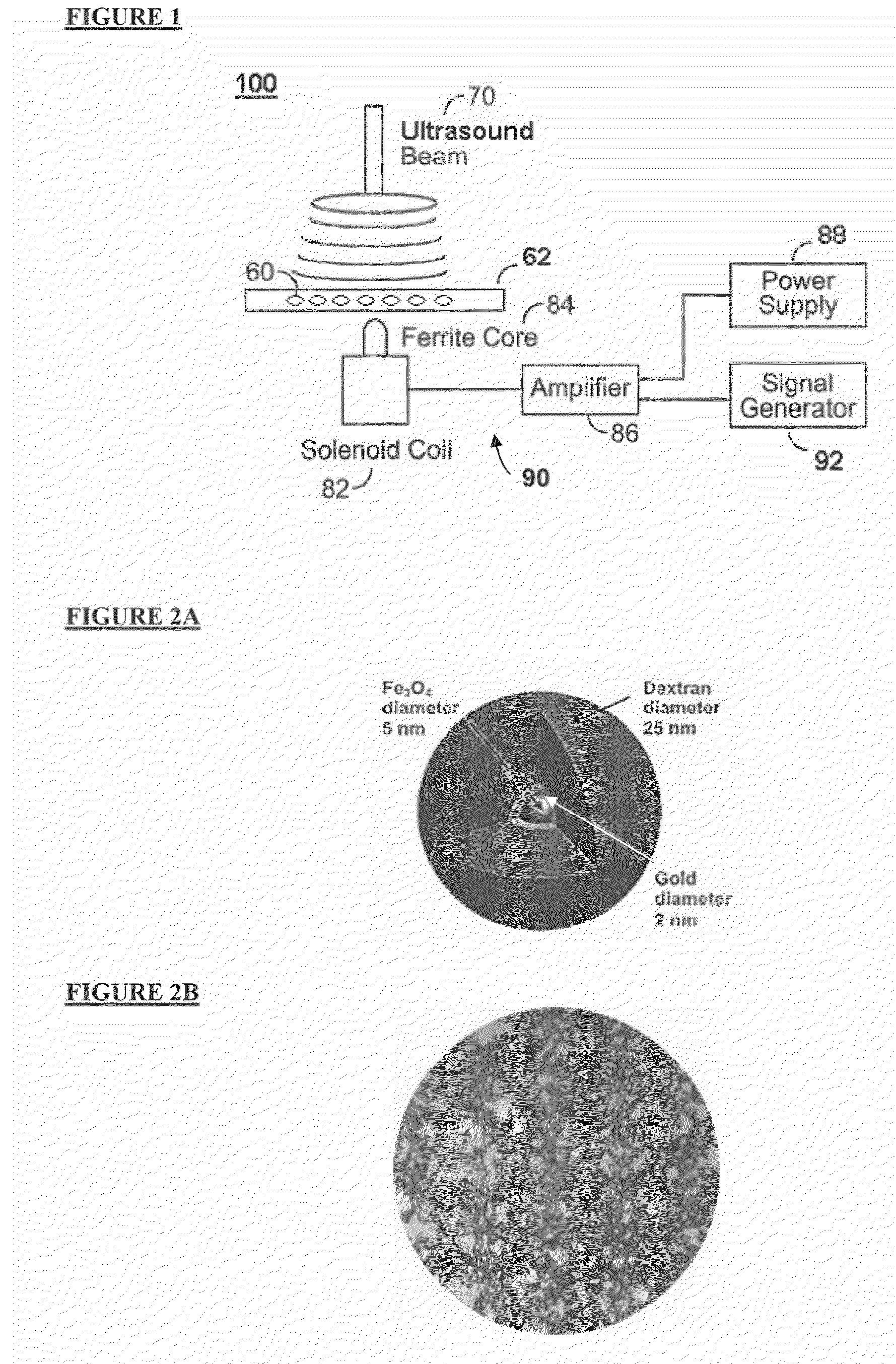
Superparamagnetic Iron Oxide (SPIO) Nanoparticles

[0095]Colloidal suspensions of SPIO nanoparticles, Ferumoxides or AMI-25 with the trade name Feridex® I.V. (Advanced Magnetics, Cambridge, Mass., USA) are approved by the United States Food and Drug Administration (FDA) for human use in 1997. The SPIO nanoparticles consisted of nonstoichiometric magnetite cores, iron, and a dextran T-10 coating added to prevent aggregation and facilitate stabilization in the liver. The mean core diameter and volume mean diameter measured by laser light scattering of these nanoparticles were 20 and 80 nm, respectively. Peak concentrations of SPIO nanoparticles in the liver were observed 1 h after an intravenous injection (18 μmol Fe / kg body weight). The uptake of SPIO nanoparticles by macrophage cells is directly proportional to the IV concentration, blood half-life, and their core size.

experimental preparation

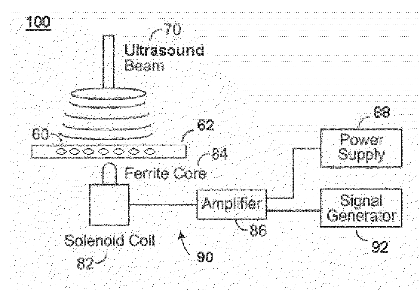
[0096]As shown in FIG. 4, a schematic diagram of the apparatus is shown. A liver sample was placed into a small rectangular plastic container (10 cm 10 cm) filled with water to provide acoustic coupling between the ultrasonic transducer and the specimen. The sample was imaged from the top using a linear array transducer (128 Channel). Magnetic excitation of the sample was provided by the solenoid positioned below at the bottom surface of the water tank. The distance between the liver specimens and iron core tip was about 1.5 mm, and the magnetic field strength at this distance was measured using a tesla-meter to observe the correlation between the magnetic field strength and the ultrasound measurement.

[0097]The magnetic generator comprises a solenoid (Ledex 6EC, Saia-Burgess Inc., USA), a function generator (HP 33120A, Hewlett Packard Inc., USA), a current amplifier, and a regulated DC power supply. A finite element method (FEM, Maxwell SV, Ansoft Inc., USA) was used to design the m...

example 2

[0103]FIG. 7 demonstrates the Doppler shift in response to an applied magnetic field in a liver specimen with a 1 mmol Fe / kg dose. The Doppler shift measurement used in this study was measured by the positive maximum frequency using Matlab software (MathWorks, USA). The frequency of the Doppler shift was exactly twice that of the modulated frequency in all data. In all experiments, the peak Doppler shift pattern exhibits a periodicity at exactly twice the frequency of the applied signal.


PUM


Abstract

Provided herein are systems, methods and compositions for the use of ultrasound for detection of cells and nanoparticles.

Description

CROSS-REFERENCE TO RELATED APPLICATIONS[0001]This application claims priority to provisional application Ser. No. 60 / 949,460, filed Jul. 12, 2007, and this application is a continuation-in part of application Ser. No. 11 / 784,477, filed Apr. 6, 2007, which is a continuation-in-part of application Ser. No. 11 / 441,824, filed May 26, 2006, which claims the benefit of U.S. Provisional Application No. 60 / 685,559, filed on May 27, 2005; and the application is also a continuation-in-part of application Ser. No. 11 / 620,562, filed on Jan. 15, 2007, which is a continuation-in-part of application Ser. No. 11 / 550,771, filed on Oct. 18, 2006. The aforementioned applications are herein incorporated by reference in their entirety.BACKGROUND[0002]Ultrasound is a broadly used tool in medical imaging and it has several advantages over other imaging techniques such as MRI and computed tomography (“CT”). Ultrasound is a real-time, nonionizing, cost effective, portable, and widely available imaging modal...

Claims


Application Information

Patent Timeline
Patent Type & Authority Applications(United States)
IPC IPC(8): A61B8/00
CPCA61B5/0048A61B5/0515A61B5/415A61B5/416A61B5/418A61B8/08A61B8/0816A61B8/12A61B8/488A61B8/13A61B8/481A61B8/486A61B8/0833
Inventor MILNER, THOMAS E.FELDMAN, MARC D.CONDIT, CHRISTOPHEROH, JUNG-HWAN
Owner BOARD OF RGT THE UNIV OF TEXAS SYST
Why Eureka
Try Eureka