Multiloop Engineered Heart Muscle Tissue

a multi-ring engineered, heart muscle technology, applied in the direction of skeletal/connective tissue cells, embryonic cells, biomass after-treatment, etc., can solve the problems of insufficient endogenous regenerative mechanisms, inability to compensate for total organ failure, and engineering is still in its infancy

Inactive Publication Date: 2009-03-05
TISSUE SYST HLDG +1
View PDF2 Cites 13 Cited by
  • Summary
  • Abstract
  • Description
  • Claims
  • Application Information

AI Technical Summary

Problems solved by technology

Myocardial infarction and heart failure represent the main cause of death in industrialized countries.
The loss of terminally differentiated cardiac myocytes which is associated with these pathologies accounts for a decrease in myocardial function which can lead to total organ failure or trigger compensatory mechanisms like hypertrophy of the remaining myocardium, activation of neurohumoral systems, and autokrine / parakrine stimulation by various growth factors / cytokines.
It has been shown that endogenous regenerative mechanisms do not suffice to compensate for cardiac myocyte death after myocardial infarction.
Despite its potential, cardiac tissue engineering is still in its infancy for several reasons: (i) Postnatal cardiac myocytes do not or not sufficiently replicate.
Given the high absolute numbers of cells needed for cardiac regeneration (Gepstein, L., Circ Res 91, 866-76 (2002)), utilisation of primary cardiac myocytes will not be feasible.
These ideal forces are unlikely to be reached in intact muscle preparations due to the lack of oxygen and metabolite supply in the absence of blood perfusion.
It has been demonstrated that a simple scale-up of the preparation approach, e.g. by use of larger casting molds than those described in International Application WO 01 / 55297 cannot solve the problem, since size of the engineered tissue constructs appears to be limited by maximum diffusion distances for nutrients and oxygen.
Indeed, none of the various tissue engineering approaches developed today generate cardiac tissue-like, contracting constructs of a thickness of more than 0.8 mm (Zimmermann, W. H. et al., Circ Res 90, 223-30 (2002)).
Yet, so far developed artificial heart tissues do not represent homogeneous myocardium, but consists of a large fraction of cell-free matrix and interconnected cardiac muscle strands that do not exceed 20-100 μm in thickness.

Method used


Image

  • Multiloop Engineered Heart Muscle Tissue
  • Multiloop Engineered Heart Muscle Tissue
  • Multiloop Engineered Heart Muscle Tissue

Examples


example 1

ETH Construction From Cardiac Myocytes of Neonatal Rats

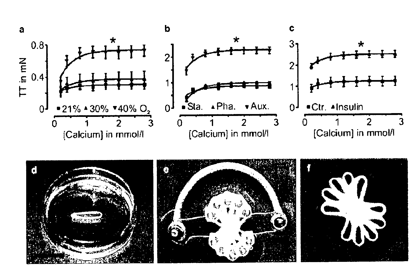
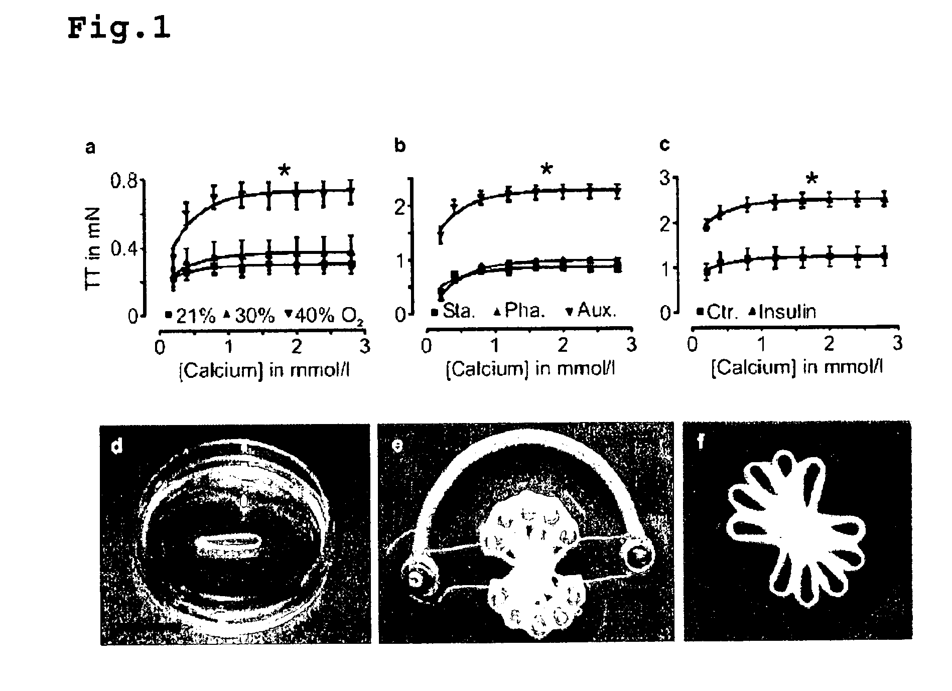
[0073]EHTs were constructed as previously described in Zimmermann, W. H. et al., Circulation 106, I 151-7 (2002) and published International Application WO 01 / 55297. Briefly, EHT rings (reconstitution volume: 0.9 ml) were prepared by mixing isolated heart cells from neonatal rats (2.5×10⁶ cells / EHT) with collagen type I from rat tails (0.8 mg / EHT; pH adjusted to physiologic values ˜7.4 with 0.1 N NaOH), serum-containing culture medium (2×DMEM, 20% horse serum, 4% chick embryo extract, 200 U / mL penicillin, and 200 mg / mL streptomycin; similar volume as neutralized collagen), and Engelbreth-Holm-Swarm tumor exudate (“Matrigel” final concentration 10% v / v; tebu, France).

[0074]EHTs were transferred after 7 days of culturing in casting molds onto custom made stretch devices to facilitate static (110% of slack length), phasic (from 100 to 110% of slack length at 2 Hz), or auxotonic (culture on deflection coils at 110% of slack length a...

example 2

Infarct Model and Grafting

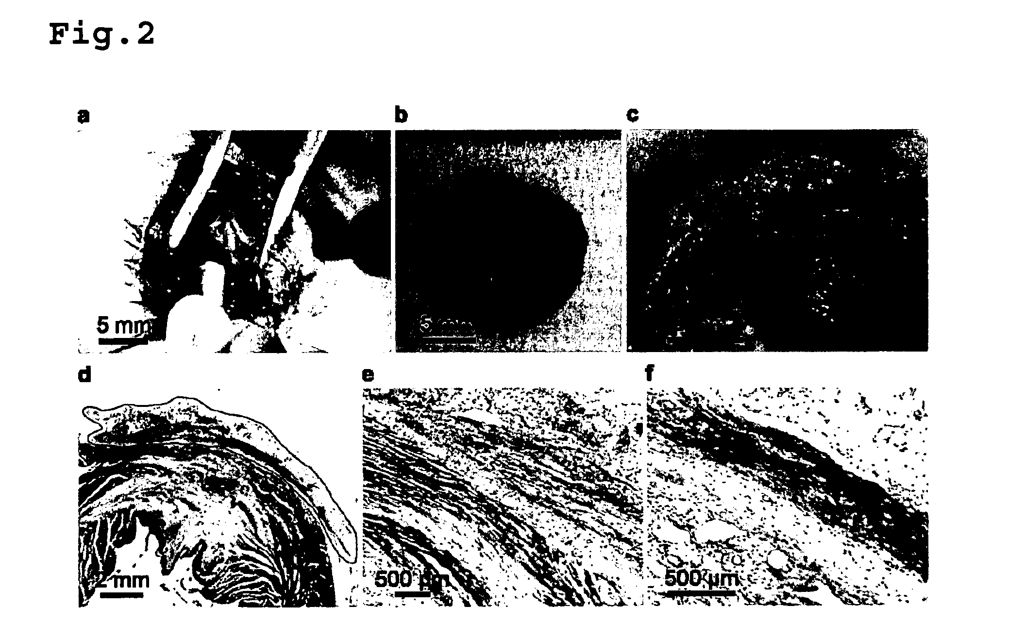
[0076]The potential of multiring EHT constructs to repair diseased hearts in male Wistar rats with myocardial infarcts was tested. Myocardial infarctions were generated in ventilated, isoflurane (2%) anesthetized male Wistar rats (318±3 g; n=121) by permanent ligation of the left anterior descending coronary artery (LAD ligation; 5-0, Prolene, Ethicon, Germany). 14 days after LAD ligation (first surgery) an independent and blinded investigator evaluated infarct localization and size by Echocardiography (ECHO).

[0077]Echocardiography was performed in volatile isoflurane (2%) anaesthesia as described previously with a HP Sonos 7500 System (Philips, Amsterdam, The Netherlands) equipped with a 15 MHz linear array transducer (Zimmermann, W. H. et al., Circulation 106, I 151-7 (2002)). To monitor changes in myocardial performance all animals were subjected to ECHO 14 days post LAD ligation and again 4 weeks after grafting or Sham-operation (longitudinal study desi...

example 3

Epicardial Mapping

[0080]Electrical coupling of multiring EHT constructs to the host myocardium was assessed 4 weeks after engraftment by high resolution epicardial mapping as described in Dhein, S. et al., Circulation 87, 617-30 (1993). Briefly, hearts were excised and Langendorff-perfused with Tyrode's solution at a constant pressure of 70 cm H2O and 37° C. Unipolar ECGs were recorded simultaneously from 256 AgCl electrodes (interelectrode distance: 1 mm; sampling rate: 4 kHz / electrode; HAL4 system; P. Rutten, Hamburg, Germany) arranged in 4 polyester blocks around the circumference of spontaneously beating hearts. Activation time was determined at each electrode and the spread of excitation was analyzed by constructing isochrones. For quantitative analysis the total activation time assessed as the delay between activation of the first and activation of the last electrode for each region under investigation was determined.

[0081]Sham animals (n=5) demonstrated the expected delay of ...


PUM

PropertyMeasurementUnit
outer diameteraaaaaaaaaa
twitch tensionaaaaaaaaaa

Abstract

The invention is directed to a method for the preparation of a multiring engineered heart tissue construct suitable for use in cardiac tissue augmentation and / or replacement therapy. The invention further refers to multiring EHT constructs which comprise at least two force-generating engineered heart tissue rings fused with each other and a device for preparing the same. Finally, the invention relates to force-generating engineered heart tissue rings derived from human cells and their use in drug screening and target validation assays.

Description

FIELD OF THE INVENTION[0001]The invention is directed to a method for the preparation of a multiring engineered heart tissue construct suitable for use in cardiac tissue augmentation and / or replacement therapy. The invention further refers to multiring EHT constructs which comprise at least two force-generating engineered heart tissue rings fused with each other and a device for preparing the same. Finally, the invention relates to force-generating engineered heart tissue rings derived from human cells and their use in drug screening and target validation assays.BACKGROUND OF THE INVENTION[0002]Myocardial infarction and heart failure represent the main cause of death in industrialized countries. The loss of terminally differentiated cardiac myocytes which is associated with these pathologies accounts for a decrease in myocardial function which can lead to total organ failure or trigger compensatory mechanisms like hypertrophy of the remaining myocardium, activation of neurohumoral s...

Claims


Application Information

Patent Timeline
Patent Type & Authority Applications(United States)
IPC IPC(8): A01N1/02C12N5/06C12M3/00C12N5/077
CPCC12M21/08C12N2506/02C12N5/0657C12M35/04
Inventor ZIMMERMANN, WOLFRAM-HUBERTUSESCHENHAGEN, THOMAS
Owner TISSUE SYST HLDG
Why Eureka
Try Eureka