Capping Bioprosthetic Tissue to Reduce Calcification

a bioprosthetic tissue and cap technology, applied in the field of bioprosthetic tissue, can solve the problems of valve failure, undesirable stiffening or degradation of bioprosthetic tissue, and calcification of the proteins of the connective tissue within it,

Active Publication Date: 2009-06-25
EDWARDS LIFESCIENCES CORP
View PDF102 Cites 320 Cited by
  • Summary
  • Abstract
  • Description
  • Claims
  • Application Information

AI Technical Summary

Problems solved by technology

One problem associated with the implantation of many bioprosthetic materials is that the connective tissue proteins (i.e., collagen and elastin) within them can become calcified following implantation in the body.
Such calcification can result in undesirable stiffening or degradation of the bioprosthesis.
This damage to the collagenous tissue leads to valve failure.
These chemicals are not permanently attached to the tissue (e.g., by addition of a reducing agent), and so diffuse out of the tissue over time, which dramatically lowers the calcium mitigation efficacy of these treatments.
The techniques in U.S. Pat. No. 5,476,516 involve the addition of polyols (e.g., glycerol) and alcohols to bioprosthetic tissues as a calcification mitigation treatment alone, but do not address any oxidation mitigation (i.e., capping) strategies.
However, neither of these addresses mitigating potential oxidation of the tissue.
This damage will lead to decreased mechanical and biochemical functionality in the tissue.
Electron beam irradiation will also cleave the collagen backbone and lead to deterioration of the tissue structure and reactivity (Grant, R A et al.
Damage from oxidation during sterilization and / or storage will contribute to valve deterioration and structural failure.
U.S. Pat. No. 6,605,667 discusses the addition of various antioxidant stabilizers to polymerizable adhesives, but does not address damage mitigation to bioprosthetic tissue by ionizing radiation or oxidation during storage.
Although these glycerol-based methods are useful as alternatives to storage in aqueous, liquid-type solutions, they do not address the fact that the post-process functional groups (i.e. aldehydes) can oxidize over time and thus increase the potential for calcification.
The prior art does not address the changes within dehydrated bioprosthetic tissue during storage that occur as a result of in vitro oxidation by air or in vivo oxidation.
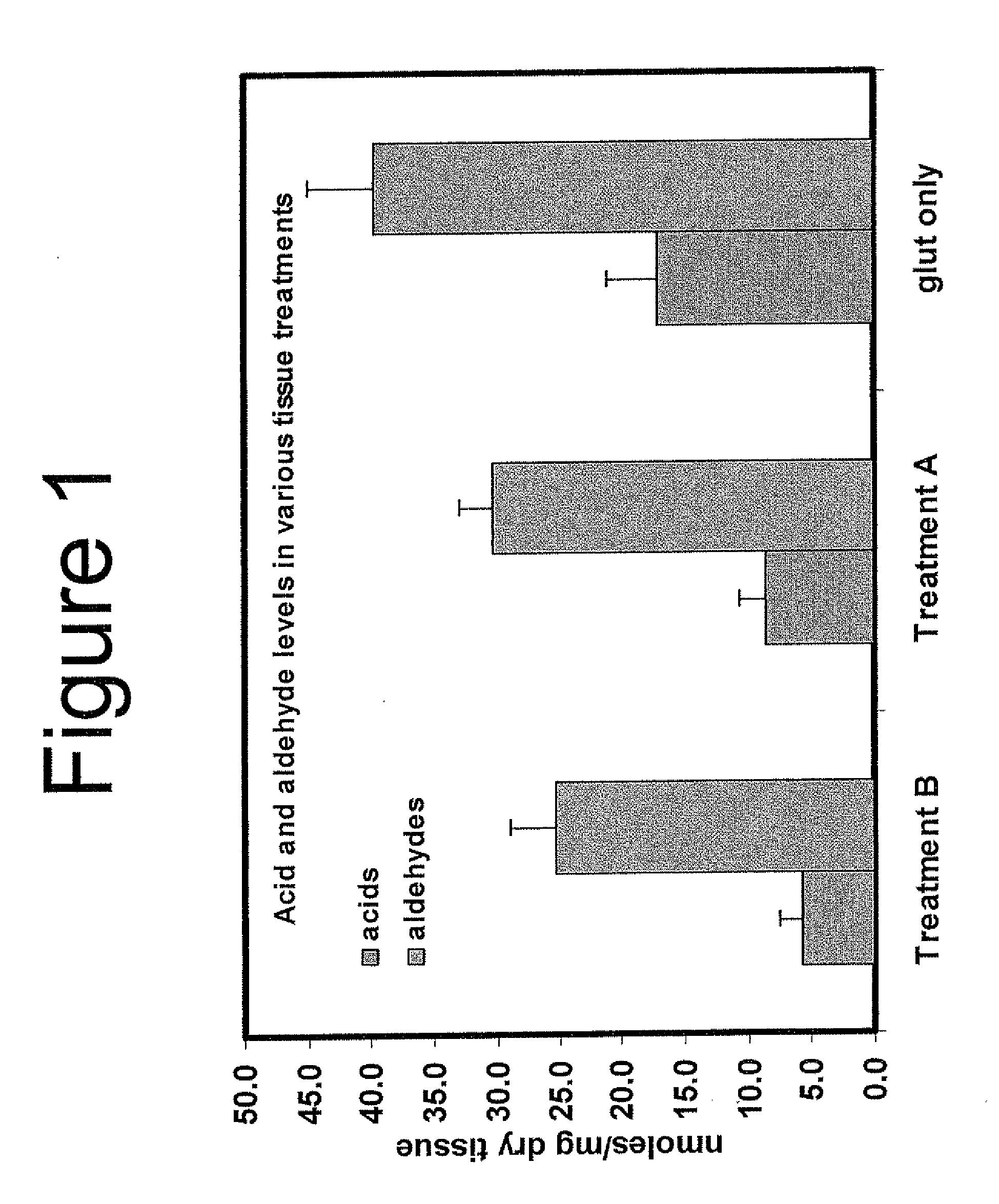
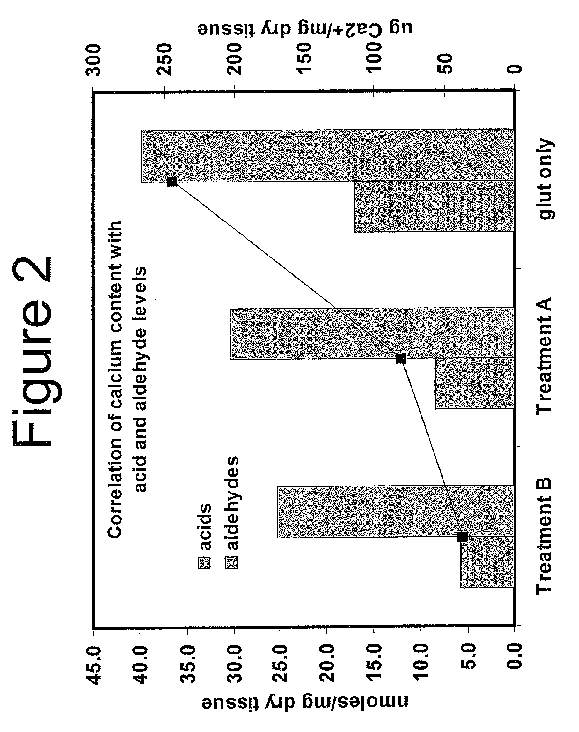
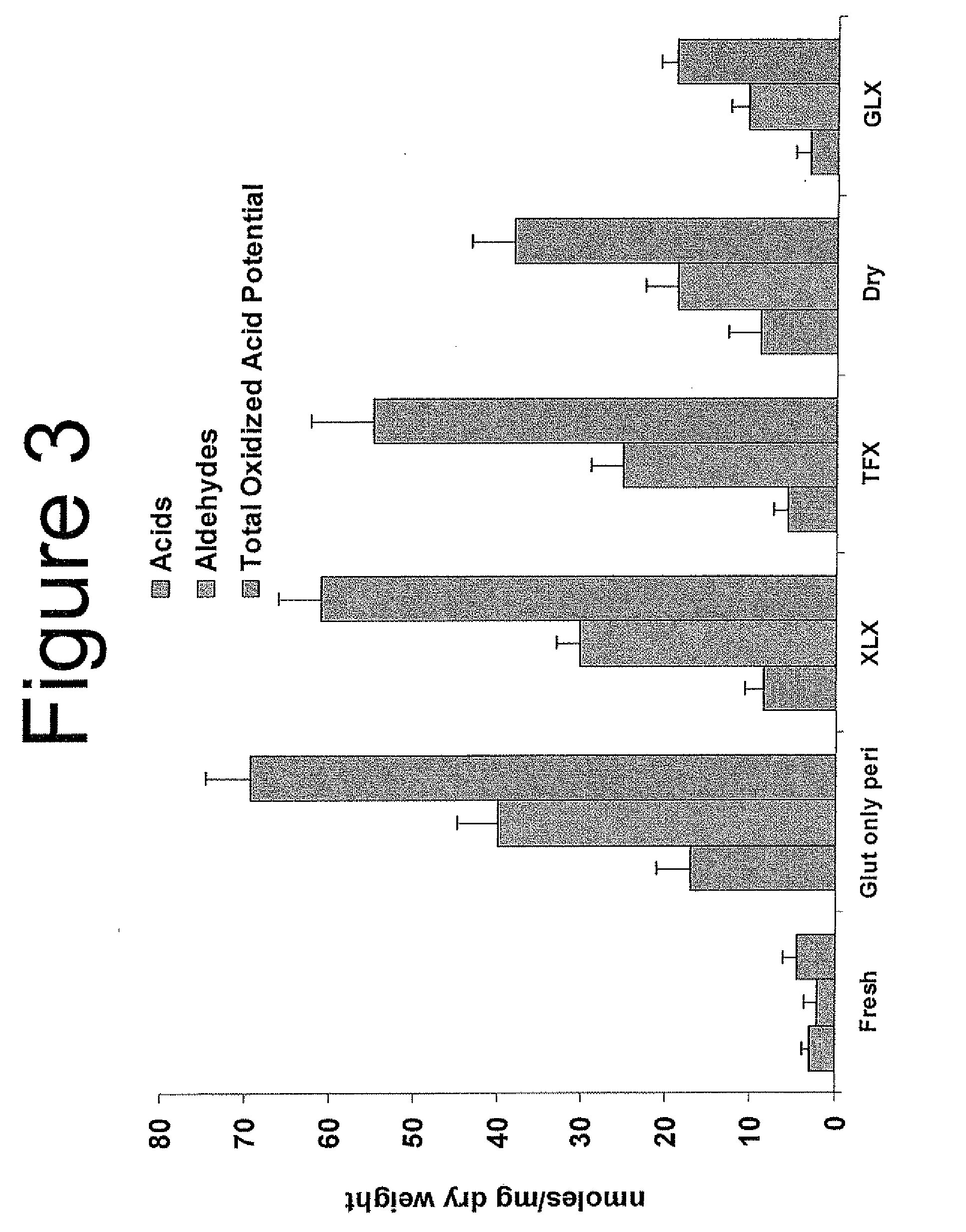
The high aldehyde content in glutaraldehyde-fixed tissue is highly susceptible to oxidation, which results in calcification and tissue failure.
In processes where the tissue is dehydrated and stored in air, the tissue is not protected from oxidation and will lead to biochemical damage from reactive oxygen species.
The resulting oxidative biomarkers, such as carboxylic acids, are likely to promote calcium binding and proceed to failure of the bioprosthesis due to calcification.
Dehydrated bioprosthetic tissue is particularly susceptible to oxidation during sterilization and storage.

Method used


Image

  • Capping Bioprosthetic Tissue to Reduce Calcification
  • Capping Bioprosthetic Tissue to Reduce Calcification
  • Capping Bioprosthetic Tissue to Reduce Calcification

Examples


example 1

[0148]Aldehyde capping using ethanolamine and sodium borohydride of glutaraldehyde-fixed tissue. Bioprosthetic tissue was removed from 0.625% glutaraldehyde just after heat treatment step and rinsed in ethanol: saline (20% / 80%) for 2 minutes. One liter of capping solution was prepared containing 10 mM ethanolamine (0.06%), and 110 mM sodium borohydride (0.42%) in 50 mM phosphate buffer (pH 7.3-7.8)

[0149]The capping solution was placed on an orbital shaker, then tissues (leaflets or valves) were placed in the solution so that they were completely submerged. The ratio of tissue to solution was 3 leaflets per 100 ml or one valve per 100 ml. The container was partly covered but not completely sealed because hydrogen gas liberated by the chemical reaction with water could cause the container to explode. The orbital shaker was operated at between 80-100 rpm for 4 hours at room temperature. The tissue was removed and rinsed in FET solution (formaldehyde, ethanol, tween-80) for three minute...

example 2

[0150]Glycerol dehydration process for pericardial valve bioprosthesis. Pericardial valves were dehydrated by holding each valve with forceps on the sewing ring of the valve and placing the valve in a glycerol / ethanol (75% / 25%) mixture. Beakers containing the valves were placed on an orbital shaker operating between 50-60 RPM for at least one (1) hour but not more than four (4) hours then immediately treated to remove excess glycerol. This was done by holding them with forceps on the sewing ring of the valve, taking the valve out of the glycerol / ethanol mixture and then placing it on an absorbent towel in a wide mouth jar. After being allowed to dry for at least 5 minutes at room temperature the jar was attached to a lyophilizer and dried for 2 hours. Valves were then transferred to ethylene oxide gas permeable packages and sterilized with ethylene oxide.

example 3

[0151]Calcification Mitigation—Small Animal Model. In order to evaluate the calcification mitigation properties of GLX treated and EO sterilized pericardial tissue, two small animal feasibility studies were conducted. These studies demonstrate that, 1) GLX is superior to TFX in mitigating the occurrence of calcification in tissue, and 2) real time aged GLX tissue is also superior to TFX in mitigating calcification. In both studies, GLX valves demonstrated reduced variability in calcification data when compared to TFX valves. Test methods and results of each are summarized below.


PUM


Abstract

A treatment for bioprosthetic tissue used in implants or for assembled bioprosthetic heart valves to reduce in vivo calcification. The method includes applying a calcification mitigant such as a capping agent or an antioxidant to the tissue to specifically inhibit oxidation in tissue. Also, the method can be used to inhibit oxidation in dehydrated tissue. The capping agent suppresses the formation of binding sites in the tissue that are exposed or generated by the oxidation and otherwise would, upon implant, attract calcium, phosphate, immunogenic factors, or other precursors to calcification. In one method, tissue leaflets in assembled bioprosthetic heart valves are pretreated with an aldehyde capping agent prior to dehydration and sterilization.

Description

RELATED APPLICATION[0001]This application claims priority to U.S. Provisional Application No. 61 / 016,263, filed Dec. 21, 2007, the entire disclosure of which is incorporated by reference herewith.FIELD OF THE INVENTION[0002]The present invention relates generally to methods for treating bioprosthetic tissue, materials, and devices to reduce post-implantation calcification, and reducing post-implantation calcification, in particular for bioprosthetic tissue used in heart valves.BACKGROUND OF THE INVENTION[0003]Heart valve replacement may be indicated when there is a narrowing of the native heart valve, commonly referred to as stenosis, or when the native valve leaks or regurgitates, such as when the leaflets are calcified. In one therapeutic solution, the native valve may be excised and replaced with either a biologic or a mechanical valve. Certain medical conditions may require grafting or suturing a tissue patch to repair physiological abnormalities. These include, but are not limi...

Claims


Application Information

Patent Timeline
Patent Type & Authority Applications(United States)
IPC IPC(8): A61F2/24A01N1/02
CPCA61L27/3604A61L27/3687A61F2/24A61L2400/02A61L27/507A61F2/2412A61L27/3691A61L2430/20A61F2/0095A61F2/2415A61F2/2418A61L27/3625A61L27/3629
Inventor DOVE, JEFFERYDOBLER, DARINDAVIDSON, JIMWRIGHT, GREGORY A.
Owner EDWARDS LIFESCIENCES CORP
Why Eureka
Try Eureka