Optical Imaging
a technology of optical imaging and optical x-ray, applied in the field of optical imaging, can solve the problems of affecting the rpe support of the photoreceptor, affecting the x-ray image quality, and affecting the image quality of the image,
- Summary
- Abstract
- Description
- Claims
- Application Information
AI Technical Summary
Benefits of technology
Problems solved by technology
Method used
Image

Examples
example 1
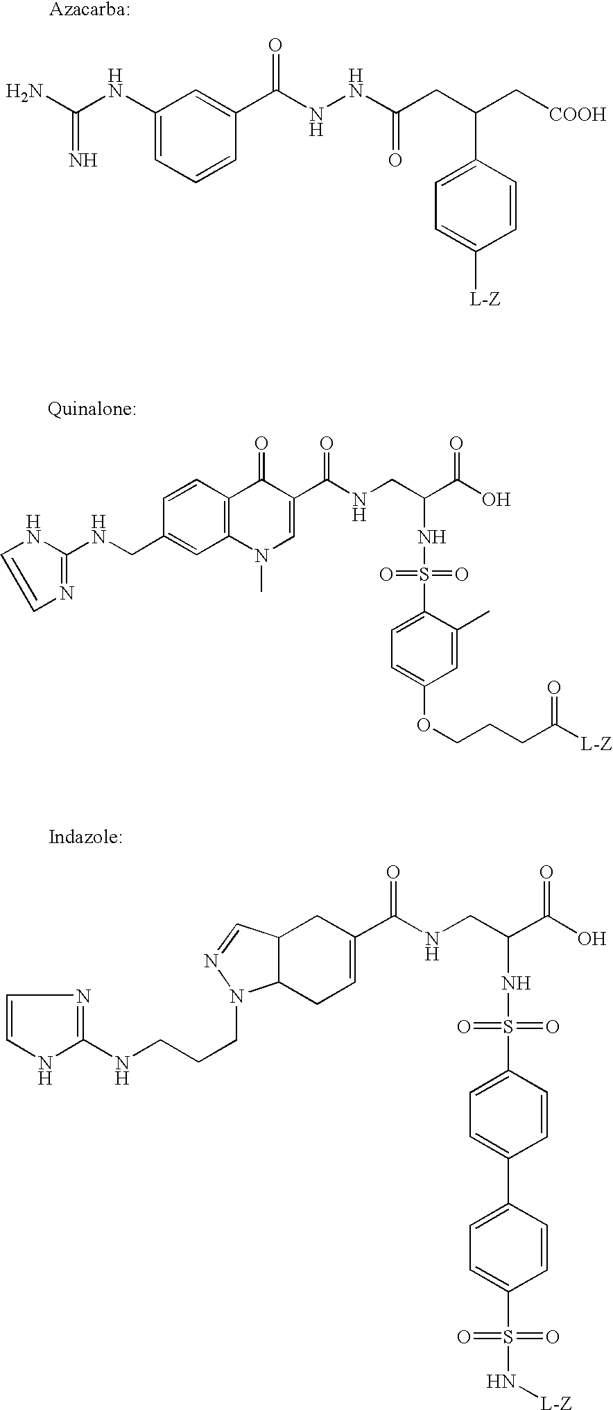
Synthesis of Cys2-6; c[CH2CO-Lys(Cy5 bis-SO3)-Cys-Arg-Gly-Asp-Cys-Phe-Cys]-(PEG)n-NH2 (n=1) (Contrast agent B)
[0098]
[0099]The peptide above was assembled using standard solid phase peptide synthesis methods. The chloroacetylated peptide was cleaved from the solid support and cyclized in solution, first forming the thioether bridge and then the disulfide bridge. The bicyclic peptide (24 mg, 0.02 mmol) was added as a solid to a solution of Cy5 mono NHS-ester bis-SO3 (7.5 mg±0.01 mmol) in DMF (2 ml), and NMM (0.01 ml, 0.09 mmol) was then added. The reaction was let proceed over night avoid form light. DMF was evaporated under reduced pressure and the crude product was purified by reverse phase preparative chromatography (Vydac C18 column, 218TP1022; solvents: A=water / 0.1% TFA and B=CH3CN / 0.1% TFA; gradient 10-30% B over 60 min; flow 10 ml / min; detection at 254 nm), affording 6.6 mg (37%) of pure product (analytical HPLC: Phenomenex Luna C18 column, 00G-4252-E0; solvents: A=water / 0.1% T...
example 2
Synthesis of disulphide [Cys2-6]thioether cyclo[CH2CO-Lys(fluorescein)-Cys2-Arg-Gly-Asp-Cys6-Phe-Cys]-PEG-NH, (Contrast agent H)
2 a) Synthesis of 17-(Fmoc-amino)-5-oxo-6-aza-3,9,12,15-tetraoxaheptadecanoic acid
[0100]
[0101]This building block is coupled to the solid-phase using Fmoc chemistry. The coupled form of this building block will be referred to in short as PEG.
1,11-Diazido-3,6,9-trioxaundecane
[0102]A solution of dry tetraethylene glycol (19.4 g, 0.100 mol) and methanesulphonyl chloride (25.2 g, 0.220 mol) in dry THF (100 ml) was kept under argon and cooled to 0° C. in an ice / water bath. To the flask was added absolution of triethylamine (22.6 g, 0.220 mol) in dry THF (25 ml) dropwise over 45 min. After 1 hr the cooling bath was removed and stirring was continued for 4 hrs. Water (60 ml) was added. To the mixture was added sodium hydrogencarbonate (6 g, to pH 8) and sodium azide (14.3 g, 0.220 mmol), in that order. THF was removed by distillation and the aqueous solution was r...
example 3
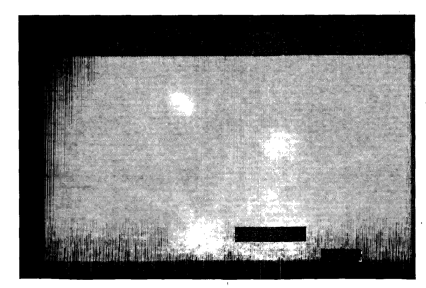
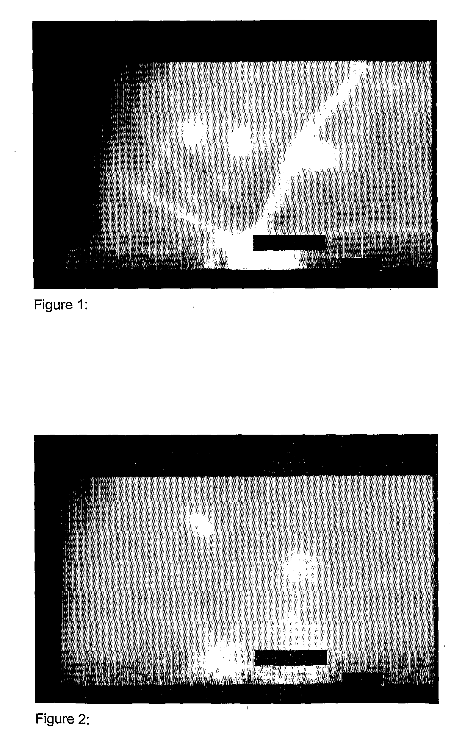
Imaging of CNV
[0115]Imaging of choroid neovascularization in rat was performed using the contrast agent from Example 2 (contrast agent H) as an optical imaging contrast agent.
[0116]Long-Evans pigmented rats, weighting between 350 g and 400 g, were used for this study. The rats were anesthetized with Ketamine (100 micro-I / 100 g) given by intramuscularly injection, and the pupils were dilated with 1% tropicamide eye drop and 0.1% phenylephrine eye drop before the photocoagulation and peptide angiographic examination. Fundus photocoagulation was performed to the right eyes of rats with a diode laser indirect opthalmoscope delivery system under the following condition: Power, 90 mW; wavelength, 810 nm; duration, 0.1 second; and spot size, 75 micro-m. 16 laser burns were delivered around the optic nerve head of each eye. A laser (IRIS Medical, OcuLight™, S / N 25745) was used to make 10 lesions in the Bruch's membrane and choroid. Each lesion was placed approximately 1 mm from the optic ne...
PUM
| Property | Measurement | Unit |
|---|---|---|
| Affinity | aaaaa | aaaaa |
Abstract
Description
Claims
Application Information