
Eureka
For R&D, Eureka makes reading and utilizing patents & technical documents easy.
Eureka AIR
Designed for self-driven R&D workflows. Generate viable solutions, solve complex R&D challenges, empower your innovation with AI.

Eureka Materials
Designed for material experts only. Revolutionize your material R&D, from search, analyze, to developing new materials.

TechResearch
Generate reliable direction feasibility study reports for your R&D in just a few steps.

TechSeek
Discover and master advanced knowledge NOW. Basics, ideas, possibilities, all at once.

TechMind
As an expert in R&D Theories, TechMind can generates customized viable solutions instantly.

TechRisk
Analyze your overall solution with one click, know your potential R&D risks in advance.

TechMonitor
Get weekly tech updates, stay abreast of the latest tech innovations and key insights.
Opsin Stabilizing Compounds and Methods of Use
- Summary
- Abstract
- Description
- Claims
- Application Information
AI Technical Summary
Benefits of technology
Problems solved by technology
Method used
Image


Examples
example 1
Use of a Crystal Structure of Rhodopsin
to Select Potential Modulators
[0184]The retinal binding pocket of a trigonal crystal form of bovine rhodopsin, PDB code 1 GZM, was used to identify small molecule modulators by a high throughput molecular docking method. The positions of each retinal atom were used to guide in the definition of the binding pocket selected for molecular docking.
[0185]Spheres were positioned at the selected site to allow the molecular docking program, DOCK 5. 1.0 (available from USCF), to match spheres with atoms in potential ligands (small molecules in this ease). During the molecular docking calculation, orientations are sampled to match the largest number of spheres to potential ligand atoms, looking for the low energy structures that bind tightly to the active site of a receptor or enzyme whose active site structure is known.
[0186]A scoring grid was calculated to estimate the interaction between potential ligands and the retinal binding pocket target site. Th...
example 2
[0194]This example describes measurement of opsin levels in HEK 293 Flp-In™ T-REX™ cell lines (Invitrogen) that are stably transformed with a mutant or wild-type opsin gene or an empty vector. Opsin expression in these cells is inducible with tetracycline. Following induction, cells are lysed in detergent buffer and cellular protein is immobilized on nitrocellulose-containing membranes. Opsin and tubulin (as a loading control) are detected with antibodies and an infrared scanner. This dot procedure can be applied to opsin-containing detergent lysates from other sources, such as mouse eyes.
Cell Growth and Plating (1.5 Days, Starting with Confluent Cells)[0195]1. The following steps must be conducted with aseptic technique in a tissue culture hood.[0196]2. Obtain confluent 10 cm plates of cell lines of Flp-In™ T-REx™ 293 containing the opsin gene and the vector control from plates grown in DMEM containing 10% fetal bovine serum, antibiotic / antimycotic solu...
example 3
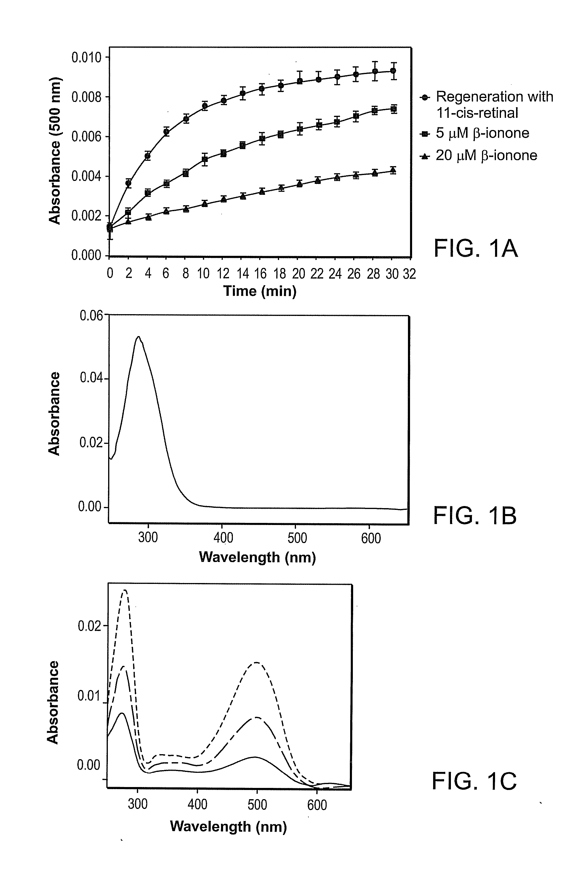
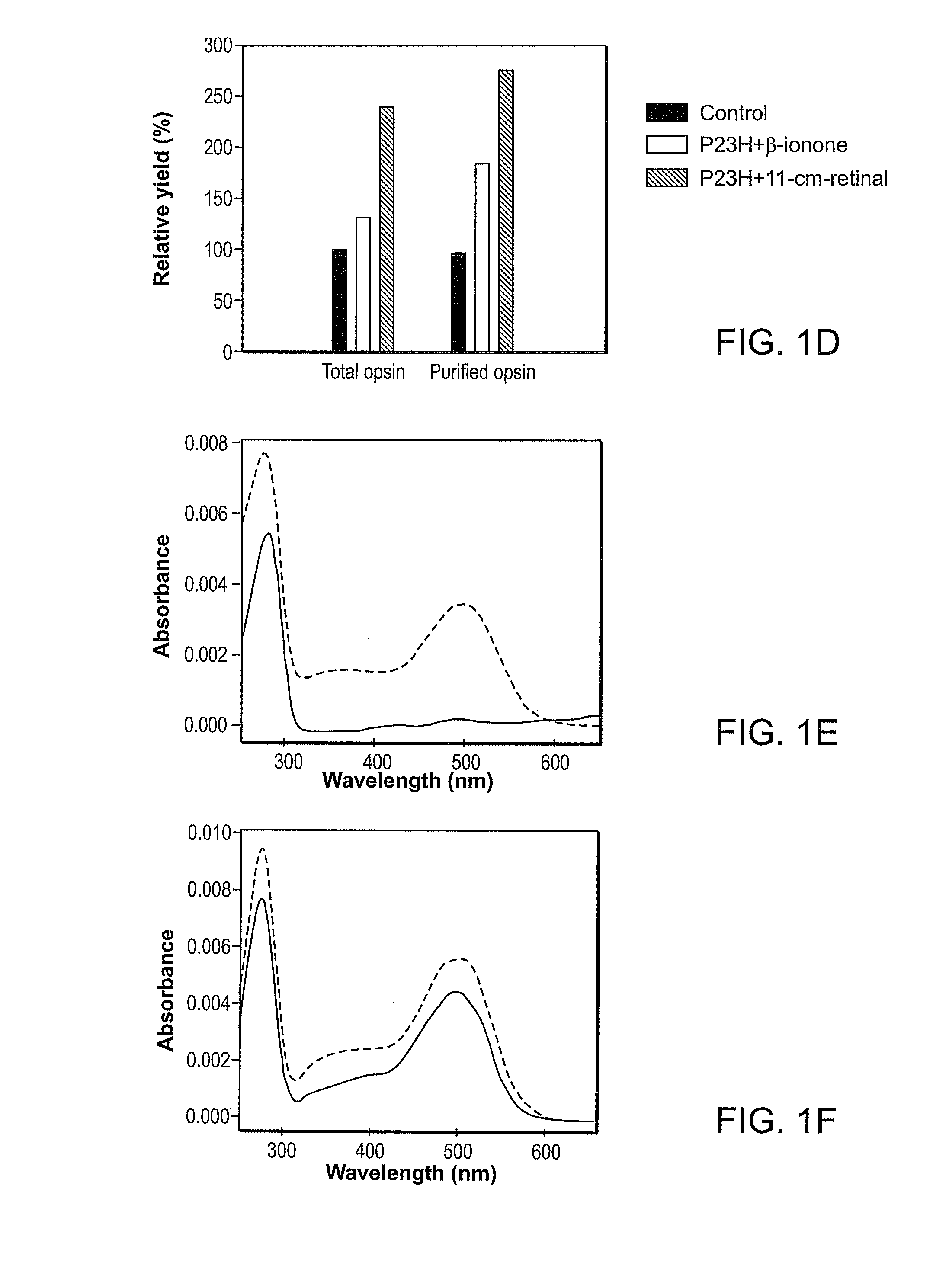
Effect of Compounds on P23H Rhodopsin Yield
[0278]The ability of candidate compounds to affect the yield of P23H rhodopsin is believed to be indicative of the ability of the compound to stabilize the mutant opsin. See generally Noorwez et al. (2004) and U.S. Patent Publication No. 2004-0242704, both of which are incorporated herein by reference.
[0279]Cell Lines and Culture Conditions.
[0280]Stable cell lines expressing the P23H opsin were generated using Flp-In T-Rex system (Invitrogen) in HEK293 cell line. To create plasmids for constructing stable cell lines, an EcoRI-NotI fragment from the wild-type or P23H mutant synthetoc bovine opsin gene in pMT4 (see Kaushal et al., Biochemistry, Vol. 33, pp. 6121-6128 (1994) was combined with the large EcoRI-NotI fragment of pcDNA5 / FRT / TO (Invitrogen). The resulting plasmids contain opsin under the control of a tetracycline-inducible human cytomegalovirus promoter and a flippase recognition sequence (FRT) for site sepecific recombination at th...
PUM
| Property | Measurement | Unit |
|---|---|---|
| Fraction | aaaaa | aaaaa |
| Fraction | aaaaa | aaaaa |
| Fraction | aaaaa | aaaaa |
Abstract
Description
Claims
Application Information
 - R&D Engineer
- R&D Manager
- IP Professional
- Industry Leading Data Capabilities
- Powerful AI technology
- Patent DNA Extraction
Browse by: Latest US Patents, China's latest patents, Technical Efficacy Thesaurus, Application Domain, Technology Topic, Popular Technical Reports.
© 2024 PatSnap. All rights reserved.Legal|Privacy policy|Modern Slavery Act Transparency Statement|Sitemap|About US| Contact US: help@patsnap.com