Real-time 3-d ultrasound guidance of surgical robotics

a robotics and real-time technology, applied in the field of real-time 3d ultrasound, can solve the problem that the laparoscopic grasper can generally only give a rudimentary feedback, and achieve the effects of improving ergonomics, increasing visibility and depth perception, and precise, dexterous control

Inactive Publication Date: 2009-11-19
THE UNIV OF NORTH CAROLINA AT CHAPEL HILL +1
View PDF28 Cites 26 Cited by
  • Summary
  • Abstract
  • Description
  • Claims
  • Application Information

AI Technical Summary

Benefits of technology

[0002]Robotic surgery technology has made recent gains as an accepted alternative to traditional instruments in cardiovascular, neurological, orthopedic, urological, and general surgery. With the da Vinci system (Intuitive Surgical, Inc., Sunnyvale, Calif.), a multi-camera endoscope is used for 3D visualization, increasing visibility and depth perception for the robot operator. The dual-lens endoscope links to two monitors, enabling 3D stereoscopic vision within the patient. The robotic arms also exhibit precise, dexterous control, eliminating tremor and improving ergonomics for the surgeon. For laparoscopic procedures, there have been published reports of using robotics in cases of splenectomy, adrenalectomy, cholecystectomy, and gastric bypass among others. In most cases, surgeons reported better visualization, increased instrument control, reduced operator fatigue, and an improved learning curve for those training to perform these procedures.
[0003]Also in recent years, the development of endoscopic transducer designs has enabled the application of B-scan laparoscopic ultrasound as a preoperative and intraoperative tool for assistance in surgical guidance and assessment. The primary advantage of laparoscopic ultrasound (LUS) is the ability to image beyond tissue boundaries. However, optical laparoscopes generally provide only views of the outer surface of organs, and laparoscopic graspers can generally only give a rudimentary feedback regarding tissue texture or underlying masses. The integration of LUS into the operating room provides visualization of most surrounding soft tissue structures, allowing access to information that might otherwise only be available in an open surgery setting. Additionally, the ability to place the transducer directly against an organ allows the use of higher frequency devices, which provide better resolution.
[0005]The application of real-time three-dimensional (RT3D) ultrasound imaging may increase the utility of laparoscopic ultrasound for these applications. While acquiring full volumes of information intraoperatively, real-time 3D ultrasound may provide improved visualization and possibly decrease procedure time and difficulty. The ability to visualize multiple planes through a volume in real-time without moving the transducer can improve determination of target geometry as well. Acquisition of volumetric data with RT3D is achieved through the use of one dimensional arrays combined with a motor or with two-dimensional transducer arrays and sector phased array scanning in both the azimuth and elevation directions. In this way, pyramidal volumes of data, as shown in FIG. 1, are acquired without the use post-acquisition reconstruction.
[0007]An additional advantage that RT3D laparoscopic ultrasound provides over conventional 2D LUS is the ability to establish a true 3D coordinate system for measurement and guidance. Traditional ultrasound scanner systems are capable of two-dimensional measurements. With volumes of data acquired in real-time, RT3D scanners can provide a surgeon with three-dimensional structural orientation within a target organ using its measurement system, providing more information than was previously available. This could be particularly useful in conjunction with recent advancements in robotic surgeries. With new equipment such as the da Vinci robotic surgical system, the integration of RT3D and its measurements can help locate targets and steer the robot's arms into position, while avoiding regions that must not be damaged.

Problems solved by technology

However, optical laparoscopes generally provide only views of the outer surface of organs, and laparoscopic graspers can generally only give a rudimentary feedback regarding tissue texture or underlying masses.

Method used


Image

  • Real-time 3-d ultrasound guidance of surgical robotics
  • Real-time 3-d ultrasound guidance of surgical robotics
  • Real-time 3-d ultrasound guidance of surgical robotics

Examples


example

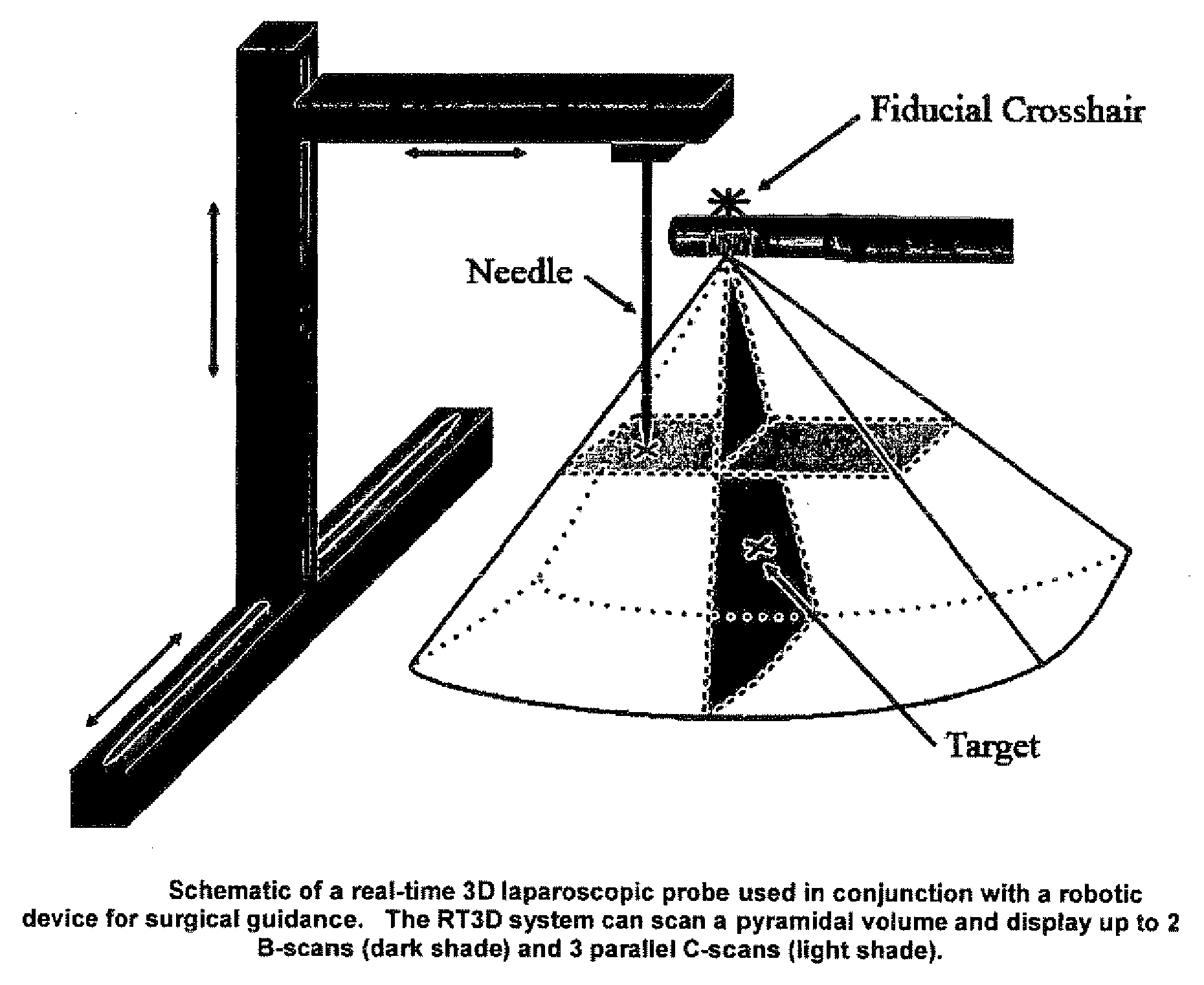
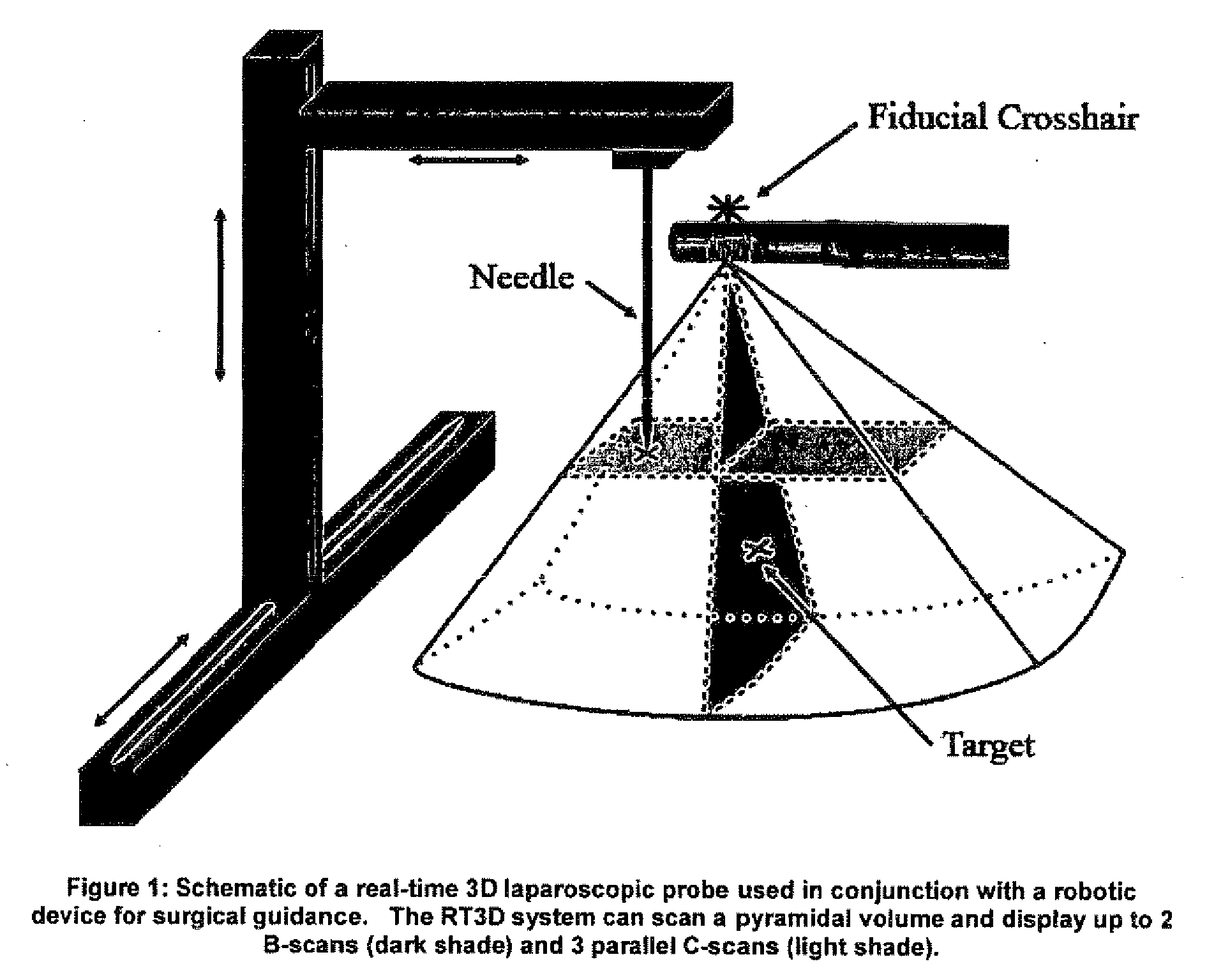
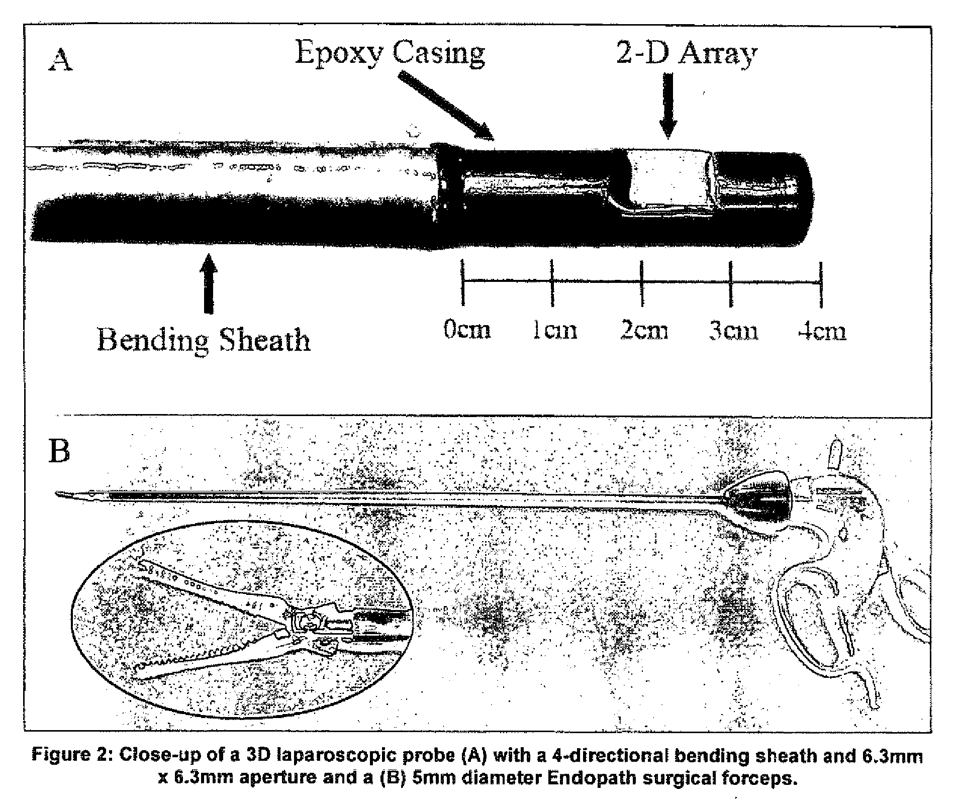
Animal Model and 3D Laparoscopic Study

[0031]The Institutional Animal Care and Use Committee approved the use of a canine model for the acquisition of in vivo 3D images, conforming to the Research Animal Use Guidelines of the American Heart Association. Ketamine hydrochloride 10-15 mg / kg IM was used to sedate the dog. An IV of 0.9% sodium chloride was established in the peripheral vein and maintained at 5 mL / kg / min. Anesthesia was induced via nasal inhalation of isoflurane gas 1-5%. An endotracheal tube for artificial respiration was inserted after oral intubation with the dog placed on its back on a water-heated thermal pad. A femoral arterial line was placed on the left side via a percutaneous puncture. Electrolyte and respirator adjustments were made based on serial electrolyte and arterial blood gas measurements. Blood pressure, electrocardiogram, and temperature were continuously monitored throughout the procedure.

[0032]After the animal preparations were complete, the dog's abdo...


PUM


Abstract

Laparoscopic ultrasound has seen increased use as a surgical aide in general, gynecological, and urological procedures. The application of real-time three-dimensional (RT3D) ultrasound to these laparoscopic procedures may increase information available to the surgeon and serve as an additional intraoperative guidance tool. The integration of RT3D with recent advances in robotic surgery can also increase automation and ease of use. In one non-limiting exemplary implementation, a 1 cm diameter probe for RT3D has been used laparoscopically for in vivo imaging of a canine. The probe, which operates at 5 MHz, was used to image the spleen, liver, and gall bladder as well as to guide surgical instruments. Furthermore, the 3D measurement system of the volumetric scanner used with this probe was tested as a guidance mechanism for a robotic linear motion system in order to simulate the feasibility of RT3D / robotic surgery integration. Using images acquired with the 3D laparoscopic ultrasound device, coordinates were acquired by the scanner and used to direct a robotically controlled needle towards desired in vitro targets as well as targets in a post-mortem canine. The RMS error for these measurements was 1.34 mm using optical alignment and 0.76 mm using ultrasound alignment.

Description

FIELD[0001]The technology herein relates to the use of real-time 3D ultrasound in a laparoscopic setting and in percutaneous procedures and as a direct guidance tool for robotic surgery.BACKGROUND AND SUMMARY[0002]Robotic surgery technology has made recent gains as an accepted alternative to traditional instruments in cardiovascular, neurological, orthopedic, urological, and general surgery. With the da Vinci system (Intuitive Surgical, Inc., Sunnyvale, Calif.), a multi-camera endoscope is used for 3D visualization, increasing visibility and depth perception for the robot operator. The dual-lens endoscope links to two monitors, enabling 3D stereoscopic vision within the patient. The robotic arms also exhibit precise, dexterous control, eliminating tremor and improving ergonomics for the surgeon. For laparoscopic procedures, there have been published reports of using robotics in cases of splenectomy, adrenalectomy, cholecystectomy, and gastric bypass among others. In most cases, surg...

Claims


Application Information

Patent Timeline
Patent Type & Authority Applications(United States)
IPC IPC(8): A61B19/00A61B8/00A61B5/05
CPCA61B8/0833A61B8/0841A61B8/12A61B8/445A61B8/4488A61B2019/5276A61B19/201A61B19/22A61B19/2203A61B2017/3413A61B2019/2211A61B8/483A61B34/70A61B2034/301A61B34/30A61B90/11A61B2090/378
Inventor PUA, ERICLIGHT, EDWARD D.VON ALLMEN, DANIELSMITH, STEPHEN W.
Owner THE UNIV OF NORTH CAROLINA AT CHAPEL HILL
Why Eureka
Try Eureka