Biologically excitable cells

a technology of excitable cells and cells, applied in the field of excitable cells, can solve the problems of confounded use of hcn genes, unpredicted consequences of hcn gene transfer in vivo, and low flexibility of use of wild-type channels in frequency tuning of engineered pacemakers

Inactive Publication Date: 2009-12-10
THE JOHN HOPKINS UNIV SCHOOL OF MEDICINE
View PDF30 Cites 0 Cited by
  • Summary
  • Abstract
  • Description
  • Claims
  • Application Information

AI Technical Summary

Benefits of technology

[0009]Yet another aspect of the invention is another method of making a biological pacemaker. An inexcitable mammalian cell is transfected with one or more nucleic acid molecules encoding a gene which depolarizes the cell membrane, a gene which repolarizes the cell membrane, and a gene which fires spontaneously. The mammalian cell thereby displays spontaneously oscillating action potentials.

Problems solved by technology

However, use of HCN genes may be confounded by unpredictable consequences of heteromultimerization with multiple endogenous HCN family members in the target cell (Ulens, C.
), HCN gene transfer in vivo may have unpredicted consequences.
Moreover, the use of wild-type channels offers little flexibility with regard to frequency tuning of the engineered pacemaker.
Such devices are expensive, and implantation involves a number of acute and chronic risks such as pulmonary collapse, bacterial infection, lead or generator failure (Bernstein, A. D. & Parsonnet, V.

Method used


Image

  • Biologically excitable cells
  • Biologically excitable cells
  • Biologically excitable cells

Examples


example 1

Materials and Methods

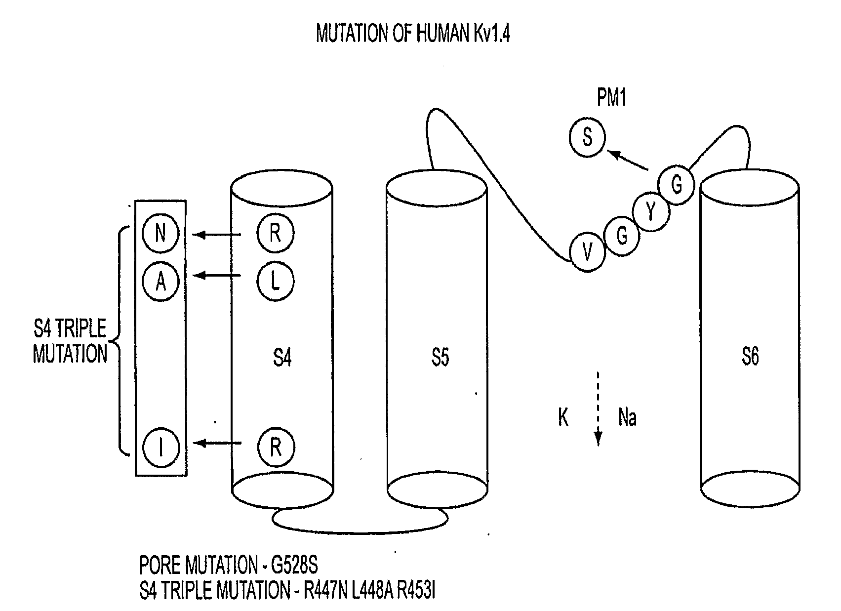
Plasmid Construction, and Adenovirus Preparation, and Mutation

[0036]Human Kv1.4 cDNA was subcloned from XL-4 vector (OriGene Technologies, Inc. Rockville, Md.) to pTracerCMV2 plasmid (Invitrogen, Carlsbad, Calif.) between EcoRI and NotI sites. The adenovirus shuttle vector pAdCGI was used for generation of adeno / S4TK1.4GYS-IRES GFP. Adenovirus was produced as previously described1. Oligonucleotide mutagenesis was performed with site-direct mutagenesis kit (Stratagene, La Jolla, Calif.).

Transient Transfections of Cultured Cell Lines

[0037]HEK293 cells were seeded at a density of 2.0×10⁵ per 35-mm2 the day before transfection. Cells were transfected with Lipofectamine 2000 (Invitrogen, Carlsbad, Calif.) according to manufacturer's protocol. Voltage- and current-clamp recording were carried out within 18-48 hours post-transfection.

Ventricular Myocyte Isolation

[0038]Guinea pig left ventricular myocytes were isolated using Langendorff perfusion, as previously describe...

example 2

Creation of a Biological Pacemaker by Cell Fusion

[0051]As an alternative strategy to electronic pacemakers or to gene therapy / stem cell approaches, we explored the feasibility of converting ventricular myocytes into pacemakers by cell fusion. The idea is to create chemically-induced fusion between ventricular myocytes and syngeneic fibroblasts engineered to express pacemaker ion channels, HCN1.

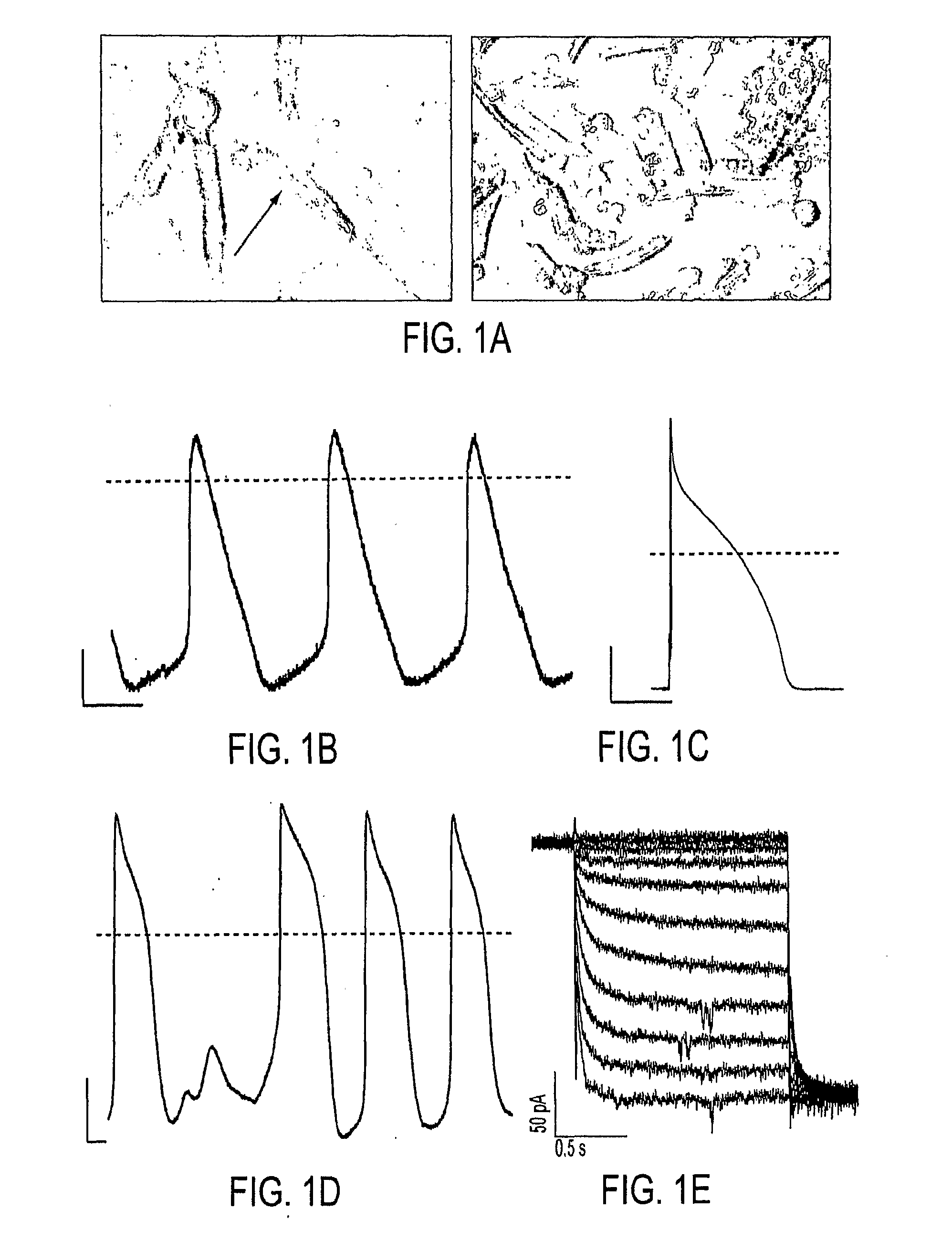
[0052]In order to examine fusion events, guinea pig lung fibroblasts stably expressing HCN1 channels (HCN1-fibroblasts) were fused with freshly-isolated guinea pig ventricular myocytes using polyethylene glycol (PEG). Within 3 minutes of dehydration and rehydration, the HCN1-fibroblasts fused with ventricular myocytes as verified by the sudden introduction of Calcein-AM fluorescence into the myocytes (FIG. 1A). Current-clamp of the myocyte / HCN1-fibroblast heterokaryon exhibited spontaneous action potentials with a slow phase-4 depolarization (FIG. 1B), suggesting the expression of pacemaker cu...

example 3

Conversion of Non-Excitable Cells to Self-Contained Biological Pacemakers

[0059]In pacemaker cells of the sinoatrial node, voltage- and time-dependent membrane ionic currents generate spontaneous action potentials (APs). We hypothesized that a non-excitable cell could be converted into a pacemaker by heterologous expression of a minimal complement of specific ion channels. To this end, HEK293 cells were engineered to express the following ionic currents: 1) an excitatory current 2) an early repolarizing current, and 3) an inward rectifier current. A Na+ channel from bacteria (NaChBac)17 (FIG. 4A, left) was chosen for the excitatory current because of its slow gating kinetics and its compact cDNA, human ether-a-go-go related gene channels (hERG)18 (FIG. 4A, middle) for repolarizing current to activate and counter the depolarizing effects of NaChBac, and Kir2.119 (FIG. 4A, right) to favor a negative diastolic potential.

[0060]In current-clamp recordings at room temperature, action poten...


PUM

PropertyMeasurementUnit
tip resistancesaaaaaaaaaa
concentrationaaaaaaaaaa
temperatureaaaaaaaaaa

Abstract

As an alternative strategy to electronic pacemaker devices, we explored the feasibility of converting normally-quiescent ventricular myocytes into pacemakers by somatic cell fusion. The idea is to create chemically-induced fusion between myocytes and syngeneic fibroblasts engineered to express HCN1 pacemaker ion channels (HCN1 fibroblasts), in normally-quiescent myocardium. HCN1-expressing fibroblasts formed stable heterokaryons with myocytes, generating spontaneously-oscillating action potentials as well as ventricular pacemaker activity in vivo and provides a platform for an autologous, non-viral, adult somatic cell therapy. We also converted a depolarization-activated potassium-selective channel, Kv1.4, into a hyperpolarization-activated non-selective channel by site-directed mutagenesis (R447N, L448A, and R453I in S4 and G528S in the pore). Gene transfer into ventricular myocardium demonstrated the ability of this construct to induce pacemaker activity, with spontaneous action potential oscillations in adult ventricular myocytes and idioventricular rhythms by in vivo electrocardiography. Given the sparse expression of Kv1 family channels in the human ventricle, gene transfer of a synthetic pacemaker channel based on the Kv1 family has therapeutic utility as a biological alternative to electronic pacemakers.

Description

[0001]This application claims the benefit of U.S. provisional application Ser. No. 60 / 726,840 filed Oct. 14, 2005, the disclosure of which is expressly incorporated herein.TECHNICAL FIELD OF THE INVENTION[0002]This invention is related to the area of excitable cells. In particular, it relates to alteration of biologically excitability of cells by changing the cell's complement of ion channel proteins.BACKGROUND OF THE INVENTION[0003]More than 250,000 people in the United States get artificial pacemakers implanted each year for the treatment of heart arrhythmias, typically slow or irregular heart beats. Biological pacemakers can be used to replace or augment the function of artificial pacemakers.[0004]In the sinoatrial node, pacemaker activity is generated by a balance of depolarizing and repolarizing currents whose gating and permeation properties, in ensemble, create a stable oscillator (DiFrancesco, D. (1995) Cardiovasc Res 29, 449-56). Hyperpolarization-activated nucleotide-gated...

Claims


Application Information

Patent Timeline
Patent Type & Authority Applications(United States)
IPC IPC(8): A61K49/00A61K35/12C12N5/00C12N15/87C12N15/00C07K14/47C12N15/11
CPCA61K48/005A61K48/0075C12N2799/04C12N5/16C12N2799/022C07K14/705A61P9/06
Inventor MARBAN, EDUARDO
Owner THE JOHN HOPKINS UNIV SCHOOL OF MEDICINE
Why Eureka
Try Eureka