
Biomaterials for regenerative medicine

a biomaterial and regenerative medicine technology, applied in the field of usable biomaterials, can solve the problems of tissue damage, unsatisfactory efficacy of symptomatic treatment, cost and risk of infection, etc., and achieve the effect of suitable properties

Inactive Publication Date: 2009-12-17
ST MARIANNA UNIV SCHOOL OF MEDICINE
View PDF6 Cites 6 Cited by
  • Summary
  • Abstract
  • Description
  • Claims
  • Application Information

AI Technical Summary

Benefits of technology

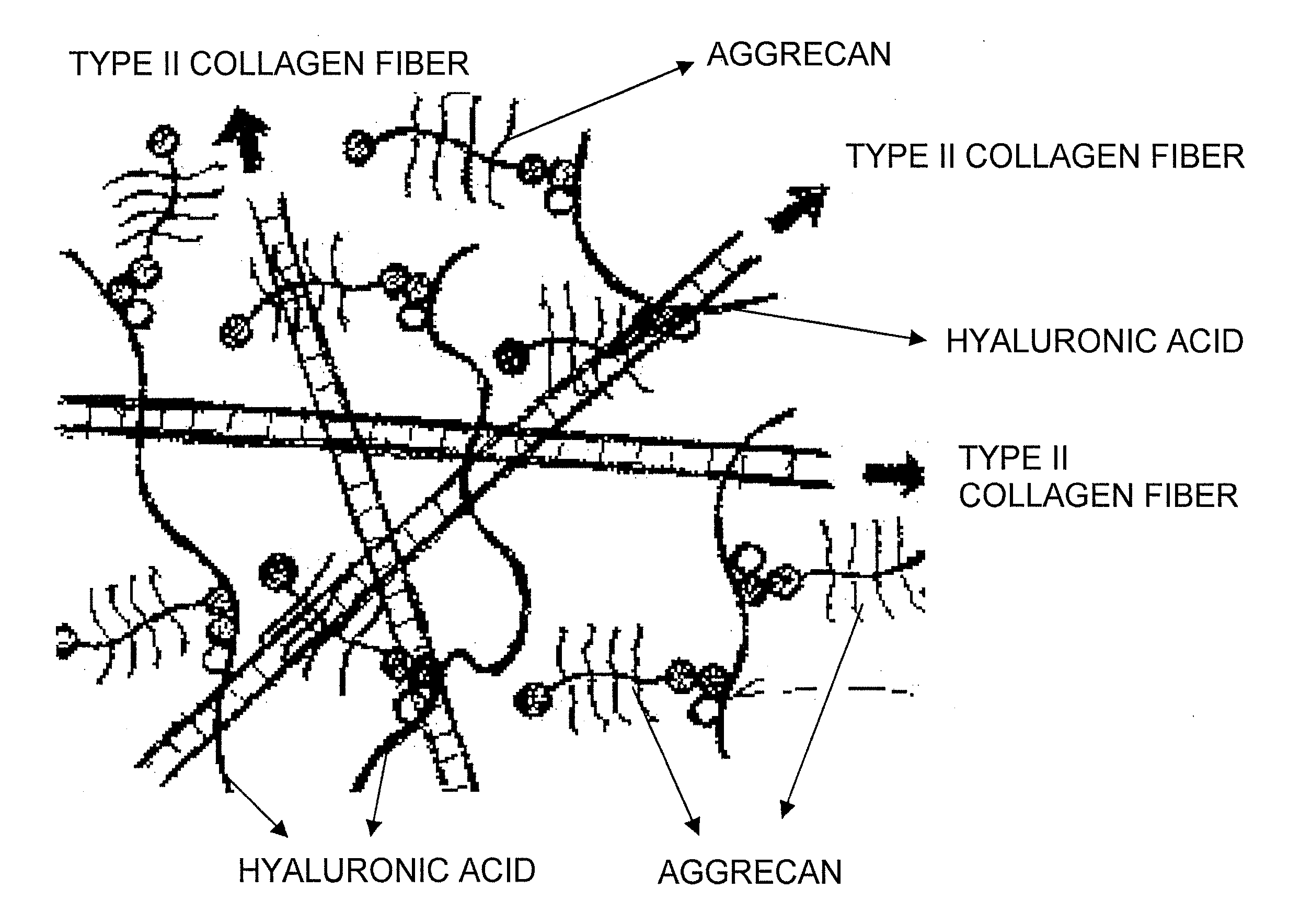
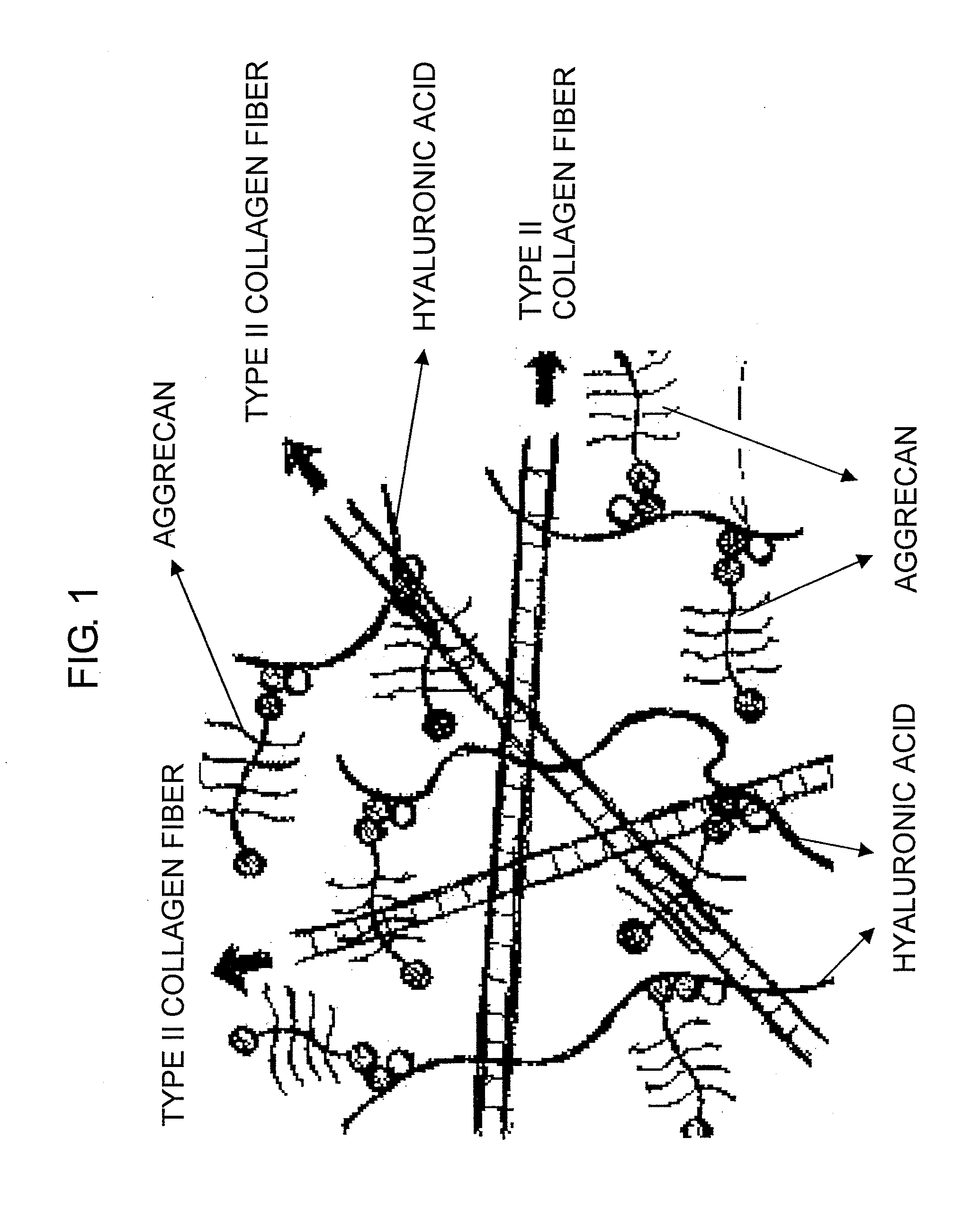
[0015]To solve the above problems, the present inventors have devoted themselves to research. The present inventors were seeking for a technique to create a cartilage-like tissue, which unlike conventional techniques, neither makes chondrocytes produce cartilage matrix to induce cartilage tissue formation, nor uses crosslinking agents and condensing agents. An attempt has been made to form cartilage-like tissues through application of self-organization techniques. Self-organization techniques use a phenomenon that, depending on environmental conditions, randomly moving molecules in a steady state may form a regularly-organized structure according to physical or chemical properties such as intermolecular bonding strength, surface modification, and orientation and ionic arrangement of covalent bonds. It is known that hydroxyapatite, collagen, hyaluronic acid, and chondroitin sulfate form a body through self-organization (Non-patent Document 3 and Patent Document 2). However, hydroxyapatite is a bone component that is intrinsically nonexistent in cartilage tissue. The formation of a cartilage-like tissue through the application of self-organization techniques has never been reported. The present inventors have examined the formation of a

Problems solved by technology

For patients, the efficacy of these symptomatic treatments is unsatisfactory.
However, surgical treatments have many issues such as cost and risk of infection, and furthermore, some patients are forced to replace their artificial joints several years to a decade later.
Preventive methods and early countermeasures are required for motor organ diseases because after disease onset, tissues become damaged with age, and articular cartilage tissues have extremely poor repairability.
However, neither effective treatments nor medical techniques have been established yet.
On the other hand, cartilage regeneration techniques have not completely reproduced cartilage-specific tissue properties (elastic deformation effect and elastohydrodynamic lubrication mechanism) in artificial cartilages.
To obtain such a large amount of cell source (number of chondrocytes) that is necessary and sufficient for the treatment, cells have to be passaged several times. However, when chondrocytes are plate-cultured like other types of cells, they lose chondrocyte-specific properties (decrease in cartilage matrix productivity and alteration in cell morphology) during passage culture, and may possibly dedifferentiate even though they show proliferation potency.
As such, when a large number of chondrocytes are required, the plate culture method has a lot of problems to solve in terms of cell source.
It requires as long as several weeks and the risk of dedifferentiation is unavoidable.
The three-dimensional culture method could provide a solution to the above issue of dedifferentiation, but similarly to the plate culture method, it require a large number of cells and a long period of time.
Besides the above time-related issue, the three-dimensional culture method is labour consuming.
However, the use of crosslinking agents and condensing agents requires washing to remove the crosslinking agents, condensing agents, and byproducts in the production process.
Moreover, transplantation of such product into the body poses a risk of residual chemical substance.
Further, since the complex prepared by using crosslinking agents and condensing agents cannot mimic a living tissue in the nano structure level, it is unclear as whether or not it can fulfil the required cartilaginous functions such as low frictionality, load resistance, and bioaffinity.
In this regard, no conventional tissue regeneration materials have been found to have sufficiently reproduced the structure and functions of a cartilage tissue.

Method used


Image

  • Biomaterials for regenerative medicine
  • Biomaterials for regenerative medicine
  • Biomaterials for regenerative medicine

Examples


example 1

Production of Hyaluronic Acid / Aggrecan / Type II Collagen Complex

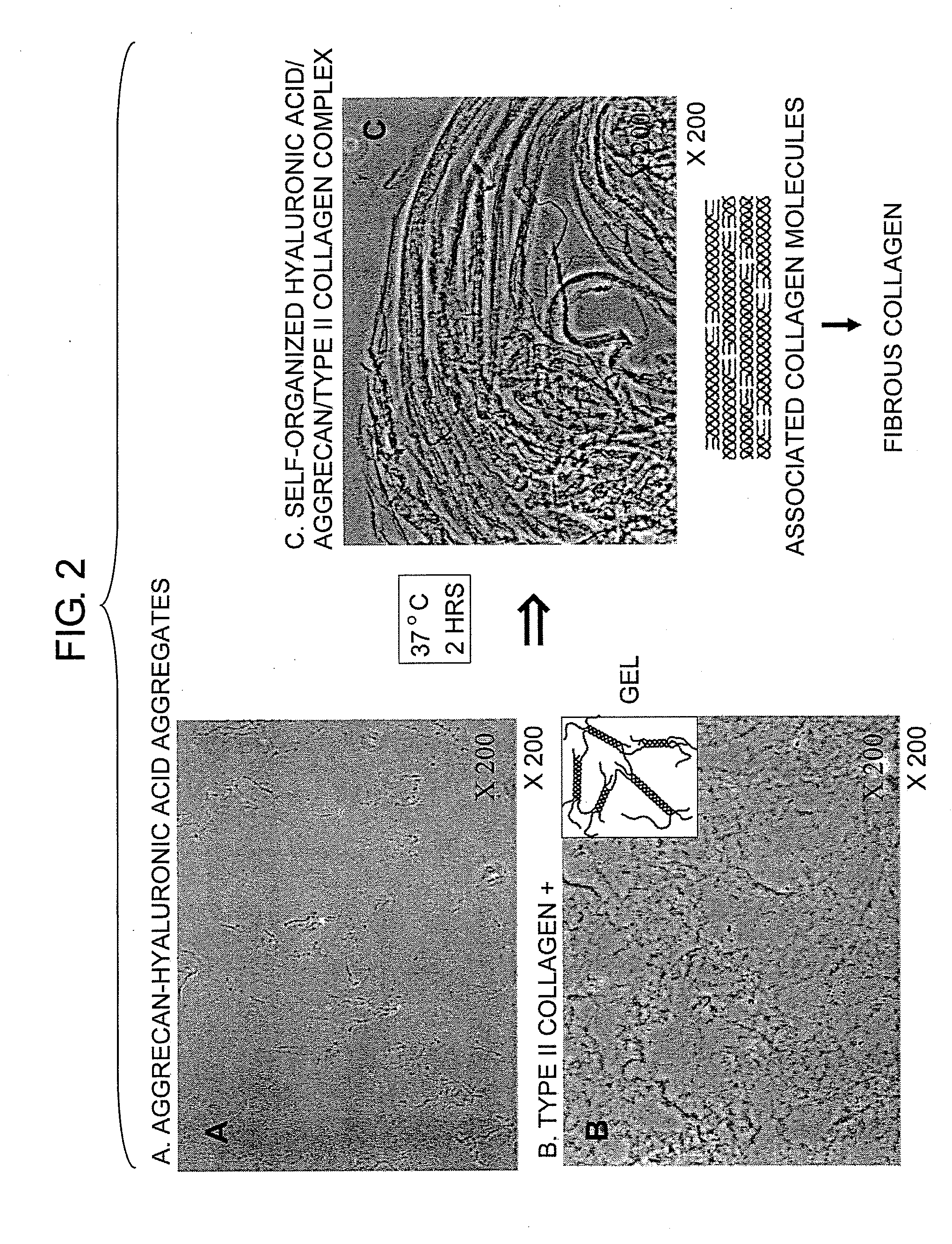
[0061]An attempt was made to produce a hyaluronic acid / aggrecan / type II collagen complex through self-organization techniques. For that purpose, the optimum conditions (concentration and pH) for hyaluronic acid, aggrecan, and collagen were examined.

[1-1] Materials and Methods

[0062]Aggrecan (hereinbelow, may be referred to as “AG”; manufactured by Sigma Co., USA) was dissolved in distilled deionized water (hereinbelow, referred to as “DDW”) as a solvent to prepare aggrecan solutions at various concentrations (the final concentration of aggrecan was 0.1, 0.25, 0.33, 0.5, or 1.0 mg / ml). Similarly, hyaluronic acid (hereinbelow, may be referred to as “HA”; manufactured by Chugai Pharmaceutical Co., Ltd., Japan; average molecular weight of 1,800,000) was dissolved in DDW to prepare hyaluronic acid solutions at various concentrations (final volume percent was 1, 2, 3, 4, or 5 volume percent). The AG solution and the HA solution...

example 2

Experiment of Cell Transfer into Self-Organized Complexes

[0072]The complex of the present invention desirably has high bioaffinity in view of its application to cartilage tissue regeneration. Specifically, it is important that an individual's chondrocytes can be engrafted when administered to a joint. Therefore, the affinity between cells and the complex of the present invention was examined by culturing chondrocytes using the complex of the present invention.

[2-1] Production of Complexes Comprising Cultured Chondrocytes

[0073]If blood serum is added to a cell culture solution for culturing chondrocytes, various factors in the blood serum would affect the formation and maintenance of the complexes and the engraftment of cells in the complexes, which may make it difficult for appropriate assessment. To avoid such effects, neutridomas were added to a serum-free medium DMEM at a final concentration of 10% to prepare a 10% neutridoma-containing DMEM solution. Rat and human chondrocytes w...

example 3

Transplantation of Self-Organized Complexes into Knee Joints of Experimental Animals

[0080]The self-organized complexes were produced using rat-derived type II collagen and aggrecan according to the method described in Example 1 above. Four 12-week-old male SD rats were anaesthetized with ether and then sterilized. Both of their knee joints were incised by aseptic techniques. The surface of articular cartilage of the medial and lateral (internal and external) condyles was pierced with an 18-gauge injection needle, to produce two articular cartilage defects per knee joint. The self-organized complexes were transplanted into one of these two articular cartilage defects (FIG. 10). The joint tissues were sutured, and the rats were grown for six weeks. In the sixth week, the mice were euthanized. The knee articular cartilage tissues were collected from them and fixed with 4% paraformaldehyde, followed by embedding in paraffin. The specimen was sliced and subjected to hematoxylin-eosin sta...


PUM

PropertyMeasurementUnit
Concentrationaaaaaaaaaa
Percent by volumeaaaaaaaaaa

Abstract

It was examined whether a cartilage-like tissue is formed under various reaction conditions using cartilage matrix components: glycosaminoglycan, proteoglycan, and collagen. The present inventors have discovered that proteoglycan bound to glycosaminoglycan through self-organization form an aggregate when the glycosaminoglycan was reacted with proteoglycan under specific concentrations and pH, and that a mesh structure composed of collagen fibers was constructed through self-organization using the aggregates as a skeleton when the aggregates were reacted with collagen molecules.

Description

CROSS-REFERENCES TO RELATED APPLICATIONS[0001]This application is a U.S. National Phase of PCT / JP2006 / 318188, filed Sep. 13, 2006, which claims priority to Japanese Patent Application No. 2005-271095, filed Sep. 16, 2005, the contents of which are herein incorporated by reference in their entirety.TECHNICAL FIELD[0002]The present invention relates to usable biomaterials for tissue regeneration, particularly to glycosaminoglycan / proteoglycan / collagen complexes formed through self-organization techniques.BACKGROUND ART[0003]With the arrival of an aging society, there is an increasing trend of patients with bone and joint diseases / motor organ diseases such as osteoporosis and osteoarthritis. In fact, the number of osteoarthritis patients in Japan is estimated to be seven to ten millions. Since bone and joint diseases / motor organ diseases pose challenges in daily life, development of countermeasures and preventive methods has called for urgent attention in the society. Most of the curre...

Claims


Application Information

Patent Timeline
IPC IPC(8): A61K35/12C07K1/107C07K14/78C12N5/00A61P19/00A61L27/00A61L27/20A61L27/22A61L27/24A61L27/38A61L27/44
CPCA61L27/26A61L27/34C08L89/06C08L5/02A61P19/00
Inventor YUDOH, KAZUO
Owner ST MARIANNA UNIV SCHOOL OF MEDICINE
Learn More