
Development of sensitive fret sensors and methods of using the same

a fret sensor and sensitive technology, applied in the field of biosensors, can solve the problems of poor spatial resolution, no currently available technology, no cellular resolution, etc., and achieve the effect of increasing rigidity

Inactive Publication Date: 2010-02-11
CARNEGIE INSTITUTION OF WASHINGTON
View PDF14 Cites 29 Cited by
  • Summary
  • Abstract
  • Description
  • Claims
  • Application Information

AI Technical Summary

Benefits of technology

[0020]The present invention therefore provides improved intramolecular biosensors and nanosensors for detecting and measuring changes in analyte concentrations, particularly transporter biosensors and biosensors constructed using bacterial periplasmic binding proteins (PBPs). In particular, the invention provides intramolecular biosensors containing at least one internally fused fluorophore moiety, as well as FRET fusion constructs encoding fluorophores with increased rigidity.

Problems solved by technology

No currently available technology addresses all these issues in a satisfactory manner.
Non-aqueous fractionation is static, invasive, has no cellular resolution and is sensitive to artifacts, while spectroscopic methods such as NMRi (nuclear magnetic resonance imaging) and PET (positron emission tomography) provide dynamic data, but poor spatial resolution.
This form of FRET is difficult to quantitate because the stoichiometry of acceptors to donors can vary with transfection efficiencies and expression levels.

Method used


Image

  • Development of sensitive fret sensors and methods of using the same
  • Development of sensitive fret sensors and methods of using the same
  • Development of sensitive fret sensors and methods of using the same

Examples


example 1

Construction of Nucleic Acids and Vectors

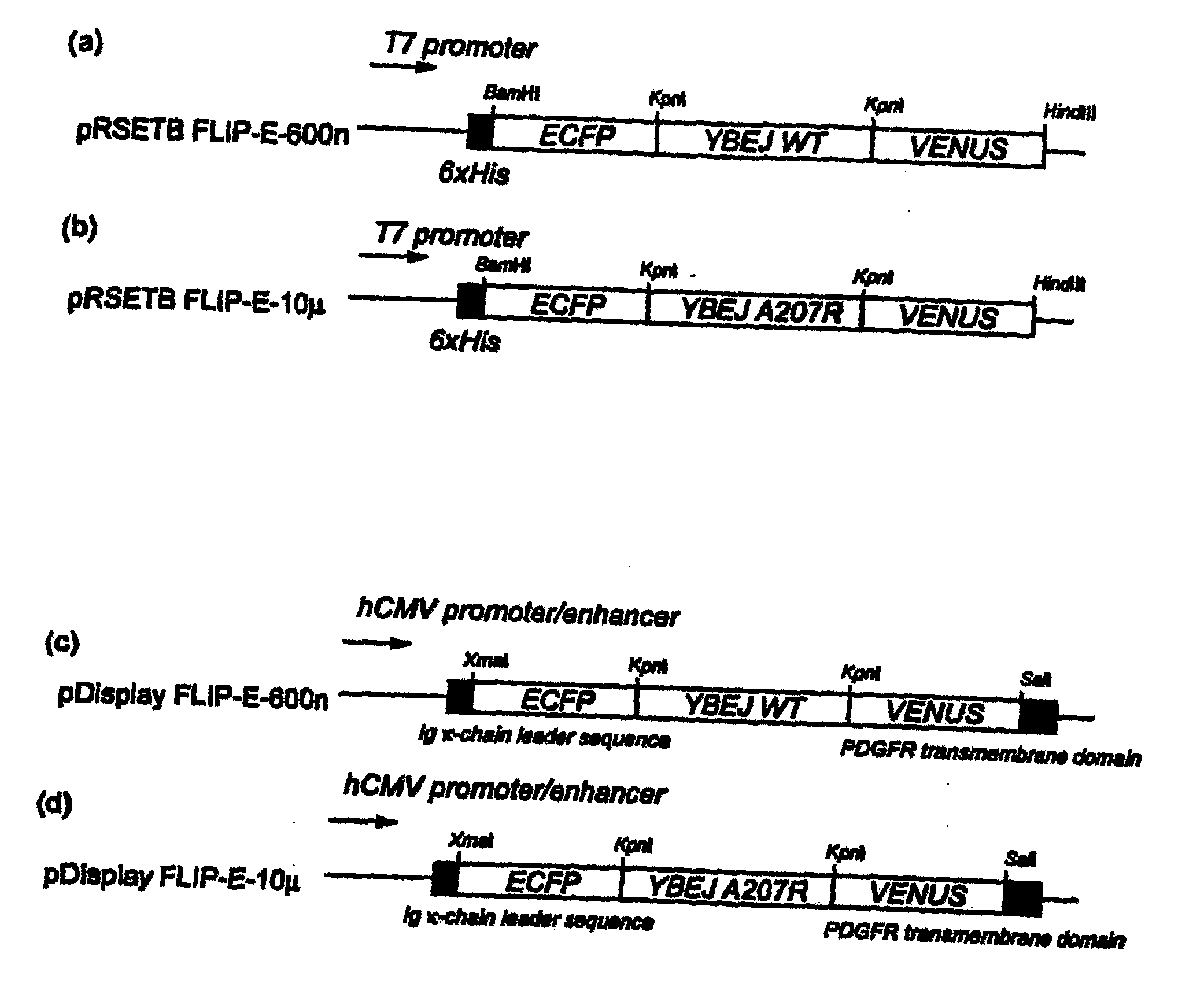
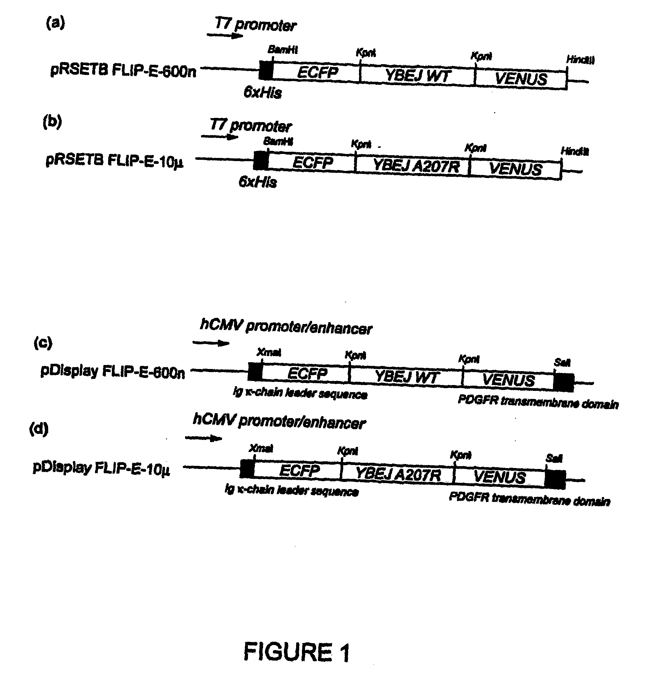
[0085]A truncated glutamate-aspartate binding protein sequence (SEQ ID No. 4), encoding mature protein without signal peptide, was amplified by PCR using E. coli K12 genomic DNA as a template. The primers used were 5′-ggtaccggaggcgccgcaggcagcacgctggacaaaatc-3′ (SEQ ID No. 5) and 5′-accggtaccggcgccgttcagtgccttgtcattcggttc-3′ (SEQ ID No. 6). The PCR fragment was cloned into the KpnI site of digested FLIPma1-25μ (Fehr et al. 2002) in pRSET vector (Invitrogen), exchanging the maltose binding protein sequence with the YbeJ sequence. The resulting plasmid was named pRSET-FLIP-E-600n (SEQ ID NO: 9).

[0086]To improve the pH and chloride tolerance and maturation of the sensor protein, the fragment containing the enhanced YFP (EYFP, CLONTECH) sequence in pRSET-FLIP-E-600n was replaced with the coding sequence of Venus, a variant of YFP with improved pH tolerance and maturation time (Nagai, T., Ibata, K., Park, E. S., Kubota, M., Mikoshiba, K., and Miyaw...

example 2

In Vitro Characterization of FLIP-E Nanosensors

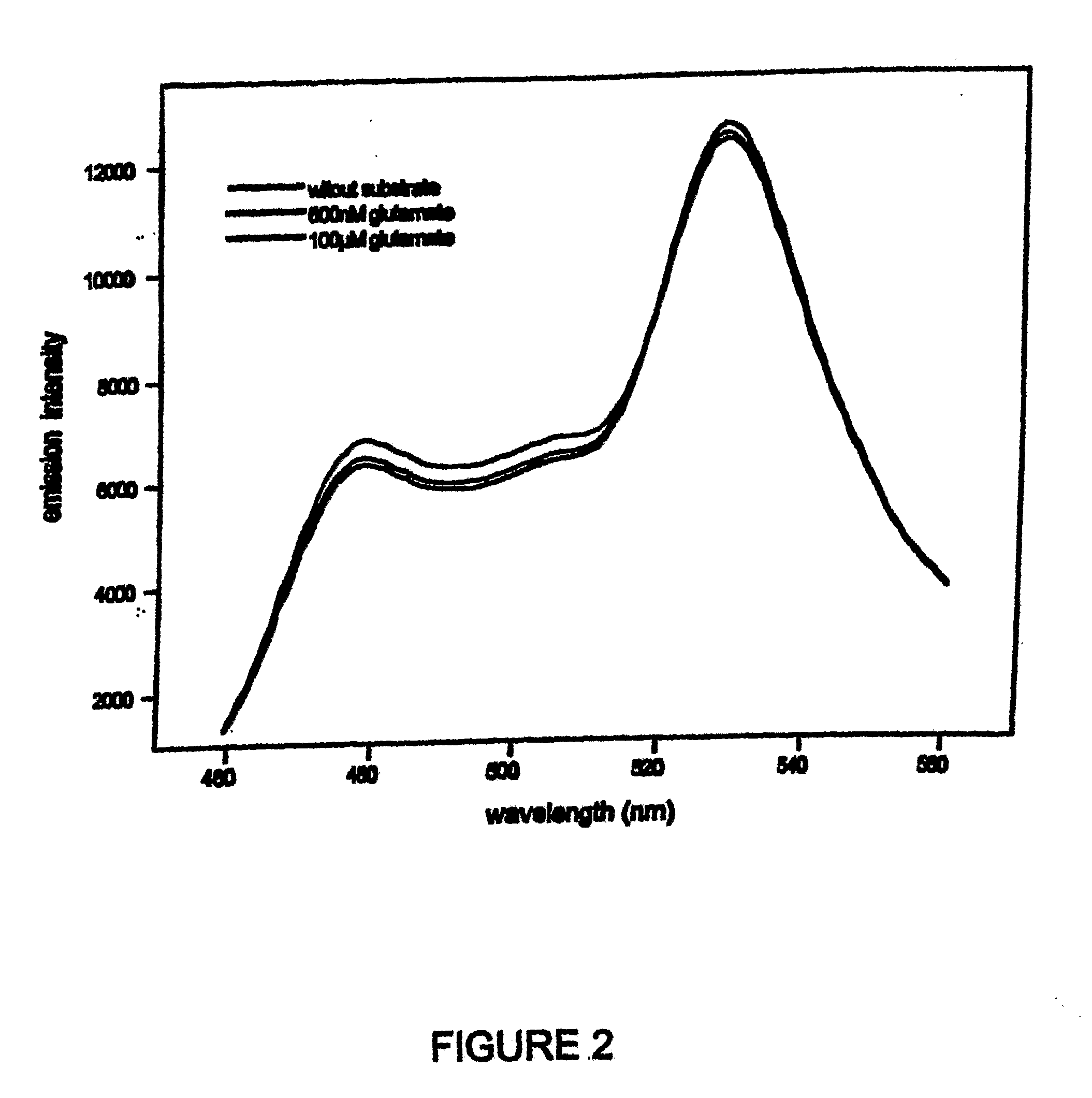
[0088]A DNA fragment encoding the mature YBEJ protein was fused to ECFP and the Venus sequence at the N- and C-termini, respectively (FIG. 1). Emission spectra and substrate titration curves were obtained by using monochromator microplate reader Safire (Tecan, Austria). Excitation filter was 433±12 nm, emission filters for CFP and YFP emission were 485±12, 528±12 nm, respectively. All analyses were done in 20 mM sodium phosphate buffer, pH 7.0.

[0089]Addition of glutamate resulted in an increase in CFP emission and a decrease in YFP emission, suggesting that binding of glutamate to YBEJ results in a conformational change of the chimeric protein potentially due to a relative change in the orientation of the dipoles of the fluorophores (FIG. 2). Since CFP and YFP moieties are assumed to be attached to the same lobe, we speculate that glutamate binding causes the change in dipole-dipole angle of two fluorophores. Interestingly, the ratio an...

example 3

In Vivo Characterization of FLIP-E

[0091]For the in vivo characterization of FLIP-E nanosensors, FLIPE-600n and FLIPE-10μ were cloned into the mammalian expression vector pDisplay (Invitrogen, USA). The pDisplay vector carries a leader sequence which directs the protein to the secretory pathway, and the transmembrane domain which anchors the protein to the plasma membrane, displaying the protein on the extracellular face. Rat hippocampal cells and PC12 cells were transfected with pDisplay FLIPE-600n (SEQ ID NO: 11) and −10μ (SEQ ID NO: 12) constructs. FRET was imaged 24-48 hours after transfection on a fluorescent microscope (DM IRE2, Leica) with a cooled CoolSnap HQ digital camera (Photometrics). Dual emission intensity ratios were simultaneously recorded following excitation at 436 nm and splitting CFP and Venus emission by DualView with the OI-5-EM filter set (Optical Insights) and Metafluor 6.1r1 software (Universal Imaging).

[0092]The expression of FLIP-E was observed on the plas...


PUM

PropertyMeasurementUnit
pHaaaaaaaaaa
pHaaaaaaaaaa
pHaaaaaaaaaa

Abstract

Intramolecular biosensors are disclosed, including PBP-based biosensors, comprising a ligand binding domain fused to donor and fluorescent moieties that permit detection and measurement of Fluorescence Resonance Energy Transfer upon binding ligand. At least one of the donor and fluorescent moieties may be internally fused to the biosensor such that both ends of the internally fused fluorophore are fixed. In addition, methods of improving the sensitivity of terminally fused biosensors are provided. The biosensors of the invention are useful for the detection and quantification of ligands in vivo and in culture.

Description

CROSS REFERENCE TO RELATED APPLICATIONS[0001]This application claims benefit of priority to U.S. Provisional application 60 / 618,179, filed Oct. 14, 2004, U.S. Provisional application 60 / 643,576, filed Jan. 14, 2005, U.S. Provisional application 60 / 654,447, filed Feb. 22, 2005, and U.S. Provisional application 60 / 658,141, filed Mar. 4, 2005, which are herein incorporated by reference in their entireties.[0002]This application is also related to provisional application Ser. No. 60 / 658,142, provisional application Ser. No. 60 / 657,702, PCT application [Attorney Docket No. 056100-5053, “Phosphate Biosensors and Methods of Using the Same”], PCT application [Attorney Docket No. 056100-5054, “Methods of Reducing Repeat-Induced Silencing of Transgene Expression and Improved Fluorescent Biosensors”], and PCT application [Attorney Docket No. 056100-5055, “Sucrose Biosensors and Methods of Using the Same”], which are herein incorporated by reference in their entireties.STATEMENT REGARDING FEDER...

Claims


Application Information

Patent Timeline
Patent Type & Authority Applications(United States)
IPC IPC(8): A01K67/00C07H21/02C12N15/74C12N5/07C12N1/21C12N1/19G01N33/566G01N33/53C07K14/00
CPCC12Q1/6818G01N33/542G01N2800/2814G01N33/566G01N2800/2857G01N2800/2871G01N2800/2835C07K14/245
Inventor FROMMER, WOLF B.OKUMOTO, SAKIKOLOOGER, LORENFEHR, MARCUS
Owner CARNEGIE INSTITUTION OF WASHINGTON
Learn More