Endoscopic biopsy apparatus, system and method

a biopsy apparatus and endoscope technology, applied in the field of endoscope biopsy systems, can solve the problems of poor diagnostic yield, uncertain patient management, and early cancer that cannot be visually identified, and achieve the effects of improving the diagnostic capability of surveillance endoscope, reducing mortality, and being safe and well tolerated

Active Publication Date: 2010-08-19
THE GENERAL HOSPITAL CORP
View PDF105 Cites 52 Cited by
  • Summary
  • Abstract
  • Description
  • Claims
  • Application Information

AI Technical Summary

Benefits of technology

[0035]According to the exemplary embodiments of the present disclosure, it is possible to screening patients for Barrett's esophagus and improving the diagnostic capabilities of surveillance endoscopy. These advances can decrease the mortality associated with esophageal adenocarcinoma.
[0036]The image-guided biopsy according to the exemplary embodiments of the present disclosure is expected to be safe and well-tolerable, detect previously unattainable subcellular and architectural information over large epithelial surfaces of the esophagus, and provide an effective method for endoscopic biopsy targeting. The impact of these exemplary embodiments c

Problems solved by technology

For a number of cases, however, metaplasia, dysplasia, and early cancer may not be visually identified.
When the disease is focal or heterogeneously distributed within a much larger suspect area, a random biopsy procedure may be analogous to “finding a needle in a haystack,” resulting in poor diagnostic yields and uncertain patient management.
Since random biopsies may only facilitate the assessment of less than 0.1% of the potentially involved tissue, these procedures are usually fraught with significant sampling error and diagnostic uncertainty.
Other tasks, such as the delineation of surgical tumor margins, can also be affected by this difficulty, resulting in all too frequent re-excisions or time-consuming frozen section analysis.
This method suffers from a low sensitivity, as it is compromised by the poor ability of endoscopists to identify SIM/dysplasia and the low fractional area of tissue sampled by biopsy.
While many of these techniques are promising, the information provided is often quite different from that conventionally used in medicine for diagnosis.
As a result, such techni

Method used


Image

  • Endoscopic biopsy apparatus, system and method
  • Endoscopic biopsy apparatus, system and method
  • Endoscopic biopsy apparatus, system and method

Examples


Embodiment Construction

[0022]Thus, at least some of these issues and / or deficiencies can be addressed with the exemplary embodiments of the apparatus, system and method according to the present disclosure.

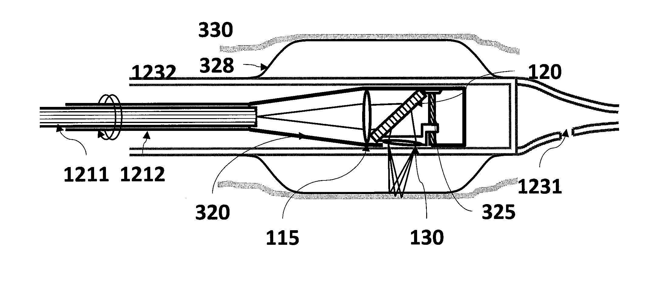
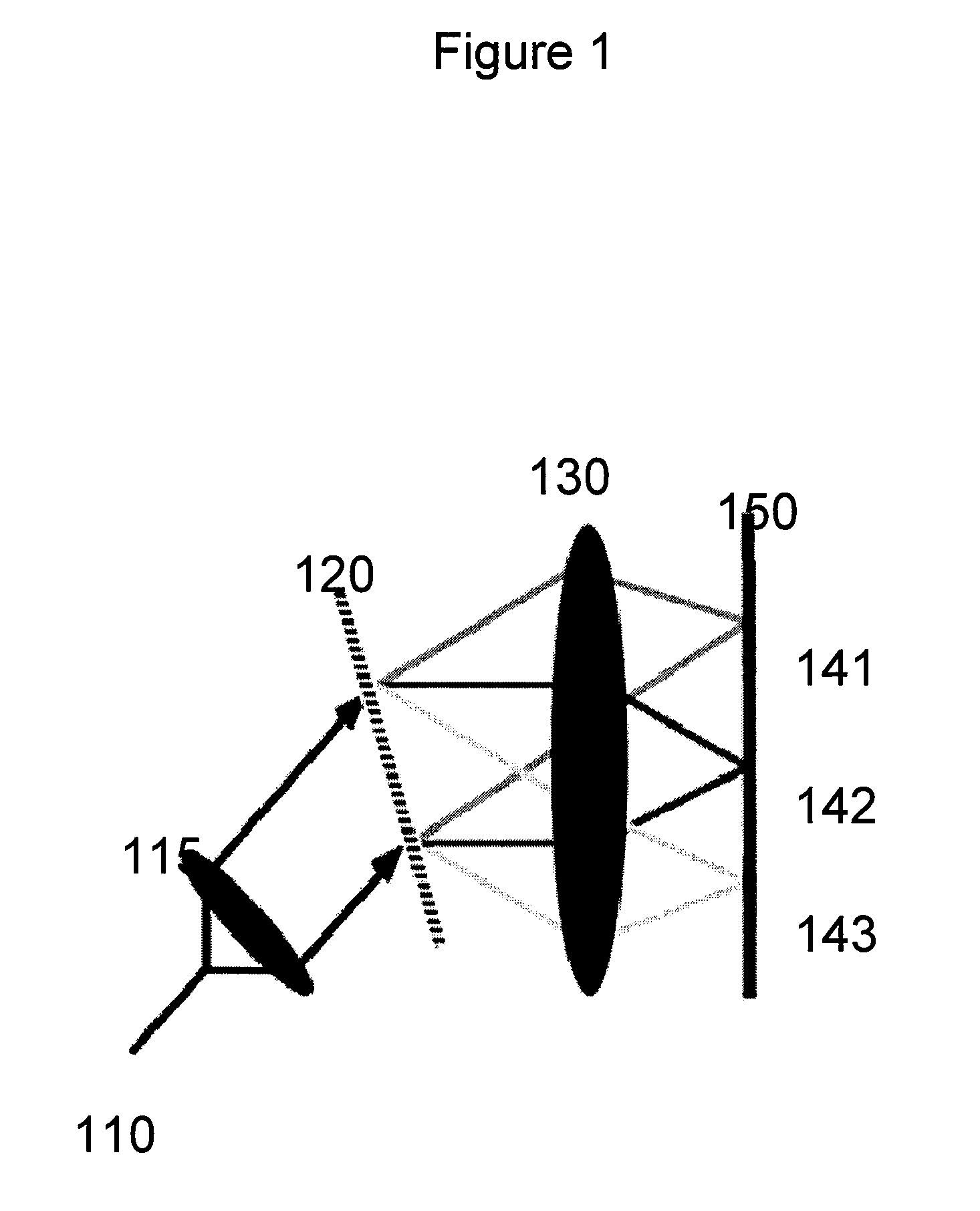
[0023]Exemplary embodiments of the present disclosure provides mechanism and a methodology for automatically maintaining the foci at a desired tissue depth while scanning the spectrally encoded line across the sample. This exemplary advancement can compensate for patient motion and enables imaging at multiple depth locations. Further, in one exemplary embodiment, it is possible to conduct a large area confocal microscopy in patients by incorporating these technologies in an endoscopic probe suitable for human use.

[0024]According to another exemplary embodiment of the present disclosure, an apparatus can be provided. The apparatus can comprise at least one dispersive first arrangement which is configured to provide data associated with a signal received from at least one region of the sample(s). The exemp...


PUM


Abstract

Exemplary embodiments of apparatus, method and system for determining a position on or in a biological tissue can be provided. For example, using such exemplary embodiment, it is possible to control the focus of an optical imaging probe. In another exemplary embodiment, it is possible to implement a marking apparatus together with or into an optical imaging probe. According to one exemplary embodiment, it is possible (using one or more arrangements) to receive information associated with at least one image of at least one portion of the biological tissue obtained using an optical imaging technique. Further, it is possible to, based on the information, cause a visible change on or in at least location of the portion(s) using at least one electro-magnetic radiation.

Description

CROSS-REFERENCE TO RELATED APPLICATION(S)[0001]This application is based upon and claims the benefit of priority from U.S. Patent Application Ser. No. 61 / 145,914, filed on Jan. 20, 2009, and from U.S. Patent Application Ser. No. 61 / 184,180, filed on Jun. 4, 2009, the entire disclosures of which are incorporated herein by reference.FIELD OF THE DISCLOSURE[0002]The present disclosure relates to exemplary embodiments of endoscopic biopsy systems that are guided by microscopic image information, and an associated method therefor.BACKGROUND INFORMATION[0003]The standard of care for the diagnosis of many epithelial precancerous and early cancer conditions is visual inspection of the patient directly or through an endoscope / laparoscope to identify abnormal tissue. Biopsies can then be obtained from these locations, processed, cut and stained with Hematoxylin and Eosin (H&E), and then observed under a microscope by a pathologist. A pathologist can view the slide at progressively increasing ...

Claims


Application Information

Patent Timeline
IPC IPC(8): A61B6/00
CPCA61B5/0066A61B5/0086A61B5/0075A61B5/0068
Inventor TEARNEY, GUILLERMO J.BOUMA, BRETT E.KANG, DONGKYUNSUTER, MELISSA J.
Owner THE GENERAL HOSPITAL CORP
Why Eureka
Try Eureka