Low Level Fluorescence Detection at the Light Microscopic Level

a fluorescence microscopy and light microscopic technology, applied in the field of light microscopic fluorescence detection, can solve the problems of lack of sensitivity of color development, lack of ability to finely discriminate subcellular localization, and limitations of each cellular target detection, so as to reduce unwanted fluorescence

Inactive Publication Date: 2010-08-26
THE TRUSTEES OF THE UNIV OF PENNSYLVANIA
View PDF7 Cites 16 Cited by
  • Summary
  • Abstract
  • Description
  • Claims
  • Application Information

AI Technical Summary

Problems solved by technology

Although both methods are highly sensitive, they each have limitations in detecting cellular target.
For example, color development (e.g. alkaline phosphates) is an excellent approach to detect low abundance targets, but it lacks the ability to finely discriminate subcellular localization.
In comparison, fluorescence microscopy lacks the sensitivity of color development predominantly because of photobleaching of target secondary fluorophores or endogenous autofluorescence of biological samples.
Unfortunately, there are several shortcomings associated with using fluorophores as reporter molecules.
Most difficulties with the technique result from the extremely limited absorptive and emissive capabilities of organic dyes.
Therefore, applications that make use of light frequencies that do not correspond to the emission peaks of preexisting organic dyes cannot be performed.
In addition, organic fluorescent dyes have a narrow absorption pattern and not always in convenient regions of the spectrum, making the excitation of various organic dyes challenging and costly.
Organic fluorescent dyes also exhibit uneven absorption and emission peaks and tend to produce ‘shoulders’ in the geometry of their emission and absorption peaks, a major disadvantage in applications that require Gaussian type emission patterns to work correctly.
One of the most problematic aspects of organic fluorescent dyes is that of stability.
In addition, all fluorescent dyes bleach over time upon observation.
Photobleaching is especially problematic with confocal microscopy due to the high intensity of the laser illumination.
When using fluorescent probes in biological tissue, a primary problem is minimizing fluorescence noise in order to maximize signal detection.
The first issue of detection is simply one of sensitivity as a result of the relative abundance of the target molecule.
Target molecules present in abundance are usually readily detectable; however molecules present at small numbers in the tissue can limit the usefulness of the technique.
Second, detection of fluorescent signal is hampered by autofluorescence.
Third, detection of a specific signal can be impeded by background fluorescence, a result of non-specific binding of an exogenously applied fluorescent probe to a tissue sample.
Similar problems are encountered when labelling DNA with fluorescent tags.
Specifically, there are two main drawbacks of the use of DNA staining agents.
The second drawback is that the presence of these dyes results in changes in the electrostatic, structural and mechanical properties of DNA which are likely to modify its interaction with proteins.
These limits constrain the use of this labeling method for DNA-protein interaction studies.
Their broad absorption spectra but very narrow emission spectra allows multiplexing of many SCN of different colors in the same sample, something that cannot be achieved with traditional fluorophores.
Although both methods are highly-sensitive, they each have limitations in detecting cellular targets.
For example, color development (eg. alkaline phosphatase) is an excellent approach to detect low abundance targets, but lacks the ability to finely discriminate subcellular localization.
In comparison, fluorescence microscopy lacks the sensitivity of color development predominantly due to photobleaching of target secondary fluorophores or endogenous autofluorescence of biological samples.

Method used


Image

  • Low Level Fluorescence Detection at the Light Microscopic Level
  • Low Level Fluorescence Detection at the Light Microscopic Level
  • Low Level Fluorescence Detection at the Light Microscopic Level

Examples


experimental examples

[0161]The invention is further described in detail by reference to the following experimental examples. These examples are provided for purposes of illustration only, and are not intended to be limiting unless otherwise specified. Thus, the invention should in no way be construed as being limited to the following examples, but rather, should be construed to encompass any and all variations which become evident as a result of the teaching provided herein.

[0162]The materials and methods employed in the experiments disclosed herein are now described.

Hippocampal Cultures

[0163]Primary cultures of hippocampal neurons from E19 rat embryos were plated on glass coverslips at 100,000 per ml in Neuralbasal media with B27 supplements (Sigma). Hippocampal neurons were dissociated in L-15 media with collagenase (20 mg / ml, Sigma) and dispase (96 mg / ml, Sigma). Enzymatic digestion was carried out at 37° C. for ˜45 min and cells triturated periodically with a fire polished pipette to facilitate diss...

example 1

Signal Intensity of Alexa 488 and Qdot-565 Before and After Photobleaching

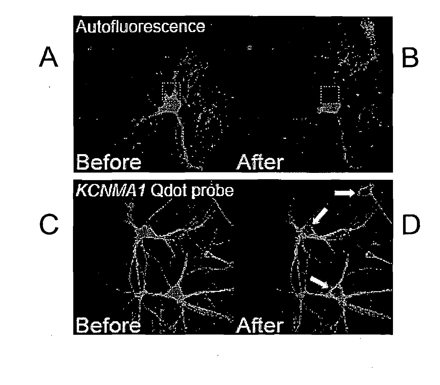
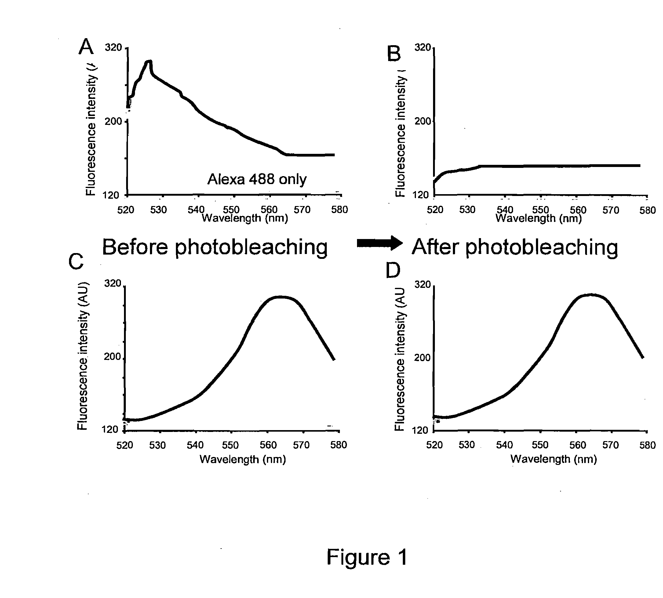
[0170]Primary rat hippocampal neurons were prepared for immunohistochemistry. Anti-digoxigenin Fab garments conjugated to Qdot 565 (Invitrogen) were used at 1:250. AlexaFluor 488 phalloidin (Molecular Probes) was used at 1:40. Images were taken with an Olympus Fluoview 1000 confocal scan head. For each cell, five randomly placed line scans were taken from three separate regions of interest and analyzed with the Metamorph image processing software. In FIG. 1, the emission spectral signature over the range from 520 to 580 nm wavelength was obtained before and after full spectral photobleaching of a sample for two seconds. In a sample stained only for Alexa 488, the photobleaching procedure abolishes Alexa 488 signal (FIG. 1B). In a second sample stained with Qdot-565, the identical full-spectrum photobleaching protocol was used, however the Qdot-565 signal was not affected (FIG. 1C and FIG. 1D).

example 2

Selective Elimination of Unwanted Fluorescent Signals in a Single Sample Using Photobleaching

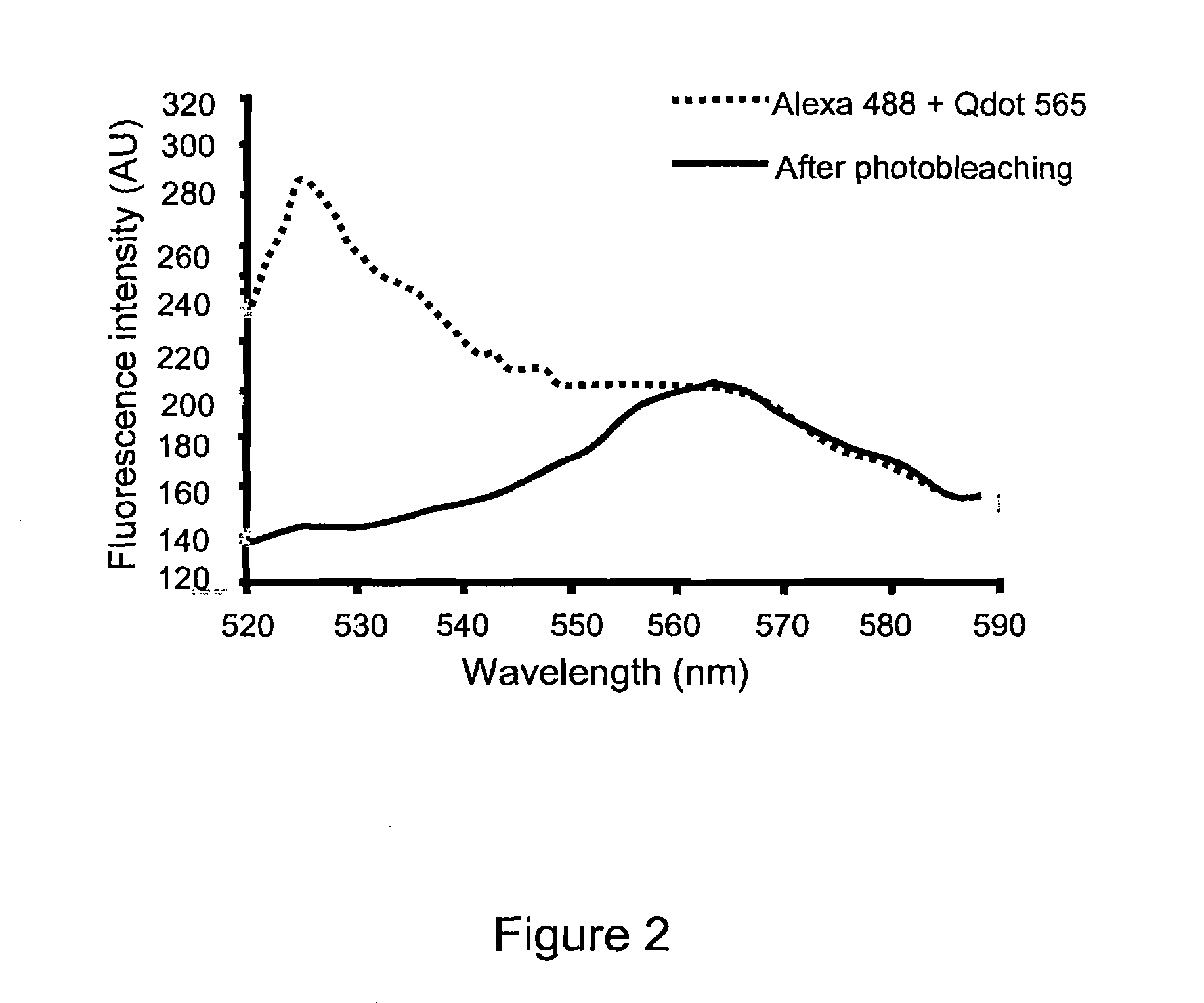
[0171]In order to confirm the specificity of the photobleaching technique and establish the utility of the present invention for use with multiple probes in a single sample, Qdot-565 and Alexa 488 were applied to a single sample at concentration of 1:250 and 1:40 respectively. The sample was photobleached using a full spectral scan for two seconds. The sample was then spectrally scanned and fluorescent intensity measured before and after photobleaching. Before photobleaching. The solid line shows the emission spectrum resulting from 458 nm excitation. The dotted line shows the remaining emission spectrum following photobleaching. These date clearly demonstrate the elimination of unwanted fluorescence signal of specific wavelength with sparing of the Qdot signal.


PUM

PropertyMeasurementUnit
characteristic wavelengthaaaaaaaaaa

Abstract

The present invention discloses methods of removing unwanted fluorescence from a sample by photobleaching said sample to enhance detection of proteins and fragments thereof, polynucleotides and fragments thereof, and biomolecules and fragments thereof in a sample by contacting said proteins, polynucleotides and biomolecules with a fluorescent reporter, wherein said fluorescent reported comprises a fluorescent semiconductor crystal or SCN, wherein said SCN further comprises a targeting moiety.

Description

STATEMENT REGARDING FEDERALLY SPONSORED RESEARCH OR DEVELOPMENT[0001]This invention was made, in part, using funds obtained from the U.S. Government (National Institutes of Health Grant No. AG9900), and the U.S. Government may therefore have certain rights in this invention.BACKGROUND OF THE INVENTION[0002]Many types of biological and industrial research rely on the ability to mark or label microscopic structures (e.g. cells, subcellular organelles) in order to track their movement, differentiation, or mark the association of a variety of components within an organism or other medium. With advances in microscopy technique, both fluorescence and chromogenic methods have become widely used though fluorescence is often the end detection point for the majority of biological measurements made in diverse disciplines—including DNA sequencing, microarray chips, neuroanatomical tracing studies, immunohistochemistry, ELISAs, and functional cellular assays such as Ca2+ imaging and voltage sens...

Claims


Application Information

Patent Timeline
Patent Type & Authority Applications(United States)
IPC IPC(8): C40B30/00C12Q1/68C12Q1/02G01N33/53G01N33/68G01N33/48G01N21/64
CPCB82Y15/00Y10T436/143333G01N33/588C12Q1/6825
Inventor EBERWINE, JAMES H.HAYDON, PHILIPSUL, JAI-YOONMIYASHIRO, KEVINBELL, THOMAS
Owner THE TRUSTEES OF THE UNIV OF PENNSYLVANIA
Why Eureka
Try Eureka