
Pcr-free sample preparation and detection systems for high speed biologic analysis and identification

Inactive Publication Date: 2010-08-26
FLUIDIGM CORP
View PDF99 Cites 16 Cited by
  • Summary
  • Abstract
  • Description
  • Claims
  • Application Information

AI Technical Summary

Benefits of technology

[0009]The present invention also provides methods of identifying one or more target biologic organisms, comprising: lysing one or more cells or spores from one or more biologic organisms in a thermal lysing chamber to give rise to a lysate, said lysing giving rise to nucleic acids from said one or more biologic organisms; filtering said nucleic acids from said lysate; transporting said nucleic acids to a porous linear microarray, said porous linear microarray comprises a plurality of spatially positioned probes capable of hybridizing at least a portion of the nucleic acids from each of said target biologic organism; hybridizing at least one nucleic acid from at least one of the target biologic organism to at least one of the probes; exciting the probes hybridized to at least one nucleic acid from at least one of the target biologic organism; detecting the spatial position of at least one of the excited probes on the linear microarray; and correlating the spatial position of the excited probe on the linear microarray to the identity of at least one of the cells or spores. In some embodiments, the linear microfluidic microarray comprises at least two distinct probes which are spatially resolved for detecting target analytes. In other embodiments, the linear microarray comprises a porous media that decreases diffusion time of the nucleic acids from the one or more target biologic organisms to hybridize with the probes. Also, a plurality of spatially positioned detection probes to test multiple analytes in a sample.

Problems solved by technology

Significant advancement of detection and identification of biological agents has been developed for centralized laboratory analysis; however, there currently exists few, if any, technologies that are cost effective for use in harsh environments.
The most commonly used tools for detection of biological agents in emergency responder situations are limited to assays, or devices, that offer only crude sample characteristics, such as pH or protein existence.
Unfortunately, each of these tasks currently require significant time and expense to complete.
While devices have been developed for field portable detection, many of these devices continue to rely on complex biochemistry systems (i.e., PCR) that lack the stability for use in harsh, point-of-incident, environments.

Method used


Image

  • Pcr-free sample preparation and detection systems for high speed biologic analysis and identification
  • Pcr-free sample preparation and detection systems for high speed biologic analysis and identification
  • Pcr-free sample preparation and detection systems for high speed biologic analysis and identification

Examples


example i

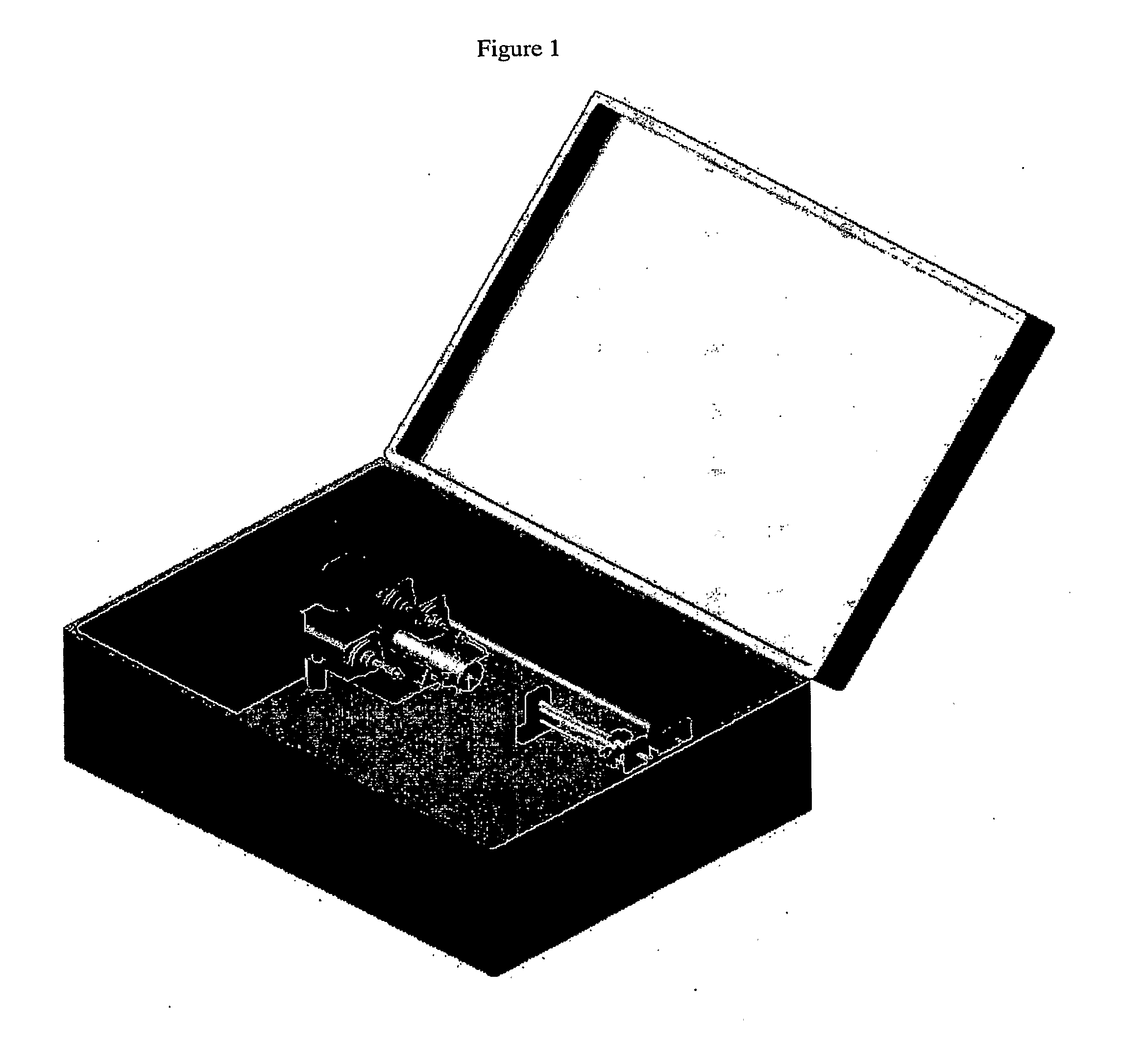
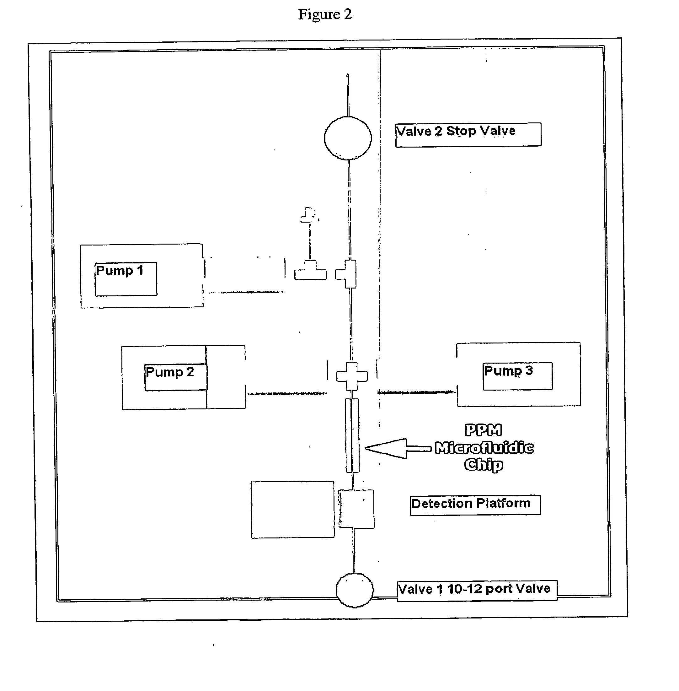
[0097]Examples for the use of sample preparation and analysis systems of the present invention are shown in the figures. An integrated detection apparatus can be used with a variety of sample matrices to perform fully automated sample preparation, hybridization and detection of sample analytes. Hybridization is accomplished using a linear porous microarray system, which dramatically reduces the diffusion distance and time to accomplish instantaneous capture and detection of the sample analyte. Detection of analytes is performed using an integrated fluorescence detection platform in combination with the use of nucleic acid and protein detection probes, including but not limited to the use of Tentacle probes as described in “Cooperative Probes and Methods of Using Them”, PCT / US07 / 63229, filed Mar. 2, 2007, the entirety of which is incorporated by reference herein. Other capture probes can be added to create higher order tentacle probes. All of these detection probes can also be substi...

example ii

Sample Preparation

Cell Suspension

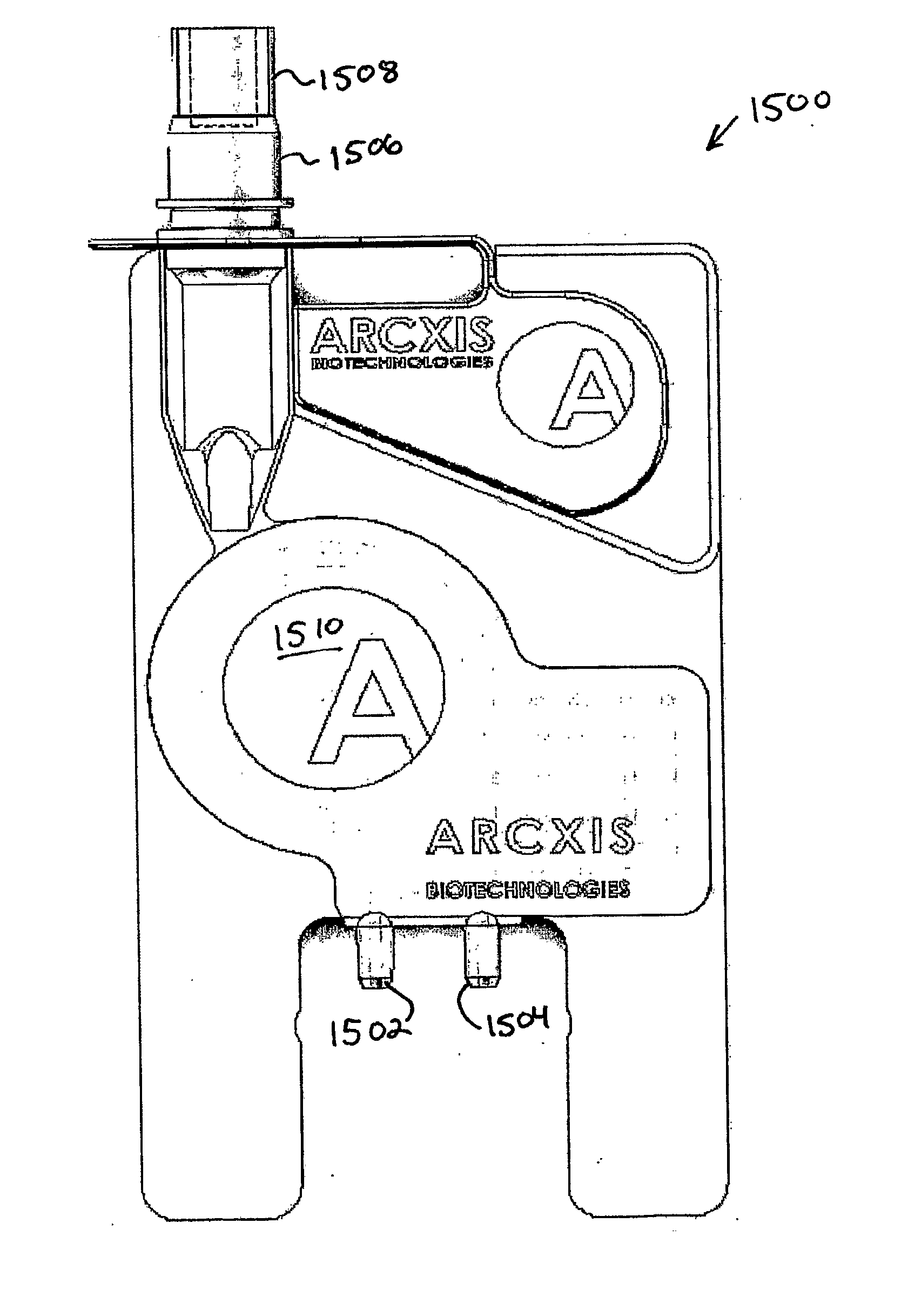
[0098]Using the sample preparation and analysis system, the time required for sample preparation is dramatically reduced. A variety of sample Matrices can be applied to the sample preparation and analysis system platform using making use of the disposable card apparatus. One such example is the use of cells in suspension. A variety of cell suspension types can be prepared for analysis using the sample preparation and analysis system card. These include, bacteria cells, virus, eukaryotic cells, plant cells. In one such example hepatocytes is suspension are centrifuged to pellet the intact cells. One pelleted the cells are resuspended in buffer containing a detergent (Triton-X100), a salt (NaPO4, TMAC, NaCL) in buffered water (pH 7.4). Once in the lysis buffer the cell suspension is deposited in the sample vial located a the top of the consumable card (i.e., disposable card apparatus) as illustrated in FIGS. 15 A-C, which depict front, exploded and bac...

example iii

Sample Preparation

Tissue Biopsy

[0099]Another example is the use of tissue biopsies. A variety of tissue biopsy types can be prepared for analysis using the sample preparation and analysis system card. In one such example a sample containing biopsy liver cells are prepared and analyzed. The biopsy liver cells are resuspended in buffer containing a detergent (Triton-X100) and a salt (NaPO4, TMAC, NaCL) in buffered water (pH 7.4). Once in the lysis buffer the tissue suspension is homogenized using a Polytron homogenizer. The homogenized sample is then deposited in the sample vial located at the top of a consumable card, such as the one depicted in FIGS. 15 A-C. The vial is then capped, a the card is inserted into to the sample preparation and analysis system instrument. Once inserted in to the instrument, an automated script actuates several pumps to transport the sample in to the lysis chamber, where the sample is heated to a temperature range between 50-110° C. During this process th...


PUM


Abstract

Provided herein are biologic sample preparation and analysis systems that are rapid, portable, robust detection system for multiplexed detection of bio-threats, and which can be ruggedized to operate in harsh environments. A new method of detection called Combinatorial Probe Analysis (CPA), which provides an exponential increase in detection reliability, has been incorporated into these systems. This type of analysis greatly reduces false positives and false negatives; in addition it is reusable and eliminates special storage requirements for reagents. Specific technical advancements in the optimization of hybridization assays for nucleic acid detection on porous polymer monoliths (PPM) are also disclosed. Performing rapid and complete solubilization of viruses, vegetative bacteria and bacterial spores with an ultra high temperature solubilization protocol is also described. The systems provided herein provides the ability to perform rapid highly multiplexed analysis of a variety of bioagents, including bacteria viruses, and protein biotoxins. The systems and assays described herein are perform completely automated sample preparation and analysis, in a time frame of five minutes or less. The assay is simple in design allowing users in personal protective equipment to easily operate the system. The disclosed systems are robust, simple to use, and address the goals of the first responder community.

Description

CROSS-REFERENCE TO RELATED PATENT APPLICATIONS[0001]This patent application also claims the benefit of priority to U.S. Provisional Application No. 60 / 747,415, “BioPhalanx, A Fully Automated Field Portable Microarray Detection System”, by West, et al., filed May 16, 2006. This patent application also claims the benefit of priority to U.S. Provisional Application No. 60 / 829,079, “Disposable Sample Preparation Cards, Methods And Systems Thereof”, by Hukari et al., filed Oct. 11, 2006. This patent application also claims the benefit of priority to International Patent Application PCT / US07 / 63229, “Cooperative Probes and Methods of Using Them” by West et al., filed Mar. 2, 2007. The entirety of each of these patent applications is incorporated by reference herein.STATEMENT OF GOVERNMENT INTERESTS[0002]Portions of this research were funded by the Department of Homeland Security under a Small Business Innovative Research grant awarded to Arcxis Biotechnologies under Contract # NBCHC060031....

Claims


Application Information

Patent Timeline
IPC IPC(8): C40B30/04C40B40/06
CPCB01L3/0275B01L3/502715B01L3/502761G01N2035/00158G01N21/6428G01N21/6456G01N2021/6482B01L2300/0636
Inventor HUKARI, KYLE WISDOMWEST, JASON ANDREW APPLETONLI, DUSTINSATTERFIELD, BRENT COLEMANMAJLOF, LARS L.
Owner FLUIDIGM CORP
Learn More