
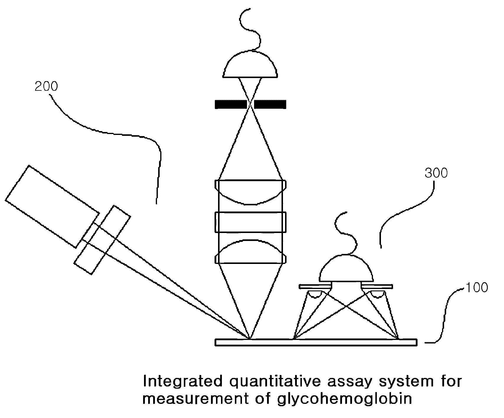
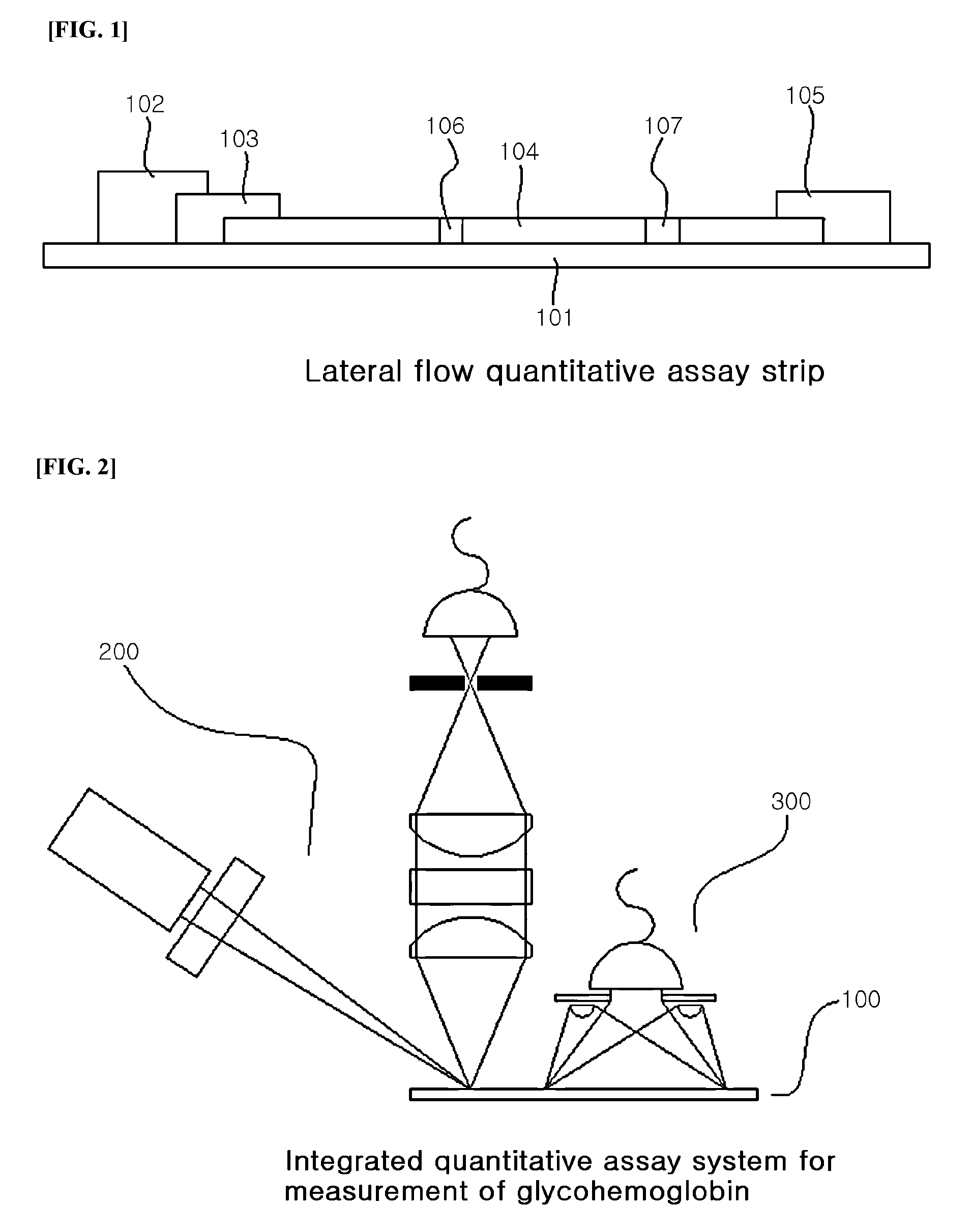
System for quantitative measurement of glycohemoglobin and method for measuring glycohemoglobin

a quantitative measurement and glycohemoglobin technology, applied in the field of quantitative measurement of glycohemoglobin and glycohemoglobin measurement system, can solve the problems of difficult practice, many complications, and inability to reliably diagnose urine glucos

Active Publication Date: 2010-12-23
BODITECHMED
View PDF7 Cites 3 Cited by
  • Summary
  • Abstract
  • Description
  • Claims
  • Application Information

AI Technical Summary

Benefits of technology

The invention provides an assay device for detecting glycohemoglobin and non-glycohemoglobin in a sample. The device includes a pad for receiving the sample and a detector with specificity for glycohemoglobin. The detector is included in the sample in a predetermined amount that is more than sufficient to bind with all the glycohemoglobin. The sample is then allowed to migrate in a chromatographic medium and the detector is captured in a detection zone. The device also includes a first detection zone and a second detection zone that provide access for detecting the non-glycohemoglobin and glycohemoglobin in the sample. The detector may be a fluorescent moiety that emits light in response to light beams irradiated to the detector. The device can be used to detect the amount of glycohemoglobin in a sample by measuring the amount of detector captured in the first detection zone. The method also includes a pre-mixed sample with a known amount of glycohemoglobin and non-glycohemoglobin, and a reference for computing the unknown amount of glycohemoglobin in the sample.

Problems solved by technology

Diabetes mellitus is a disease of abnormal carbohydrate metabolism in which glucose cannot enter the body's cells and be utilized by the body, and therefore remains in the blood in high concentrations to cause many complications.
Diabetes can be diagnosed by measuring urine or blood glucose levels, but urine glucose is not reliable for diagnosis.
These methods are difficult to practice, and thus require advanced skills and equipment.
However, since quantification cannot be performed with the naked eye and hence, an exact amount of an analyte cannot be determined, its application is restricted.
However, this method is disadvantageous in that a specialized analytical laboratory and automated equipment are required, since it comprises several processing steps.
Unfortunately, an exact amount of glycohemoglobin cannot be determined, since the kit is a semiquantitative assay system, in which a dye is added to the antibodies against glycohemoglobin, and color change caused by the presence of HbA1c is detected by the naked eye or by visual comparison against a color chart.

Method used


Image

  • System for quantitative measurement of glycohemoglobin and method for measuring glycohemoglobin
  • System for quantitative measurement of glycohemoglobin and method for measuring glycohemoglobin
  • System for quantitative measurement of glycohemoglobin and method for measuring glycohemoglobin

Examples


example 1

Preparation of Protein-Fluorescent Material Conjugate

[0111]A fluorescent material as a signal generating source was ligated to the mouse monoclonal antibody against an antigen analyte of interest, glycohemoglobin (HbA1c), as follows. Proteins to be used in binding of the fluorescent material were purified to a purity of at least 95%. The proteins were used at a concentration of at least 1 mg / ml for optimal binding. The purified proteins were dialyzed against a buffer solution (0.1 M sodium bicarbonate, pH 8.5) not containing ammonia or amine ions in a refrigerator at 4° C. for 12 to 24 hours in order to facilitate the reaction with the fluorescent material. The proteins dialyzed were kept in a freezer at −20° C. until use. The proteins dialyzed in the buffer solution were directly but slowly added to powdered Alexa 647 fluorescent material (Molecular Probes, USA) and the mixture was stirred for 1 to 2 hours in a refrigerator at 4° C.

example 2

Purification of Protein-Fluorescent Material Conjugate

[0112]Excess fluorescent material that did not react with the protein / fluorescent material conjugate was removed using a distribution column packed with Sephadex G-25. As a purifying buffer solution, 0.1 M sodium carbonate (pH 8.5) was used. The purified protein / fluorescent material conjugates were kept in a refrigerator or -20° C. freezer until use.

example 3

Immobilization of Captor on Nitrocellulose Membrane

[0113]The captor, HbA1c was immobilized on a nitrocellulose membrane in a thin line shape using a Biodot dispenser while varying the concentration and amount. The membrane with immobilized proteins was stored in a dehumidifier kept at 25° C. and a humidity of 35 to 50% for 2 hours. Then, in order to stabilize the protein and prevent non-specific reactions between reagents, the membrane was treated with a stabilizing solution (1% BSA, 0.05% Tween20, 1% sucrose, 0.1% PVA) and equilibrated for 5 minutes. As the components of the stabilizing solution, BSA may be substituted with gelatin, Tween 20 may be substituted with Triton X-100, sucrose may be substituted with trehalose, PVA (polyvinylalcohol) may be substituted with PEG or PVP (polyvinylpyrrolidone). After removing excess solution, the membrane was dried at 40° C. for 30 minutes. The dry membrane was stored in an appropriate container kept at 25° C. and RH of 35 to 50% until use.


PUM

PropertyMeasurementUnit
pHaaaaaaaaaa
concentrationaaaaaaaaaa
pHaaaaaaaaaa

Abstract

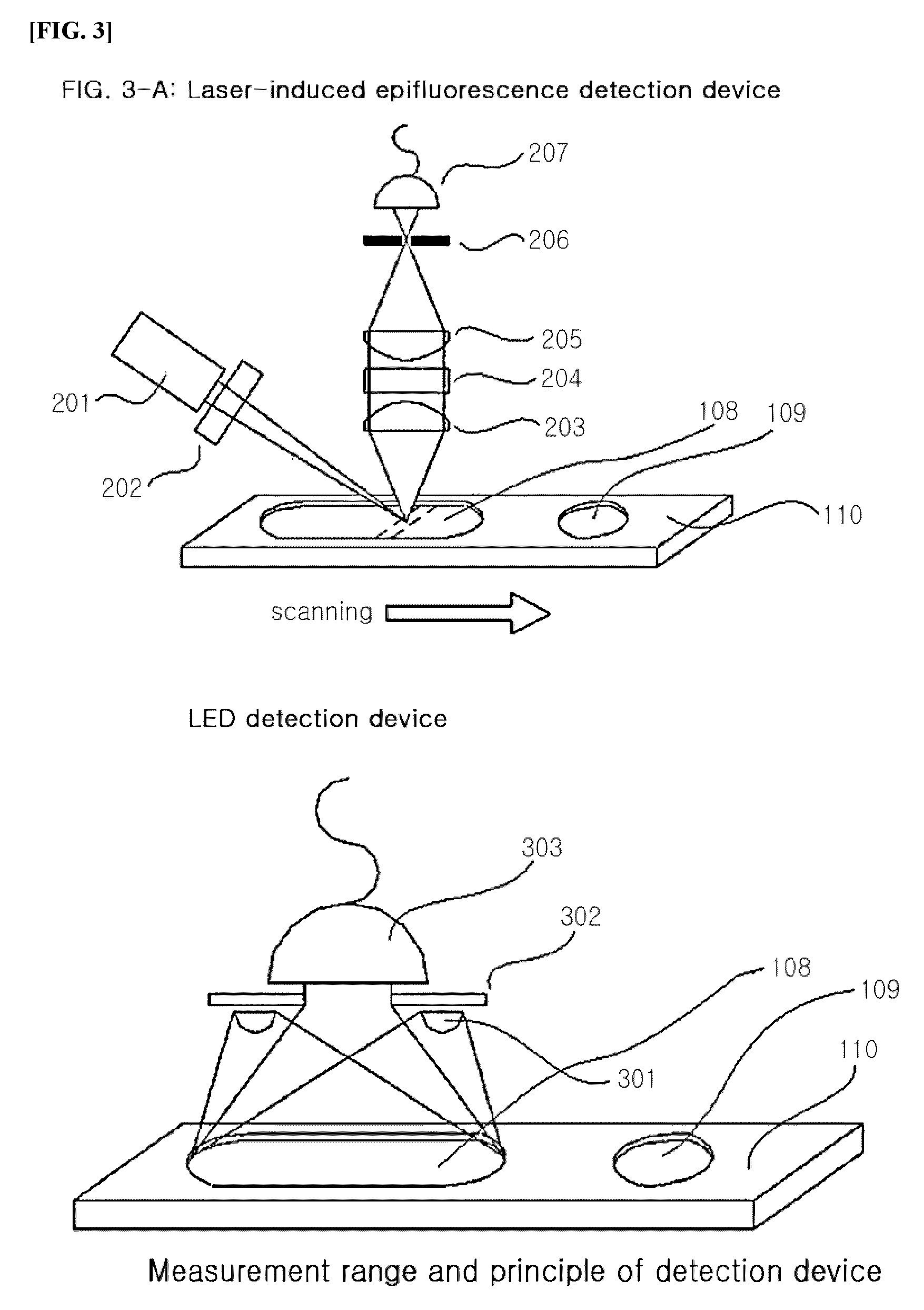
The present invention provides a device for simultaneously detecting hemoglobin and glycohemoglobin in blood, comprising a lateral flow assay strip, a laser-induced epifluorescence detection device, and an LED detection device, and a simple method for simultaneously quantifying hemoglobin and glycohemoglobin in blood by an immunological method using the same.

Description

CROSS-REFERENCE TO RELATED APPLICATIONS[0001]This application is an application under 35 U.S.C. §365(c) of International Application No. PCT / KR2008 / 006306, filed Oct. 24, 2008 designating the United States. The International Application No. PCT / KR2008 / 006306 was published in English as WO2009 / 099269 Al on Aug. 13, 2009. This application incorporates herein by reference the International Application No. PCT / KR2008 / 006306 including the International Publication No. WO2009 / 099269 Al in their entirety.BACKGROUND[0002]1. Field[0003]The present invention relates to a lateral flow quantitative assay system capable of quantifying glycohemoglobin in blood with high sensitivity and a method thereof, in particular, an integrated quantitative assay method and system capable of simultaneously quantifying glycohemoglobin and total hemoglobin.[0004]2. Description of the Related Art[0005]Diabetes mellitus is a disease of abnormal carbohydrate metabolism in which glucose cannot enter the body's cell...

Claims


Application Information

Patent Timeline
Patent Type & Authority Applications(United States)
IPC IPC(8): G01N33/72
CPCG01N33/726G01N33/723
Inventor CHOI, EUI YULNAHM, KIE BONGKIM, JAE HOONJEONG, DONG SEOKPARK, SANG YEOLMOON, JOUNG DAEJUNG, JIN HAKIMJUNG, SO YOUNGPARK, AE KYUNGKIM, BYEONG CHULKIM, SUNG JOONG
Owner BODITECHMED
Learn More