Biodegradable stent formed with polymer-bioceramic nanoparticle composite and method of making the same

Inactive Publication Date: 2011-05-19
DR TIM WU
View PDF0 Cites 19 Cited by
  • Summary
  • Abstract
  • Description
  • Claims
  • Application Information

AI Technical Summary

Benefits of technology

[0021]Preferably, the biodegradable stent made from invented polymeric composite has at least 10% improvement of stent's biocompatibility, material degradation duration and mechanic property increase than that made from no nanoparticle encapsulated polymer only. More preferably, 50% improvement of stent's biocompatibility, mate

Problems solved by technology

The total direct cost for these life-saving procedures is over $2 billion annually.
Despite the prevalent use of DES, there are significant drawbacks, including the need for costly, long-term anti-platelet therapy, as well as the metal artifact remaining in the vessel.
Coronary stents are only required to provide scaffolding for up to six months following the procedure, however, since the stent remains in the vessel, potential long term complications may arise.
In addition, the remaining metal scaffolding precludes the vessel from returning to its natural state and prevents true endothelial repair and arterial remodeling.
Those drawbacks had caused two major issues in current DESs including in-stent restenosis and late stage thrombosis.
ISR has been the biggest problem in PCI until the recently successful development of DESs.
However, ISR in patients with high risk such as small vessels, diabetes, and long diffusion diseased arteries still remains unacceptably high (30%-60% in bare metal stents and 6%-18% in DESs).
The greatest concern, however, has been of stent thrombosis which is associated with a high rate of myocardial i

Method used


Image

  • Biodegradable stent formed with polymer-bioceramic nanoparticle composite and method of making the same
  • Biodegradable stent formed with polymer-bioceramic nanoparticle composite and method of making the same
  • Biodegradable stent formed with polymer-bioceramic nanoparticle composite and method of making the same

Examples


example 1

PLGA / ACP Composite Preparing and Tube Extrusion

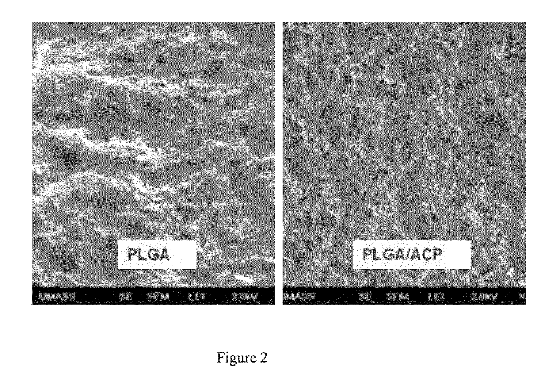
[0092]In the study, all PLGA pellet were first grinded to the particle size of 200 nm with an electrical grinder at 25,000 RPM. 4 grams of ACP (size: 100-150 nm) were mixed with 200 gram grinded PLGA nanoparticle and continuously blended with the same electrical grinder for another ten minutes for uniformly mixing. Both the PLGA and the PLGA / ACP composite mixture were then extruded through a signal screw extruder with a puller at 200 degree C. The extruded tubing has an outside diameter of 1.8 mm and wall thickness of 150 um. Microscopic observation showed that the tube extruded with PLGA only material is clear, colorless, while the tube extruded with PLGA / ACP composite (ACP:PLGA=2:98) is bone-white and the ACP particles were uniformly dispersed among PLGA polymers.

examples 2

Stent Fabrication from PLGA / ACP Composite

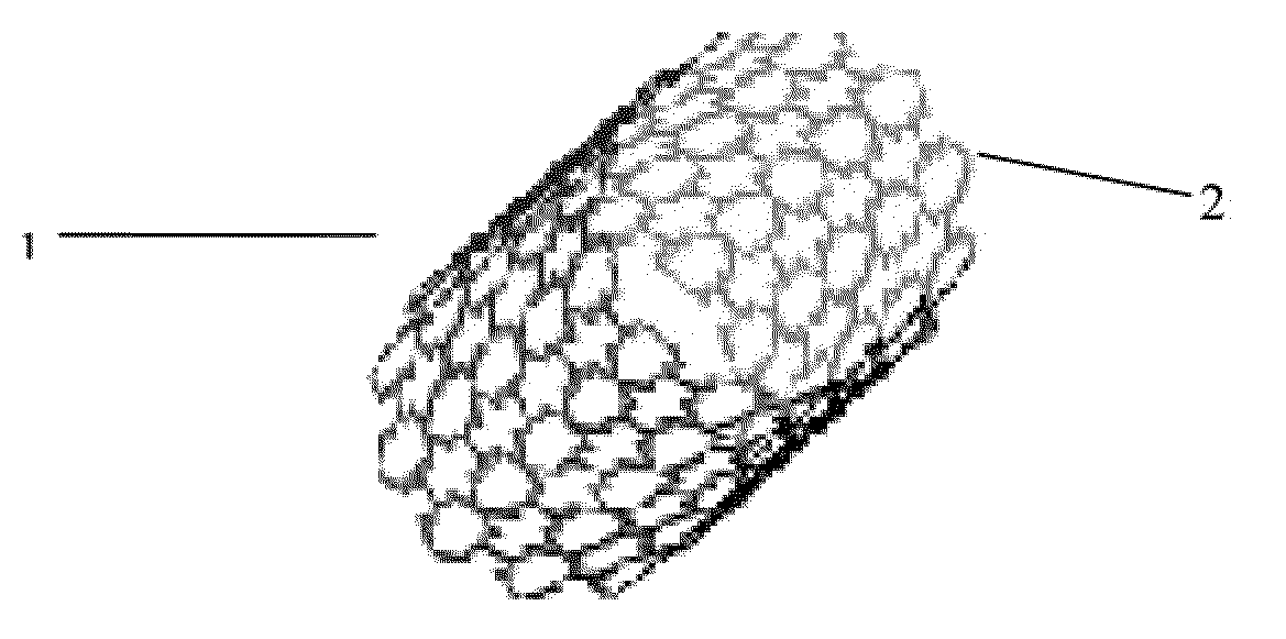
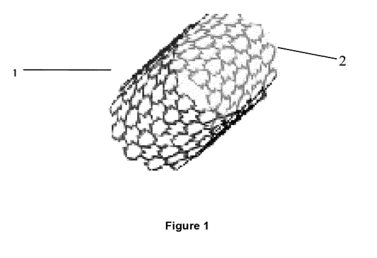
[0093]Tubes extruded from Example 1 study were cut from a femtosecond laser according to design specification. The stent strut thickness is 150 um which is the same as the tube thickness. FIG. 1 is the stent image made from PLGA / ACP composite.

examples 3

Mechanic Property Measurement of Tube Extruded from PLGA / ACP Composite

[0094]Tubes extruded from both PLGA and PLGA / ACP composite using a plastic-extruder during Example 1 study were further subjected for mechanical properties test. In the study, the tensile strength and radial strength of both tubes were measured with a catheter tensile / radial strength testing machine (Model 4400R, Instron, Inc. Nonvood, Mass.). As shown in FIG. 6, the tube made from PLGA / ACP composite has a significantly higher maximum tensile-load-at-break (A, 96.29±2.15N vs. 71.11±3.21N, n=6, P<0.001) and maximum radial-strength (load)-at-crush (B, 470±3.20N vs. 400±2.09N, N=6, P<0.001) than that in the PLGA only tube.


PUM

PropertyMeasurementUnit
Timeaaaaaaaaaa
Adhesion strengthaaaaaaaaaa
Mechanical propertiesaaaaaaaaaa

Abstract

The present invention relates to biodegradable medical devices such as stents manufactured from biodegradable polymeric-bioceramic nanoparticle composites. The invented medical devices include at least one bioceramic nanoparticle dispersed in at least one biodegradable polymer, wherein the said biodegradable polymers include biodegradable polyesters. The device and methods to disperse one or more bioceramic nanoparticle, wherein the said bioceramic nanoparticle include, but are not limited to, amorphous calcium phosphate (ACP), dicalcium phosphate (DCP), tricalcium phosphate (TCP), pentacalcium hydroxyl Apatite (HAp), tetracalcium phosphate monoxide (TTCP) and combinations or analogues thereof. Other embodiments include methods of fabricating biodegradable stent with said polymeric-nanoparticle composites.

Description

CROSS-REFERENCE TO RELATED APPLICATIONS[0001]This application is a continuation-in-part of the U.S. patent application Ser. No. 11 / 843,528, filed on Aug. 22, 2007, which claims the benefit of U.S. provisional patent application No. 60 / 823,168, filed on Aug., 22, 2006. This application is also a continuation-in-part of the U.S. patent application Ser. No. 12 / 209,104, filed on Sep. 11, 2008, which claims the benefit of U.S. provisional patent application No. 60 / 578,219, filed on Jun. 8, 2004. This application also claims the benefit of the U.S. provisional application No. 61 / 368,833, filed on Jul. 29, 2010 and U.S. provisional patent application No. 61 / 427,141 filed on Dec. 24, 2010. The disclosures of all of which are hereby incorporated by reference in their entireties.FIELD OF THE INVENTION[0002]The present invention relates to a biodegradable stent comprising at least one bioceramic nanoparticle encapsulated inside at least one biodegradable polymer wherein the encapsulated biocer...

Claims


Application Information

Patent Timeline
IPC IPC(8): A61F2/82B29C47/20B29C48/32B82Y5/00
CPCA61B17/06166A61F2/82A61B2017/00004A61F2/0077A61F2/28A61F2/30767A61F2/3094A61F2002/30062A61F2002/30064A61F2002/30677A61F2210/0004A61F2240/001A61F2250/0067A61F2310/00011A61F2310/00293A61F2310/0097A61L27/46A61L27/58A61L31/127A61L31/148A61L2400/12A61B17/86C08L67/04
Inventor WU, TIM
Owner DR TIM WU
Why Eureka
Try Eureka