Method for identifying individual viruses in a sample

Inactive Publication Date: 2011-07-07
FRIEDRICH SCHILLER UNIV JENA
View PDF0 Cites 5 Cited by
  • Summary
  • Abstract
  • Description
  • Claims
  • Application Information

AI Technical Summary

Benefits of technology

[0015]The object of the invention is to identify individual viruses in a solid, liquid or gaseous sample quickly, unambiguously and reliably, and with the least possible preparation-related and technology-related expenditure, without necessitating immobilization by using antibodies and without requiring an indication or at least a suspicion of potentially present viruses.
[0016]The method of the invention has been developed to detect individual viruses in any type (solid, liquid or gaseous) of sample and to identify the specific type of these viruses or bacteriophages without time-consuming and material-intensive preparations. Thanks to the invention it will be possible to get reliable information about the type and composition of the virus particles in a sample thus allowing the exact and unambiguous identification of these particles. This method can be universally applied to all viruses independently of the cells that are attacked by the viruses and the type of the individual virus. As the method can determine the viruses regardless of their origin it can also be used in other fields of application (e.g. for the detection of tobacco mosaic viruses in plants, for the detection of virus particles in the air, or for the detection of viruses and bacteriophages in biotechnological production). The term “virus” as used herein includes all viruses, that is, bacteriophages or “phages” as well as all other viruses. Therefore, when “viruses” or a “virus” is referred to herein without specific mention of “bacteriophages” or “phages”, it is always intended that bacteriophages or phages be included as well.
[0020]Compared to the conventional methods described above, the inventive method offers many advantages: It does not require expensive molecular-biological reagents and allows an unambiguous and comparatively quick identification of viruses and even individual viruses, and the time-expensive and complex pre-cultivation and sample preparation are not necessary any longer. Thus, this invention is considerably more exact and reliable and requires less time and effort than the methods of virus detection mentioned before. Moreover, even individual viruses are not only analyzed by their shape as described but additionally by vibrational spectroscopy as recommended. Apart from data about size and structure, exact information about the chemical composition of the analyzed particles is also gathered in this method so that it allows the type-specific and unambiguous determination of these viruses for the first time.
[0021]Unlike immunological or PCR virus detection methods, the inventive method does not use molecular-biological detection reactions so that the complex sample pre-treatment and preparation of these molecular-biological processes is not necessary. Furthermore, the frequently high costs of the primers, antibodies, enzymes and other reagents required for these detection methods are saved.
[0022]Contrary to the just imaging procedures (AFM, TEM) the user can obtain very detailed information about the material composition of the scanned sample for high detection sensitivity by coupling an imaging technique with a vibrational spectroscopic method thus also allowing a very detailed structure comparison with reference data and thus an extremely precise and unambiguous virus identification and determination.
[0024]According to this invention even the detection of individual viruses is possible so that a minimum concentration is not required for the reliable identification and determination and the corresponding pre-cultivations can be omitted. Only sizing by pre-treatment of the sample, for example by filtration or homogenization, is an advantageous sample preparation for the application of the invention in order to separate the large particles from the viruses and thus to purify the sample (i.e., increase the concentration of viruses in the sample) and simplify the analysis, i.e. simplify the selection of the scanning sites on the basis of the height profile of the carrier surface as well as the virus detection and determination. For this purpose, for example, a filter array with decreasing pore size is used to filter the sample. Then, the viruses in enriched form are located on a filter with the corresponding pore size and are separated from larger particles such as dust or bacteria. The sample which is filtered may be, for example a liquid or a gas. The sample may be filtered directly or, for example, a gaseous sample may first be dissolved in a liquid and the liquid solution be filtered. An example of sizing by homogenization of the sample is mechanical comminution of a solid sample.

Problems solved by technology

But this detection requires a lot of time and only allows a general virus test but not the exact determination of the virus type.
It may be that the selected test conditions are not optimal for the viral proliferation or even inhibit it or the virus is in a so called lysogenic cycle and the DNA of the virus or phage is integrated in the DNA of the host cell so that a lytic scheme will not be developed although viruses have really attacked the cell and then that virus attack cannot be detected or determined in this way.
But it is a disadvantage of these methods that the detection of individual viruses is very difficult, if not possible at all.
This cultivation requires much time and effort, too.
This sample preparation is very time and labor consuming.
For the PCR, expensive reagents and specific primers must be added to the isolated hereditary material.
That means that if the PCR preparation contains the wrong primers the amplification of DNA does not occur and the viruses cannot be detected.
But slight changes in the hereditary material of the viruses can prevent that the primers bind to the DNA and thus the DNA does not amplify either.
But viruses in low concentrations can only be detected after an incubation and amplification.
If antibodies do not exist or wrong antibodies are used, the virus detection will not be possible.
Furthermore, the production of the antibodies requires much time and effort and an additional immobilization step is necessary in which either the viruses or the antibodies are bound to a solid substrate.
Moreover, this method requires much apparatus-related and preparation-related expenditure.
But the obtained data can be easily misinterpreted (false-positive results), in particular if the sample is contaminated with particles of other origin that have a similar size and shape and therefore cause confusion.
A serious disadvantage of all imaging techniques is the fact that information about the composition of the imaged particles cannot be obtained.
Thus, an assignment and determination of the particles is only realized via their shape and size so that confusion and consequently misinterpretations are particularly caused by spherical viruses.
But the Raman effect on which this technique is based is very weak and thus the Raman spectroscopy method can only be used for bulk material (G. J. Thomas Jr.
The determination of individual viruses is impossible in this method.
However, this method cannot be employed in routine diagnostics because the enhancement of the Raman effect considerably varies in dependence on the nano-structures and -particles used and thus a reliable detection is not possible.
Furthermore, this method does not allow the identification of individual viruses either (S. Shanmukh, L. Jones, J. Driskell, Y. Zhao, R. Dluhy, R. A. Tripp: Rapid and Sensitive Detection of Respiratory Virus Molecular Signatures Using a Silver Nanorod Array SERS Substrate, Nano Letters 2006, 6, (11), 2630-2636).
Therefore, these tests have not shown encouraging results either.
The detected TERS spectra are not reproducible among each other, a fact that is indicates a mobility in the outer cell wall so that experts consider this method unsuitable for identifying bacteria (U. Neugebauer, P. Rösch, M. Schmitt, J. Popp, C. Julien, A. Rasmussen, C. Budich, V. Deckert: On the Way to Nanometer-Sized Information of the Bacterial Surface by Tip-Enhanced Raman Spectroscopy, ChemPhysChem 2006, 7, 1428-1430).

Method used


Image

  • Method for identifying individual viruses in a sample
  • Method for identifying individual viruses in a sample

Examples


embodiment 1

Detection of Tobacco Mosaic Virus (TMV) in a Plant Sample

[0029]The TMV leads to the economically important mosaic disease of tobacco, but it can also infest other plant families. The virus is particularly stable and can be easily transmitted, e.g. by direct contact between the plants, by plant sap, in some plants by seed and most of all by agricultural methods for handling infected plants.

[0030]To avoid more serious economic damage an exact and quick identification of the virus is necessary.

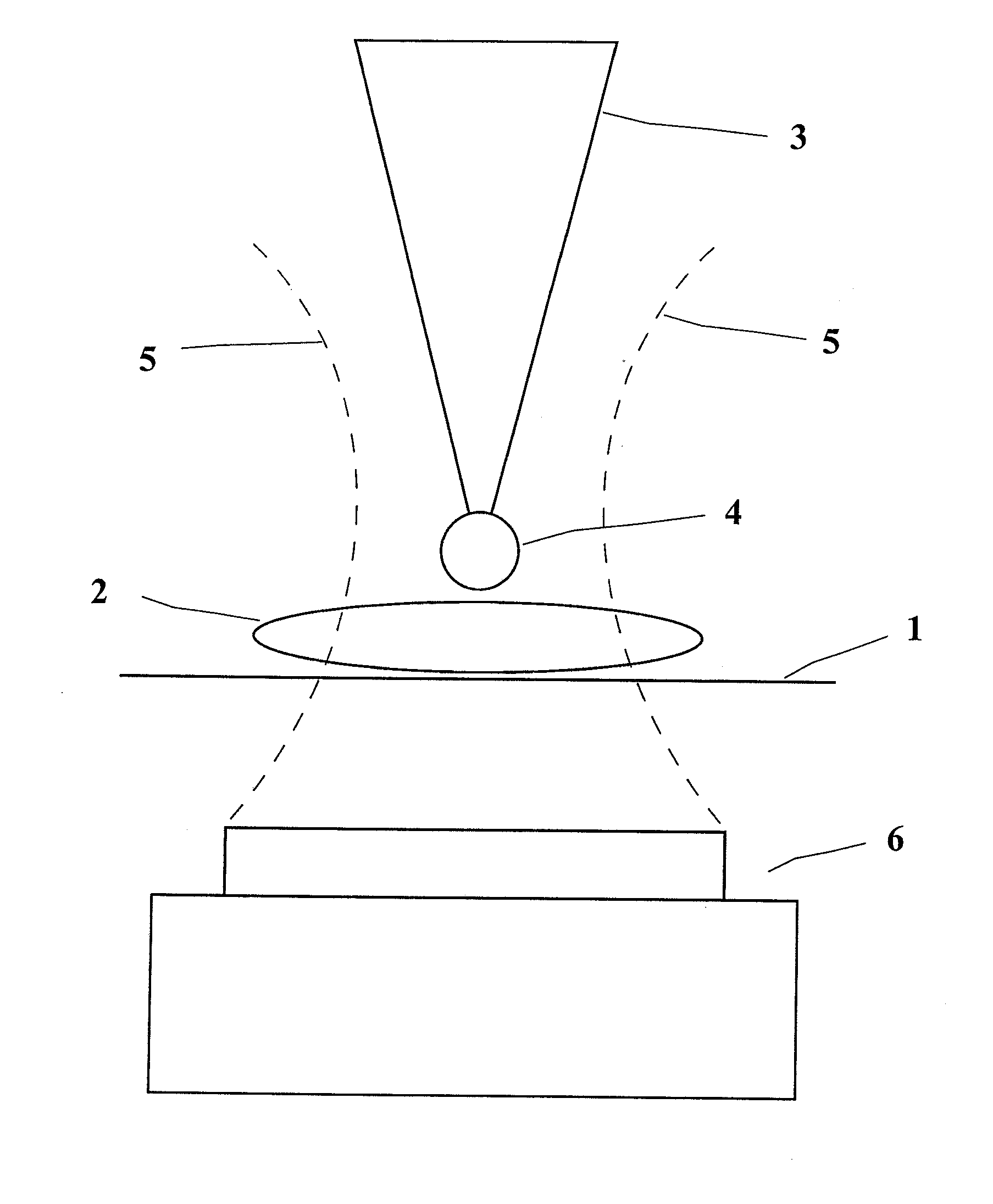
[0031]For doing this, either pressed plant juice or plant parts (leaves, buds, fruit, trunks, stalks, roots, or similar parts) are used. If plant parts are used, they are lysed mechanically or chemically in a suitable buffer in the first step to release the viruses from the cell structure. Then, the obtained liquid is guided (like the pressed plant juice) through a filter array with decreasing pore size. Afterwards only the filters that catch the virus particles with a size from 15 nm to 400 nm a...

embodiment 2

Detection of the Foot-and-Mouth-Disease Virus

[0032]Foot-and-mouth disease is a highly contagious and compulsorily notifiable disease of cattle and pigs; but goats, sheep and other even-toed ungulates can also be infected. Infections of elephants, hedgehogs, rats and of men are described in the literature, too.

[0033]For example, aphtha liquid, organ homogenates, pharynx mucus samples (probang sample), secretions and cell culture supernatants can be used for identifying the virus. They are transferred to a suitable lysis buffer which leads to release of the viruses from the cells. Afterwards, the liquid obtained in this way is guided through the filter array mentioned in embodiment 1 and the filters of interest are analyzed as described.

embodiment 3

Detection of Influenza Viruses in Air Samples

[0034]In humans, influenza is caused by the influenza virus of type A or B. The infection is often a result of a so called droplet or smear infection. The droplet infection is the medical term for the direct inhalation of expiration droplets (exhalation droplets) of infected persons.

[0035]Contact infection or smear infection with the viruses is caused by highly infectious expiration droplets that have fallen on objects or body surfaces or it is caused, for example, by smeared nasal secretion.

[0036]To identify viruses in an air sample a pre-defined volume of air is filtered in the already described filter array (cf. embodiment 1). Then, the filters are analyzed by means of the method explained above.


PUM


Abstract

Individual viruses in any type of sample are identified quickly, unambiguously and reliably, and with the least possible preparation-related and technology-related expenditure, without necessitating immobilization using antibodies and without requiring an indication or at least a suspicion of potentially present viruses. This is accomplished by scanning the height profile of the sample, from which scanning sites suspected of containing viruses are selected, exposing those cites to monochromatic excitation light and spectroscopically analyzing the resulting Raman scattered light.

Description

BACKGROUND OF THE INVENTION[0001]The invention relates to a method for identifying individual viruses in a sample quickly and reliably and with the least possible effort and expenditure.[0002]Infection tests can be considered the oldest methods applied for the detection of virus contaminations. In these tests, bacteria and cell cultures are infected with potentially virus-containing material. After a defined time of incubation the virus infection can be shown by a visually detectable change (lytic scheme) in the cell or bacteria culture. But this detection requires a lot of time and only allows a general virus test but not the exact determination of the virus type. Moreover, a virus attack does not always result in the lysis of the infected host cells. It may be that the selected test conditions are not optimal for the viral proliferation or even inhibit it or the virus is in a so called lysogenic cycle and the DNA of the virus or phage is integrated in the DNA of the host cell so t...

Claims


Application Information

Patent Timeline
IPC IPC(8): C12Q1/70
CPCB82Y15/00B82Y35/00G01N1/286G01N21/658G01N33/48G01N2001/4088H01J2237/2814G01Q60/40H01J37/28H01J2237/24578H01J2237/2583H01J2237/2802G01Q30/02
Inventor POPP, JURGENDECKERT, VOLKERNAUMANN, DIETERMOLLER, ROBERTCIALLA, DANA
Owner FRIEDRICH SCHILLER UNIV JENA
Why Eureka
Try Eureka