Pacap as a marker for cancer

a technology of pacap and cancer, applied in the field of pacap as a cancer marker, can solve the problems of increasing the concentration of pacap protein and/or fragments in the test sample, and is associated with the occurrence of cancer

Inactive Publication Date: 2011-09-01
ROCHE DIAGNOSTICS OPERATIONS INC
View PDF2 Cites 21 Cited by
  • Summary
  • Abstract
  • Description
  • Claims
  • Application Information

AI Technical Summary

Benefits of technology

[0072]In a preferred embodiment the present invention relates to a method for assessing cancer in vitro comprising measuring in a sample the concentration of a) a PACAP protein and / or fragments thereof, b) optionally one or more other marker of cancer, and (c) using the measurement results of step (a) and optionally of step (b) in the assessment of cancer, wherein an increased concentration of PACAP protein and / or fragments thereof is indicative for cancer.

Problems solved by technology

Surprisingly, it was found that an increased concentration of a PACAP protein and / or fragments thereof in the test sample is associated with the occurrence of cancer.

Method used


Image

  • Pacap as a marker for cancer
  • Pacap as a marker for cancer
  • Pacap as a marker for cancer

Examples


example 1

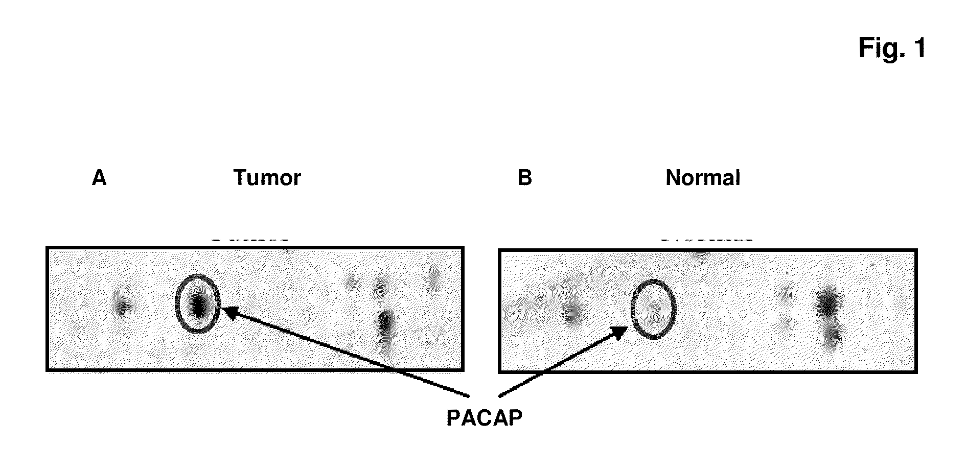
Identification of PA CAP as a Marker for Lung Cancer

[0176]Sources of Tissue:

[0177]In order to identify tumor-specific proteins as diagnostic markers for lung cancer, analysis of two different kinds of tissue using proteomics methods is performed.

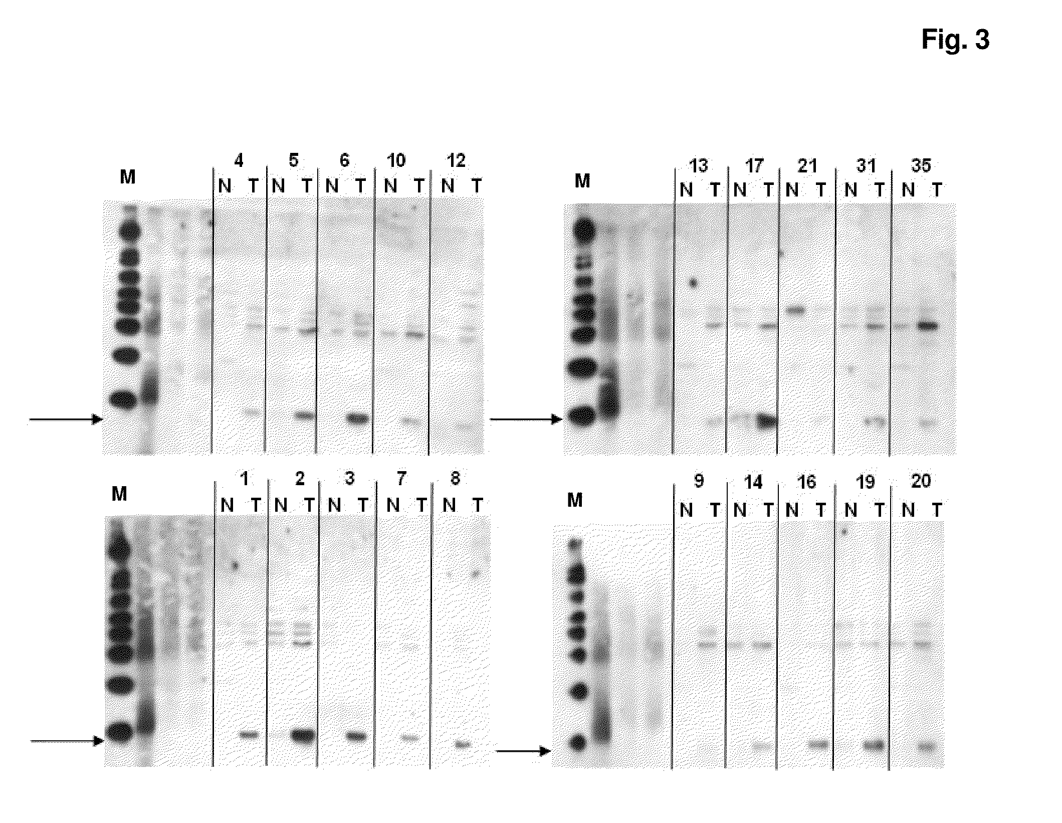
[0178]In total, tissue specimen from 20 patients suffering from lung cancer (10 adeno-CA and 10 squamous cell-CA) are analyzed. From each patient two different tissue types are collected from therapeutic resections: tumor tissue (>80% tumor) (T) and adjacent healthy tissue (N). The latter one serves as matched healthy control samples. Tissues are immediately snap frozen after resection and stored at −80° C. before processing. Tumors are diagnosed by histopathological criteria.

[0179]Tissue Preparation:

[0180]0.8-1.2 g of frozen tissue are cut into small pieces, transferred to the chilled grinding jar of a mixer ball mill and completely frozen by liquid nitrogen. The tissue is pulverized in the ball mill, dissolved in the 10-fold volume (w / v) o...

example 2

Generation of Antibodies to the Lung Cancer Marker Protein PACAP

[0188]Polyclonal antibody to the lung cancer marker protein PACAP is generated for further use of the antibody in the measurement of serum and plasma and blood levels of PACAP by immunodetection assays, e.g. Western Blotting and ELISA.

[0189]Recombinant Protein Expression in E. coli:

[0190]In order to generate antibodies against PACAP, the recombinant antigen is produced in E. coli: Therefore, the PACAP coding region is PCR amplified from the full-length cDNA clone IRALp962G1939Q obtained from the German Resource Center for Genome Research (RZPD, Berlin, Germany) using the primers:

Forward primer LC13Bfor-EcoRI:(SEQ ID NO: 2 / EcoRI-start codon underlined)5′ aCGTACGTgaattcattaaagaggagaaattaact atgagaggatcgcatcaccatcaccatcacattgaaggccgtagg_ctgtcactgccactgctgc,Reverse primer LC13Brev-HindIII:(SEQ ID NO: 3)5′ acgtacgtaa gctttcatta gagctcttct cttgtggctg.

[0191]The forward primer features (besides the EcoRI cloning and ribosomal ...

example 3

ELISA for the Measurement of PACAP in Human Serum and Plasma Samples

[0204]For detection of PACAP in human serum or plasma, a sandwich ELISA is developed. For capture and detection of the antigen, aliquots of the anti-PACAP polyclonal antibody (see Example 2) are conjugated with biotin and digoxigenin, respectively.

[0205]Streptavidin-coated 96-well microtiter plates are incubated with 100 μl biotinylated anti-PACAP polyclonal antibody for 60 min at 10 μg / ml in 10 mM phosphate, pH 7.4, 1% BSA, 0.9% NaCl and 0.1% TWEEN 20 (ICI Americas Inc.). After incubation, plates are washed three times with 0.9% NaCl, 0.1% TWEEN 20. Wells are then incubated for 2 h with either a serial dilution of the recombinant protein (see Example 2) as standard antigen or with diluted plasma samples from patients. After binding of PACAP, plates are washed three times with 0.9% NaCl, 0.1% TWEEN 20. For specific detection of bound PACAP, wells are incubated with 100 μl of digoxygenylated anti-PACAP polyclonal ant...


PUM

PropertyMeasurementUnit
molecular massaaaaaaaaaa
volumeaaaaaaaaaa
pHaaaaaaaaaa

Abstract

The present invention relates to a method aiding in the assessment of cancer. It discloses the use of the proapoptotic caspase adaptor protein (=PACAP) as a universal marker of different types of cancer. PACAP aids in the assessment of pulmonary or lung cancer (LC), particularly of non-small cell lung carcinoma (NSCLC), but also of other specific types of cancer. Such specific types of cancer are e.g. colon, bladder, cervix, ovary, endometrial, head and neck, breast, melanoma, pancreas, kidney, prostate, esophagus, stomach or bile duct cancer. Furthermore, the present invention especially relates to a method for assessing cancer from a liquid sample, derived from an individual by measuring PACAP in said sample. Measurement of PACAP can, e.g., be used in the early detection of cancer or in the surveillance of patients who undergo surgery.

Description

RELATED APPLICATIONS [0001]This application is a continuation of PCT / EP2009 / 008006 filed Nov. 10, 2009 and claims priority to EP 08019731.2 filed Nov. 12, 2008.SEQUENCE LISTING [0002]The instant application contains a Sequence Listing which has been submitted in ASCII format via EFS-Web and is hereby incorporated by reference in its entirety. Said ASCII copy, created on Apr. 29, 2011, is named 25485US.txt, and is 4,540 bytes in size.DESCRIPTION [0003]The present invention relates to a method aiding in the assessment of cancer. It discloses the use of the proapoptotic caspase adaptor protein (=PACAP) as a universal marker of different types of cancer. PACAP aids in the assessment of pulmonary or lung cancer (LC), particularly of non-small cell lung carcinoma (NSCLC), but also of other specific types of cancer. Such specific types of cancer are e.g. colon, bladder, cervix, ovary, endometrial, head and neck, breast, melanoma, pancreas, kidney, prostate, esophagus, stomach or bile duct ...

Claims


Application Information

Patent Timeline
Patent Type & Authority Applications(United States)
IPC IPC(8): G01N33/68C12Q1/527C12Q1/02
CPCG01N33/57423G01N33/57407
Inventor HAGMANN, MARIE-LUISEKARL, JOHANNKLOECKNER, JULIAROESSLER, MARKUSTACKE, MICHAEL
Owner ROCHE DIAGNOSTICS OPERATIONS INC
Why Eureka
Try Eureka